[Preprint]. 2024 Dec 4:2024.12.04.626854.
doi: 10.1101/2024.12.04.626854.

Deep mutationally scanned (DMS) CHIKV E3/E2 virus library maps viral amino acid preferences and predicts viral escape mutants of neutralizing CHIKV antibodies

Affiliations

Deep mutationally scanned (DMS) CHIKV E3/E2 virus library maps viral amino acid preferences and predicts viral escape mutants of neutralizing CHIKV antibodies

Megan M Stumpf et al. bioRxiv. .

Update in

Abstract

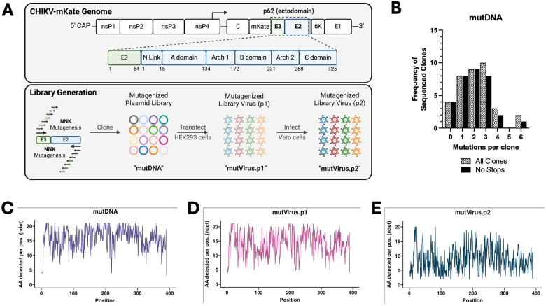
As outbreaks of chikungunya virus (CHIKV), a mosquito-borne alphavirus, continue to present public health challenges, additional research is needed to generate protective and safe vaccines and effective therapeutics. Prior research has established a role for antibodies in mediating protection against CHIKV infection, and the early appearance of CHIKV-specific IgG or IgG neutralizing antibodies protects against progression to chronic CHIKV disease in humans. However, the importance of epitope specificity for these protective antibodies and how skewed responses contribute to development of acute and chronic CHIKV-associated joint disease remains poorly understood. Here, we describe the deep mutational scanning of one of the dominant targets of neutralizing antibodies during CHIKV infection, the E3/E2 (also known as p62) glycoprotein complex, to simultaneously test thousands of p62 mutants against selective pressures of interest in a high throughput manner. Characterization of the virus library revealed achievement of high diversity while also selecting out non-functional virus variants. Furthermore, this study provides evidence that this virus library system can comprehensively map sites critical for the neutralization function of antibodies of both known and unknown p62 domain specificities.


Figures

Figure 1.
Figure 1.. Generation of a deep mutationally scanned CHIKV p62 full-length virus library.
(A) Schematic of the CHIKV genome (box indicates mutagenized region), procedure for generation of the mutagenized libraries, and naming scheme for each of the library iterations. (B) Sangar sequencing results for individual plasmid DNA clones. The lined black bars represent the number of nonsynonymous mutations per full-length CHIKV p62 clone (Sangar primers available in Materials and Methods). The solid black bars exclude any sequences containing stop codons. Average number of mutations per clone for the lined and solid black bars are 2.2 and 2.0, respectively. Distribution of these mutations across the CHIKV p62 region are detailed in Figure S1C. (C-E) Following deep sequencing, total number of detected amino acids per codon position (‘ndet’), for the (C) mutDNA, (D) mutVirus.p1, and (E) mutVirus.p2 libraries are plotted for the entire mutagenized CHIKV p62 region. Results for the wtDNA sequencing control are shown in Figure S1D.
Figure 2.
Figure 2.. Deep mutational scanning of CHIKV p62 reveals mutational tolerance of the different p62 domains.
(A) Logoplot showing the diversity of amino acids per codon position for the mutVirus.p2 virus library. WT residues are shown above each codon position and colored bars for each p62 domain below (E3: light blue, E2 N Link: light yellow, E2 A domain: light red, E2 Arch 1: light green, E2 B domain: light orange, E2 Arch 2: light purple, E2 C domain: light brown). Size of each letter is normalized to the number of amino acids detected for that codon position. (B) Using dms-viz (61), heatmaps were generated for the trimeric E2/E1 CHIKV envelope glycoproteins cryo-em structure (PDB: 3J2W) and the mature envelope glycoprotein complex (p62/E1; PDB: 3N42). For the trimeric structure, the heatmap represents E2 diversity from a top-down view. For the p62/E1 complex, the heatmap represents E3 diversity from a side view (with E2 colored in blue to highlight E3). E1 is shown in gray. (C) Violin plots showing the relative diversity at each site of each mutagenized domain are plotted. Colors are matched to colors shown in logoplot annotations in Panel 2A. *The E2 C domain region only includes the ectodomain portion of the C domain. One-way ANOVA with Tukey’s test for multiple comparisons. ** p< 0.01, *** p< 0.001.
Figure 3.
Figure 3.. Escape mutant profile for CHK-152 monoclonal antibody reveals modest escape from selected panel of positive selection mutants.
(A) Total site positive differential selection scores for CHK-152 were plotted via heatmap on the trimeric E2/E1 CHIKV envelope glycoproteins (PDB: 3J2W) and the mature envelope glycoprotein complex (p62/E1; PDB: 3N42). For the trimeric structure, the heatmap represents E2 positive site selection from a top-down view. For the p62/E1 complex, the heatmap represents E3 positive site selection from a side view (with E2 colored in blue to highlight E3). E1 is shown in gray for both structures. (B) Sites selected for validation of their sensitivity to neutralization by CHK-152. (C) FRNT curves for CHK-152 against WT CHIKV and the indicated mutant virus. Dotted line represents the FRNT50 threshold. (D) PRNT curve for CHK-152 against WT and E2 N219P CHIKV. Dotted line represents the PRNT50 threshold.
Figure 4.
Figure 4.. Escape mutant profile for CHK-265 monoclonal antibody reveals modest escape from selected panel of positive selection mutants.
(A) Total site positive differential selection scores for CHK-265 were plotted via heatmap on the trimeric E2/E1 CHIKV envelope glycoproteins (PDB: 3J2W) and the mature envelope glycoprotein complex (p62/E1; PDB: 3N42). For the trimeric structure, the heatmap represents E2 positive site selection from a top-down view. For the p62/E1 complex, the heatmap represents E3 positive site selection from a side view (with E2 colored in blue to highlight E3). E1 is shown in gray for both structures. (B) Sites selected for validation of their sensitivity to neutralization by CHK-265. (C) FRNT curves for CHK-265 against WT CHIKV and the indicated mutant virus. Dotted line represents the FRNT50 threshold. (D) PRNT curve for CHK-265 against WT and E2 N219P CHIKV. Dotted line represents the PRNT50 threshold.
Figure 5.
Figure 5.. Escape mutant profile for CHK-11 monoclonal antibody reveals modest escape from selected panel of positive selection mutants.
(A) Total site positive differential selection scores for CHK-11 were plotted via heatmap on the trimeric E2/E1 CHIKV envelope glycoproteins (PDB: 3J2W) and the mature envelope glycoprotein complex (p62/E1; PDB: 3N42). For the trimeric structure, the heatmap represents E2 positive site selection from a top-down view. For the p62/E1 complex, the heatmap represents E3 positive site selection from a side view (with E2 colored in blue to highlight E3). E1 is shown in gray for both structures. (B) Sites selected for validation of their sensitivity to neutralization by CHK-11. (C) FRNT curves for CHK-11 against WT CHIKV and the indicated mutant virus. Dotted line represents the FRNT50 threshold. (D) PRNT curve for CHK-11 against WT and E2 N219P CHIKV. Dotted line represents the PRNT50 threshold.
Figure 6.
Figure 6.. Validation of CHK-11 and CHK-265 similarity.
(A) Competition ELISA against CHIKV virions (strain 181/25) with competitor antibodies added prior to the addition of biotinylated CHK-11. Percent binding (Absorbance @ 450 nm) relative to BT-CHK-11 alone (no competitor) was determined. (B) PRNT curves against CHIKV, Ross River virus (RRV), o’nyong’nyong virus (ONNV), and Mayaro virus (MAYV) on Vero cells. DENV E51 was included as an isotype, neutralizing antibody negative control. Dotted line represents the PRNT50 threshold. (C) Calculation of the PRNT50 in μg/mL for each virus-antibody condition, ordered by relatedness to CHIKV. (D) Correlation of shared positively selected mutants between CHK-11, CHK-152, and CHK-265 was evaluated to compare mutation effect sizes (log2 differential selection score) using linear regression in R v4.2.1. Top ten mutants for each mAb pair are colored blue.

References

    1. Borgherini G, Poubeau P, Jossaume A, Gouix A, Cotte L, Michault A, Arvin-Berod C, Paganin F. 2008. persistent arthralgia associated with chikungunya virus: a study of 88 adult patients on Reunion Island. Clin Infect Dis 47:469–475. - PubMed
    1. Couturier E, Guillemin F, Mura M, Léon L, Virion J-M, Letort M-J, De Valk H, Simon F, Vaillant V. 2012. Impaired quality of life after chikungunya virus infection: a 2-year follow-up study. Rheumatology 51:1315–1322. - PubMed
    1. Gérardin P, Fianu A, Malvy D, Mussard C, Boussaïd K, Rollot O, Michault A, Gaüzere B-A, Bréart G, Favier F. 2011. Perceived morbidity and community burden after a chikungunya outbreak: the TELECHIK survey, a population-based cohort study. BMC Med 9:5. - PMC - PubMed
    1. Schilte C, Staikovsky F, Couderc T, Madec Y, Carpentier F, Kassab S, Albert ML, Lecuit M, Michault A. 2013. Chikungunya virus-associated long-term arthralgia: a 36-month prospective longitudinal study. PLoS Negl Trop Dis 7:e2137. - PMC - PubMed
    1. Sissoko D, Malvy D, Ezzedine K, Renault P, Moscetti F, Ledrans M, Pierre V. 2009. Post-Epidemic Chikungunya disease on Reunion Island: course of rheumatic manifestations and associated factors over a 15-month period. PLoS Negl Trop Dis 3:e389. - PMC - PubMed

Publication types

LinkOut - more resources