[Preprint]. 2024 Dec 4:2024.12.03.626538.
doi: 10.1101/2024.12.03.626538.

Trehalose supports the growth of Aedes aegypti cells and modifies gene expression and dengue virus replication

Affiliations

Trehalose supports the growth of Aedes aegypti cells and modifies gene expression and dengue virus replication

Andrew D Marten et al. bioRxiv. .

Update in

Abstract

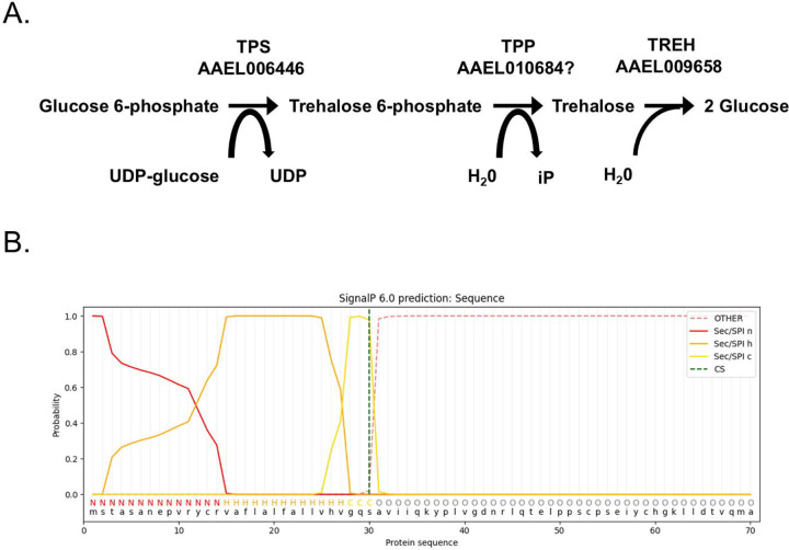
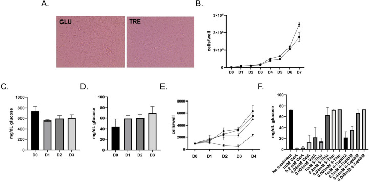
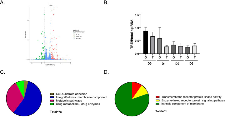
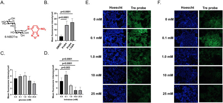
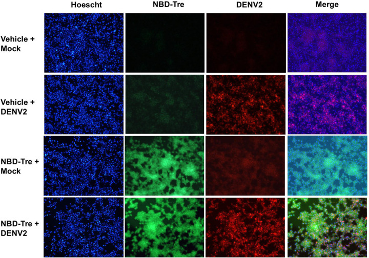
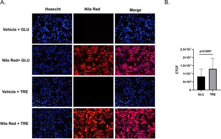
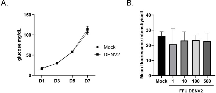
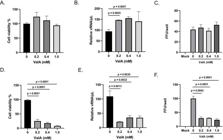
Trehalose is a non-reducing disaccharide that is the major sugar found in insect hemolymph fluid. Trehalose provides energy, and promotes growth, metamorphosis, stress recovery, chitin synthesis, and insect flight. Trehalase is the only enzyme responsible for the hydrolysis of trehalose, which makes it an attractive molecular target. Here we show that Aedes aegypti (Aag2) cells express trehalase and that they can grow on trehalose-containing cell culture media. Trehalase activity was confirmed by treating Aag2 cells with trehalase inhibitors, which inhibited conversion of trehalose to glucose and reduced cell proliferation. Cell entry of a fluorescent trehalose probe was dependent on trehalose concentration, suggesting that trehalose moves across the cell membrane via passive transport. Culturing Aag2 cells with trehalose-containing cell culture media led to significant changes in gene expression, intracellular lipids, and dengue virus replication and specific infectivity, and increased their susceptibility to trehalase inhibitors. These data describe an in vitro model that can be used to rapidly screen novel trehalase inhibitors and probes and underscores the importance of trehalose metabolism in Ae. aegypti physiology and transmission of a mosquito-borne virus.


Figures

Figure 1.
Figure 1.. Trehalose metabolism genes expressed in Aag2 cells.
(A) Trehalose pathway showing Ae. aegypti enzymes involved in synthesis: TPS; AAEL006446 and TPP; AAEL010684 and catabolism: TREH; AAEL009658. (B) SignalP 6.0 prediction of a signal peptide in TREH; AAEL009658.
Figure 2.
Figure 2.. Trehalose supports the growth of Aag2 cells.
(A) Representative image of Aag2 cells at 20X magnification maintained in either glucose (GLU) or trehalose (TRE)-containing cell culture media. (B) Cell proliferation assay of Aag2 cells maintained in either glucose (circles) or trehalose (squares)-containing cell culture media was performed for 7 days (D0-D7). (C) Glucose assay of Aag2 cell supernatant at days 0, 1, 2, and 3 post-feeding (D0-D3) with glucose-containing cell culture media. (D) Glucose assay of Aag2 cell supernatant at days 0, 1, 2, and 3 (D0-D3) post-feeding with trehalose-containing cell culture media. (E) Cell proliferation assay of Aag2 cells grown in either glucose-containing cell culture media with (square) or without (circle) 1.0 mM ValA or trehalose-containing cell culture media with (upside down triangle) or without (triangle) 1.0 mM ValA. (F) Glucose assay of Aag2 cell supernatants from cells maintained in trehalose-containing cell culture media and treated with 0.008–1 mM ValA, 5-ThioTre, and 6-TreNH2. Assays were performed in triplicate. Unpaired Student’s t tests were performed between groups to assess statistical significance. Standard deviations are shown.
Figure 3.
Figure 3.. RNA-Seq analysis and differentially expressed genes.
(A) Volcano plot of upregulated (157) and downregulated (118) differentially expressed genes present in Aag2 cells that were maintained in trehalose-containing cell culture media. (B) RT-qPCR analysis of TREH expression in Aag2 cells that were maintained in either glucose (G) or trehalose (T)-containing cell culture media on 0–3 dpt (D0-D3). (C) Functional annotation categories present in upregulated genes. (D) Functional annotation categories in downregulated genes.
Figure 4.
Figure 4.. NBD-Tre probe uptake in Aag2 cells.
(A) Chemical structure of fluorescently labeled trehalose probe NBD-Tre. (B) Aag2 cells were treated with either vehicle control (DMSO), 1.0 mM NBD-Tre probe, or with 1.0 mM NBD-Tre probe and 1.0 mM ValA in glucose free cell culture media for 4 hours and then mean fluorescence intensity/cell was determined. (C-D) Aag2 cells were treated with 1.0 mM NBD-Tre probe in the presence of (C) 0, 0.1, 1, 10, and 25 mM glucose and (D) 0, 0.1, 1, 10, and 25 mM trehalose for 4 hours and then mean fluorescence intensity/cell was determined. (E-F) Representative images of Aag2 cells that were treated with 1.0 mM NBD-Tre probe in the presence of (E) 0, 0.1, 1, 10, and 25 mM glucose and (F) 0, 0.1, 1, 10, and 25 mM trehalose with a Hoescht nuclear counterstain. Assays were performed in triplicate. Unpaired Student’s t tests were performed between groups to assess statistical significance. Standard deviations are shown.
Figure 5.
Figure 5.. Co-localization of NBD-Tre and DENV2 in Aag2 cells.
Aag2 cells were uninfected (Mock) or infected with DENV2 for 5 days and then treated with no glucose cell culture media alone (Vehicle) or 1 mM NBD-Tre probe for 4 hours. Aag2 cells were then fixed and stained with anti-DENV2 Mab and digital images were obtained at 20X magnification. Hoescht nuclear stain was added to each condition.
Figure 6.
Figure 6.. Trehalose promotes DENV2 cell entry and virus shedding.
Aag2 cells were Mock infected or infected with DENV in either glucose (GLU) or trehalose (TRE)-containing cell culture media. (A) Trehalose-containing cell culture media was only present during inoculation and was then exchanged for glucose-containing cell culture media and focus-forming units were quantified 3 dpi. (B) Trehalose-containing cell culture media was present during inoculation and for the three days prior to a focus-forming unit assay. (C) The total number of DENV2 positive cells per well were quantified from (B). (D) Representative images from (B) showing distinct foci are shown. (E) An attachment assay was performed by incubating DENV2 with Aag2 cells at 4°C for 1 hour. Unbound virus was removed, cells were washed with PBS, and total RNA was extracted and quantified by RT-qPCR. DENV2 viral RNA (vRNA) was normalized by total nanograms of cellular RNA (F) A cell entry/replication assay was performed by incubating DENV2 with Aag2 cells at 25°C for 1 hour. Unbound virus was removed and replaced with either glucose or trehalose-containing cell culture media. Total RNA was extracted 3 dpi, quantified by RT-qPCR, an DENV2 vRNA was normalized using total nanograms of cellular RNA. (G) A virus shedding assay was performed by collecting cell free supernatants at 0, 1, and 3 dpi from Aag2 cells that were inoculated with DENV2 in either glucose or trehalose-containing cell culture media. Cell free supernatants were used to inoculate monolayers of Aag2 cells grown in glucose-containing cell culture media and focus forming unit assays were used to quantify the infectious units present in each sample. (H) DENV2 stocks were generated from Aag2 cells that were grown in either glucose or trehalose-containing cell culture media. Focus forming unit assays were performed to quantify the infectivity of each stock. Total RNA was also extracted from virus stocks and DENV2 vRNA was quantified using RT-qPCR. Specific infectivity was derived by normalizing the total number of focus forming units in a sample by its vRNA content (i.e., FFU/vRNA). Assays were performed in triplicate. Unpaired Student’s t tests were performed between groups to assess statistical significance. Standard deviations are shown.
Figure 7.
Figure 7.. Trehalose promotes intracellular lipid accumulation in Aag2 cells.
(A) Intracellular lipids were labeled in Aag2 cells maintained in either glucose (GLU) or trehalose (TRE) cell culture media using either vehicle control (DMSO) or Nile Red. Cells were fixed and digital images were obtained at 20X magnification. Hoescht nuclear stain was added to each condition. (B) The corrected total cell fluorescence (CTCF) was quantified using the integrated density of 60 individual cells per condition. Assays were performed in triplicate. Unpaired Student’s t tests were performed between groups to assess statistical significance. Standard deviations are shown.
Figure 8.
Figure 8.. DENV2 infection does not influence trehalase activity or trehalose uptake.
(A) A glucose assay was performed on cell-free supernatants taken from Aag2 cells maintained in trehalose-containing cell culture media that were either uninfected (Mock) or infected with DENV2 for 1, 3, 5, and 7 dpi. (B) A trehalose probe uptake assay was performed on Aag2 cells that were either uninfected (Mock) or infected with 1, 10, 100, and 500 FFUs of DENV2 5 dpi. 1.0 mM NBD-Tre probe was added to cells in glucose free cell culture media for 4 hours. Unbound probe was removed, and then digital images were obtained at 20X magnification. Mean fluorescence intensity was determined for 50 individual cells per condition. Assays were performed in triplicate. Unpaired Student’s t tests were performed between groups to assess statistical significance. Standard deviations are shown.
Figure 9.
Figure 9.. Validamycin A reduces cell viability and DENV2 productivity when Aag2 cells are maintained in trehalose-containing cell culture media.
Vali (A) Subconfluent Aag2 cells maintained in glucose-containing cell culture media were treated with 0, 0.2, 0.4, and 1.0 mM ValA for 5 days and then trypan blue exclusion assays were performed to quantify percent cell viability. (B) Subconfluent Aag2 cells maintained in glucose-containing cell culture media were inoculated with 20 FFUs of DENV for 1 hour, followed by treatment with 0, 0.2, 0.4, and 1.0 mM ValA for 5 days. vRNA was extracted from equivalent volumes of cell free supernatants and quantified by RT-qPCR. (C) Subconfluent Aag2 cells maintained in glucose-containing cell culture media were inoculated with 20 FFUs of DENV for 1 hour, followed by treatment with 0, 0.2, 0.4, and 1.0 mM ValA for 3 days. FFU assays were performed to quantify the total FFUs present in each well. (D) Subconfluent Aag2 cells maintained in trehalose-containing cell culture media were treated with 0, 0.2, 0.4, and 1.0 mM ValA for 5 days and then trypan blue exclusion assays were performed to quantify percent cell viability. (E) Subconfluent Aag2 cells maintained in trehalose-containing cell culture media were inoculated with 20 FFUs of DENV for 1 hour, followed by treatment with 0, 0.2, 0.4, and 1.0 mM ValA for 5 days. vRNA was extracted from equivalent volumes of cell free supernatants and quantified by RT-qPCR. (F) Subconfluent Aag2 cells maintained in trehalose-containing cell culture media were inoculated with 20 FFUs of DENV for 1 hour, followed by treatment with 0, 0.2, 0.4, and 1.0 mM ValA for 3 days. Cell free supernatants were harvested and used for a downstream FFU assay performed on Aag2 cells maintained on glucose-containing cell culture media, which quantified the total FFUs present in each well. Assays were performed in triplicate. Unpaired Student’s t tests were performed between groups to assess statistical significance. Standard deviations are shown.

Similar articles

References

    1. Paul LM, Carlin ER, Jenkins MM, Tan AL, Barcellona CM, Nicholson CO, et al. Dengue virus antibodies enhance Zika virus infection. Clin Transl Immunology. 2016;5(12):e117. - PMC - PubMed
    1. Masel J, McCracken MK, Gleeson T, Morrison B, Rutherford G, Imrie A, et al. Does prior dengue virus exposure worsen clinical outcomes of Zika virus infection? A systematic review, pooled analysis and lessons learned. PLoS Negl Trop Dis. 2019;13(1):e0007060. - PMC - PubMed
    1. Guzman MG, Alvarez M, Halstead SB. Secondary infection as a risk factor for dengue hemorrhagic fever/dengue shock syndrome: an historical perspective and role of antibody-dependent enhancement of infection. Arch Virol. 2013;158(7):1445–59. - PubMed
    1. Obi JO, Gutiérrez-Barbosa H, Chua JV, Deredge DJ. Current Trends and Limitations in Dengue Antiviral Research. Trop Med Infect Dis. 2021;6(4). - PMC - PubMed
    1. Dighe SN, Ekwudu O, Dua K, Chellappan DK, Katavic PL, Collet TA. Recent update on anti-dengue drug discovery. Eur J Med Chem. 2019;176:431–55. - PubMed

Publication types

LinkOut - more resources