[Preprint]. 2024 Dec 5:2024.11.29.625575.
doi: 10.1101/2024.11.29.625575.

Cutaneous human papillomavirus E6 impairs the cGAS-STING pathway

Affiliations

Cutaneous human papillomavirus E6 impairs the cGAS-STING pathway

Emily Tolbert et al. bioRxiv. .

Abstract

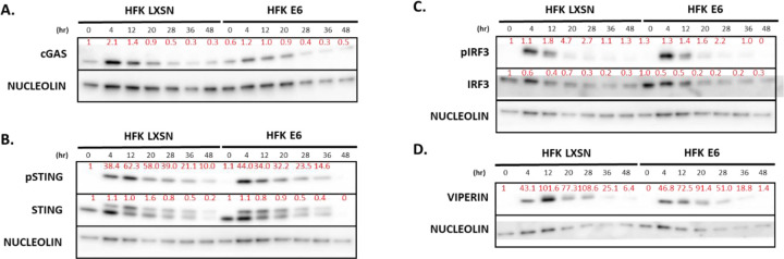
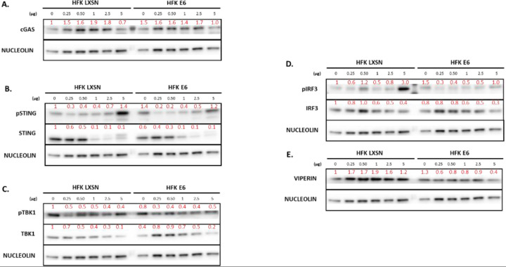
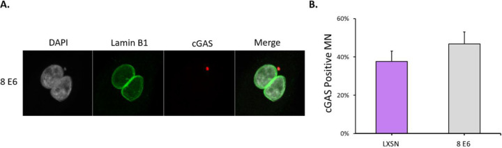
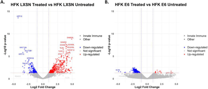
Beta genus human papillomaviruses (β-HPVs) are ubiquitous double stranded DNA (dsDNA) viruses that may promote skin cancers by destabilizing the host genome. Supporting this, expression of the E6 gene from a β-HPV (β-HPV 8 E6) results in increased micronuclei that should induce an innate immune response that eliminates these cells. Yet, β-HPV 8 E6 promotes rather than restricts proliferation. We hypothesize that β-HPV 8 E6 accomplishes this by attenuating the cyclic GMP-AMP synthase-stimulator of interferon genes (cGAS-STING) pathway, an innate immune pathway that becomes activated in response to cytosolic micronuclear dsDNA. Here, we show that in response to stimulation by transfection of pLVX-GFP plasmid, β-HPV 8 E6 reduced the magnitude and intensity of cGAS-STING pathway activation in immunoblot experiments. These data also demonstrate that impairment of the cGAS-STING pathway is strongest downstream of STING phosphorylation. Further, RNA-sequencing suggests that β-HPV 8 E6 downregulates other innate immune pathways. We also show that cGAS is recruited to micronuclei induced by β-HPV 8 E6. These data suggest a mechanism by which β-HPV 8 E6 facilitates proliferation of cells destabilized by micronuclei and support the hypothesis that the prevalence of β-HPV infections is in part due to the impairment of the cGAS-STING innate immune response.


Figures

Figure 1.
Figure 1.. E6 truncates the response of cGAS-STING signaling.
Representative immunoblots of time-course transfected hTERT HFK LXSN (HFK LXSN) and hTERT HFK 8 E6 (HFK E6) cell lines. NUCLEOLIN (C23) was used as a loading control for all immunoblots. Cells were transfected with 0.5ug of pLVX-GFP plasmid then harvested 0–48 hours post transfection. Immunoblot experiments were repeated in n=2. Red numbers above bands show densitometry quantification normalized to HFK LXSN 0 hour treatment and NUCLEOLIN loading control. (A) Representative immunoblot of cGAS abundance. (B) Representative immunoblot of STING (total and phosphorylated) abundance. (C) Representative immunoblot of IRF3 (total and phosphorylated) abundance. (D) Representative immunoblot of VIPERIN abundance.
Figure 2.
Figure 2.. E6 decreases abundance of cGAS-STING pathway in response to dose gradient.
Representative immunoblots of dsDNA-dose response transfected hTERT HFK LXSN (HFK LXSN) and hTERT HFK 8 E6 (HFK E6) cell lines. Immunoblot experiments were repeated in n=2. NUCLEOLIN (C23) was used as a loading control for all immunoblots. Cells were transfected with 0–5ug of pLVX-GFP plasmid then harvested 20 hours post transfection. Red numbers above bands show densitometry quantification normalized to HFK LXSN 0ug treatment and NUCLEOLIN loading control. (A) Representative immunoblot of cGAS abundance. (B) Representative immunoblot of STING (total and phosphorylated) abundance. (C) Representative immunoblot of TBK1 (total and phosphorylated) abundance (D) Representative immunoblot of IRF3 (total and phosphorylated) abundance. (E) Representative immunoblot of VIPERIN abundance.
Figure 3.
Figure 3.. Functional cGAS detection in hTERT HFK E6 cells.
(A) Representative immunofluorescent images of cGAS localization to micronuclei in hTERT HFK E6 cells. (B) Quantification of cGAS positive micronuclei in hTERT HFK LXSN and hTERT HFK E6 cells. Results were averaged between three independent experiments. At least 250 micronuclei/cell line were quantified for cGAS frequency across the three experiments. Graph depicts ± the standard errors of the mean.
Figure 4.
Figure 4.. E6 impairs innate immune responses.
hTERT HFK LXSN (HFK LXSN) and hTERT HFK E6 (HFK E6) cells were transfected with 1ug of pLVX-GFP plasmid and harvested 20 hours post-transfection. RNA was isolated and purified then submitted for Illumina sequencing. Differentially expressed genes were identified using DESeq2 software. Significant differentially expressed genes were identified as having p-values less than 0.05. (A) Volcano plot of significant differentially expressed genes between hTERT HFK LXSN treated and hTERT HFK LXSN untreated cells. (B) Volcano plot of significant differentially expressed genes between hTERT HFK E6 treated and hTERT HFK E6 untreated cells.
Figure 5.
Figure 5.. Summary Figure of Changes Induced by Beta HPV 8 E6.
Red arrows denote proteins that display decreased abundance in immunoblotting experiments or downregulation in RNA-sequencing experiments.

References

    1. Albertini S., Lo Cigno I., Calati F., De Andrea M., Borgogna C., Dell’Oste V., Landolfo S., & Gariglio M. (2018). HPV18 Persistence Impairs Basal and DNA Ligand–Mediated IFN-β and IFN-λ1 Production through Transcriptional Repression of Multiple Downstream Effectors of Pattern Recognition Receptor Signaling. The Journal of Immunology, 200(6), 2076–2089. 10.4049/jimmunol.1701536 - DOI - PubMed
    1. Antonsson A., Karanfilovska S., Lindqvist P. G., & Hansson B. G. (2003). General Acquisition of Human Papillomavirus Infections of Skin Occurs in Early Infancy. Journal of Clinical Microbiology, 41(6), 2509–2514. 10.1128/JCM.41.6.2509-2514.2003 - DOI - PMC - PubMed
    1. Babraham Bioinformatics—FastQC A Quality Control tool for High Throughput Sequence Data. (n.d.). Retrieved September 4, 2024, from https://www.bioinformatics.babraham.ac.uk/projects/fastqc/
    1. Bouwes Bavinck J. N., Feltkamp M., Struijk L., & ter Schegget J. (2001). Human papillomavirus infection and skin cancer risk in organ transplant recipients. The Journal of Investigative Dermatology. Symposium Proceedings, 6(3), 207–211. 10.1046/j.0022-202x.2001.00048.x - DOI - PubMed
    1. Chiang C., Pauli E.-K., Biryukov J., Feister K. F., Meng M., White E. A., Münger K., Howley P. M., Meyers C., & Gack M. U. (2018). The Human Papillomavirus E6 Oncoprotein Targets USP15 and TRIM25 To Suppress RIG-I-Mediated Innate Immune Signaling. Journal of Virology, 92(6), 10.1128/jvi.01737–17. 10.1128/jvi.01737-17 - DOI - PMC - PubMed

Publication types