

Bilateral Middle Cerebellar Peduncle Infarction Presenting With Vertigo and Hearing Impairment Mimicking Peripheral Vestibulopathy
- PMID: 39678002
- PMCID: PMC11646164
- DOI: 10.7759/cureus.75623
Bilateral Middle Cerebellar Peduncle Infarction Presenting With Vertigo and Hearing Impairment Mimicking Peripheral Vestibulopathy
Abstract
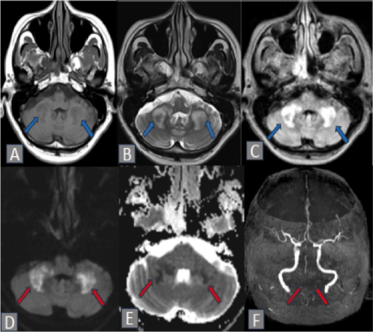
Bilateral middle cerebellar peduncle (MCP) infarction is a rare manifestation of ischemic stroke. We report a middle-aged male patient who presented with acute onset of vertigo, left ear deafness, and severe ataxia. Magnetic resonance imaging (MRI) of the brain confirmed the presence of infarction in the bilateral middle cerebellar peduncles due to stenosis of the posterior circulation arteries. This case highlights the importance of recognizing this clinical syndrome, as the symptoms may resemble those of peripheral vestibulopathy. Timely recognition is of paramount importance, given that untreated posterior circulation stroke can result in poor neurological outcomes.
Keywords: acute hearing loss; aica infarct; middle cerebellar peduncle; stroke; vertigo.
Copyright © 2024, Ramli et al.
Conflict of interest statement
Human subjects: Consent for treatment and open access publication was obtained or waived by all participants in this study. Conflicts of interest: In compliance with the ICMJE uniform disclosure form, all authors declare the following: Payment/services info: All authors have declared that no financial support was received from any organization for the submitted work. Financial relationships: All authors have declared that they have no financial relationships at present or within the previous three years with any organizations that might have an interest in the submitted work. Other relationships: All authors have declared that there are no other relationships or activities that could appear to have influenced the submitted work.
Figures
References
-
- Evaluation of clinical features and stroke etiology in patients with bilateral middle cerebellar peduncle infarction. Zhou C, Fan H, Chen H, et al. Eur Neurol. 2020;83:271–278. - PubMed
-
- Bilateral middle cerebellar peduncle infarction. Juneja A, Anand KS, Goyal H. Indian Journal of Medical Specialities. 2021;12:2.
-
- The anterior inferior cerebellar artery infarcts: a clinical-magnetic resonance imaging study. Roquer J, Lorenzo JL, Pou A. Acta Neurol Scand. 1998;97:225–230. - PubMed
-
- Bilateral middle cerebellar peduncle infarction caused by traumatic vertebral artery dissection. Akiyama K, Takizawa S, Tokuoka K, Ohnuki Y, Kobayashi N, Shinohara Y. Neurology. 2001;56:693–694. - PubMed
-
- Cerebellar infarction in the territory of the anterior and inferior cerebellar artery. A clinicopathological study of 20 cases. Amarenco P, Hauw JJ. Brain. 1990;113:139–155. - PubMed
Publication types
LinkOut - more resources
Full Text Sources
Miscellaneous