. 2024 Dec 8:19:13217-13233.
doi: 10.2147/IJN.S489872. eCollection 2024.

Fabrication of Biomimetic Hybrid Liposomes via Microfluidic Technology: Homotypic Targeting and Antitumor Efficacy Studies in Glioma Cells

Affiliations

Fabrication of Biomimetic Hybrid Liposomes via Microfluidic Technology: Homotypic Targeting and Antitumor Efficacy Studies in Glioma Cells

Ilaria Arduino et al. Int J Nanomedicine. .

Abstract

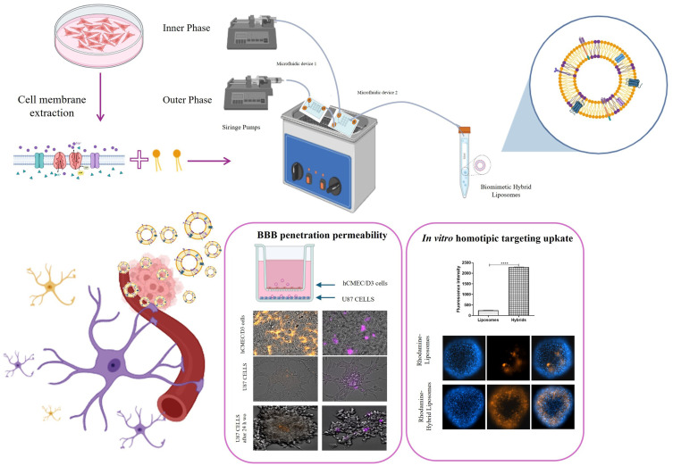
Introduction: The treatment of glioblastoma is hindered by the blood-brain barrier (BBB) and rapid drug clearance by the immune system. To address these challenges, we propose a novel drug delivery system using liposomes modified with cell membrane fragments. These modified liposomes can evade the immune system, cross the BBB, and accumulate in tumor tissue through homotypic targeting, thereby delivering drugs like paclitaxel and carboplatin more effectively.

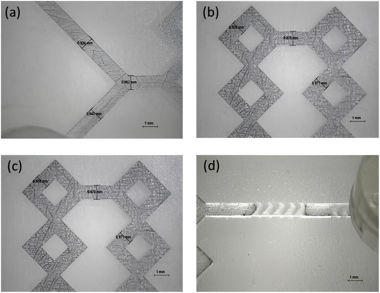
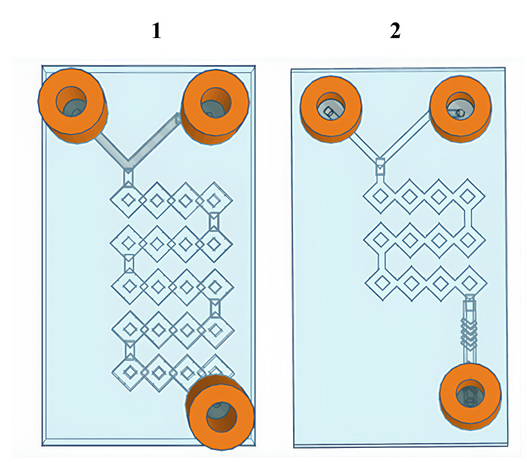
Methods: In this work, the hybrid liposomes were synthesized using microfluidics and integrating 3D printing to produce the microfluidic devices. In vitro, we explored the homotypic targeting capability, BBB passing ability, and therapeutic efficacy of paclitaxel and carboplatin.

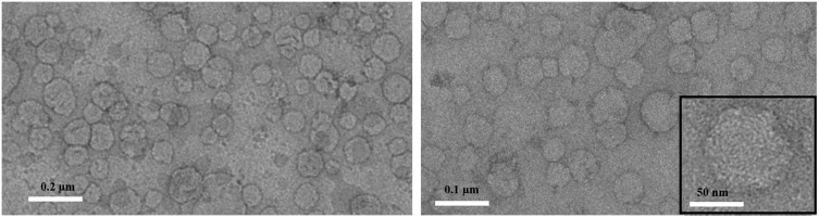
Results: The production of hybrid liposomes by microfluidics has been key to creating high-quality biomimetic nanoparticles, and the integration of 3D printing has simplified the production of microfluidic devices, making the process more efficient and economical. In vitro experiments have shown that these drug-loaded biomimetic hybrid liposomes are able to reach the homotypic target, cross the BBB, and maintain the efficacy of paclitaxel and carboplatin.

Conclusions: The development of biomimetic hybrid liposomes represents a promising approach for the treatment of glioblastoma. By combining the advantages of liposomal drug delivery with the stealth properties and targeting capabilities of cell membrane fragments, these nanoparticles can potentially overcome the challenges associated with traditional therapies.

Keywords: bioinspired materials; biomimetic nanoparticles; drug delivery system; emerging technology; glioblastoma cells; microfluidics.


Conflict of interest statement

The authors of this paper declare no competing financial or other interests that could affect the work they describe here.

Figures

None
Graphical abstract
Figure 1
Figure 1
Representative images of device 1 (a and b), and device 2 (c and d). Both devices have rectangular shape (28.00 × 50.00×2.70 mm) and same dimensions as the micromixing path (1.0 × 1.0×2.30 mm). Additionally, the device 2 is decorated with a modified herringbone geometry (0.8 × 1.94×1.22 mm).
Figure 2
Figure 2
Graphical representation of 3D-printed microfluidic devices: Microfluidic device 1 and microfluidic device 2.
Figure 3
Figure 3
Representative TEM micrographs obtained with staining for hybrid liposomes. Scale bar: 0.2 µm (left panel), 0.1 µm (right panel), 50 nm (inset in the right panel).
Figure 4
Figure 4
Validation of hybrid liposome formation. (A) FRET analysis illustrating the successful fusion of CMs and liposomes. The FRET analysis was performed using the fluorescent donor NBD (λem = 525 nm) and the fluorescent acceptor RhB (λem = 595 nm) at an excitation wavelength of 470 nm. (B) Measurement of FRET efficiency and the decrease in FRET efficiency following the hybridization of CMs. All bars represent mean values ± standard deviation; n = 3. (C) Confirmation of liposome-CMs fusion via FCM by determining the percentage of nanoparticles with overlapping red and green fluorescence in hybrid liposomes. (D) Western blot protein analysis of: 1. cancer cell membrane, 2. hybrid liposomes, 3. bare liposomes.
Figure 5
Figure 5
2D and 3D in vitro uptake studies (A) Uptake experiment results by flow cytometry analysis after 2 hours and 24 hours of incubation. The histograms represent average internalized fluorescence values (37°C - 4°C values). The tested concentration of liposomes or hybrid liposomes, in terms of fluorescent lipids, was 0.3 μM. Differences were considered very significant with **** p<0.0001, *** p<0.0005. (B) Representative images showing cell uptake of fluorescent hybrid liposomes and liposomes in a 3D U87 model after 2 hours and 24 hours of incubation. The upper panel shows the fluorescence intensities of Rhodamine-Liposomes (orange), Hoechst (blue, nuclei), and their merge after 2 hours; the bottom panel shows the fluorescence intensities of Rhodamine-Hybrid Liposomes (orange), Hoechst (blue, nuclei), and their merge after 24 hours. Scale bar: 100 μm. The histograms show the quantified fluorescence intensities detected in U87 spheroids after 2 hours and 24 hours of incubation with liposomes and hybrid liposomes. Differences were considered very significant with **** p<0.0001.
Figure 6
Figure 6
In vitro BBB penetration ability of hybrid liposome and liposomes and homotypic U87 tumor cell uptake. (A) Schematic illustration of the in vitro BBB model (Transwell™) for evaluating the potential BBB penetration ability of the hybrid liposomes and liposomes. (B) Representative FI images of the hCMEC/D3 BBB layer (top panel) and U87 cells (second and third panels) showing the penetration and targeting ability of liposomes and hybrid liposomes after 2h and 24 h after washing out. Orange: rhodamine-labelled liposomes; Violet: rhodamine-labelled hybrid liposomes. Scale bar: 25μm/100μm for hCMEC/D3 and U87, respectively. (C) Fluorescence intensity quantification of liposomes and hybrid liposomes internalized by U87 cells after crossing the BBB. Data in C were presented as the mean ± SD, n = 3 independent experiment. (D) Apparent permeability (Papp), in units of cm/second for the Liss Rhod PE labelled liposomes and hybrid liposomes.
Figure 7
Figure 7
In vitro antitumor efficacy. Histogram plot reporting the results of MTT assay performed in U87 cell line treated with paclitaxel, carboplatin, hybrid liposomes-PCX and hybrid liposomes-CBP for 24 h, showed as mean±SD of three experiments (***p<0.001).

References

    1. Zhang G, Yao M, Ma S, et al. Application of cell membrane-functionalized biomimetic nanoparticles in the treatment of glioma. J Mat Chem B. 2023;11(30):7055–7068. doi: 10.1039/D3TB00605K - DOI - PubMed
    1. Kang W, Xu Z, Lu H, et al. Advances in biomimetic nanomaterial delivery systems: harnessing nature’s inspiration for targeted drug delivery. J Mat Chem B. 2024;12(29):7001–7019. doi: 10.1039/D4TB00565A - DOI - PubMed
    1. Arduino I, Di FR, Tiboni M, et al. Microfluidic development and biological evaluation of targeted therapy-loaded biomimetic nano system to improve the metastatic melanoma treatment. Int J Pharm. 2024;650:123697. doi: 10.1016/j.ijpharm.2023.123697 - DOI - PubMed
    1. Li H, Zhu L, Zhang Y, Yang L, Wu W, Yang D. Biomimetic nanotherapeutics for homotypic-targeting photothermal/chemotherapy of oral cancer. J Control Release. 2024;366:28–43. doi: 10.1016/j.jconrel.2023.12.039 - DOI - PubMed
    1. Arduino I, Liu Z, Iacobazzi RM, et al. Microfluidic preparation and in vitro evaluation of iRGD-functionalized solid lipid nanoparticles for targeted delivery of paclitaxel to tumor cells. Int J Pharm. 2021;610:121246. doi: 10.1016/j.ijpharm.2021.121246 - DOI - PubMed

MeSH terms