[Preprint]. 2025 Jul 9:arXiv:2412.04639v2.

Multi-dynamic deep image prior for cardiac MRI

Affiliations

Multi-dynamic deep image prior for cardiac MRI

Marc Vornehm et al. ArXiv. .

Update in

  • Multi-dynamic deep image prior for cardiac MRI.
    Vornehm M, Chen C, Sultan MA, Arshad SM, Han Y, Knoll F, Ahmad R. Vornehm M, et al. Magn Reson Med. 2025 Jul 22. doi: 10.1002/mrm.70000. Online ahead of print. Magn Reson Med. 2025. PMID: 40692503

Abstract

Purpose: Cardiovascular magnetic resonance imaging is a powerful diagnostic tool for assessing cardiac structure and function. However, traditional breath-held imaging protocols pose challenges for patients with arrhythmias or limited breath-holding capacity. This work aims to overcome these limitations by developing a reconstruction framework that enables high-quality imaging in free-breathing conditions for various dynamic cardiac MRI protocols.

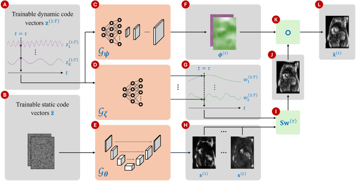
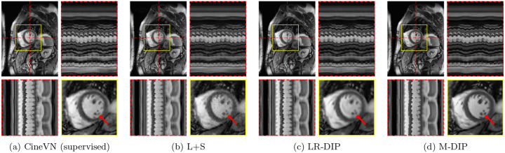
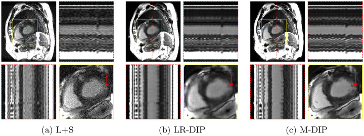
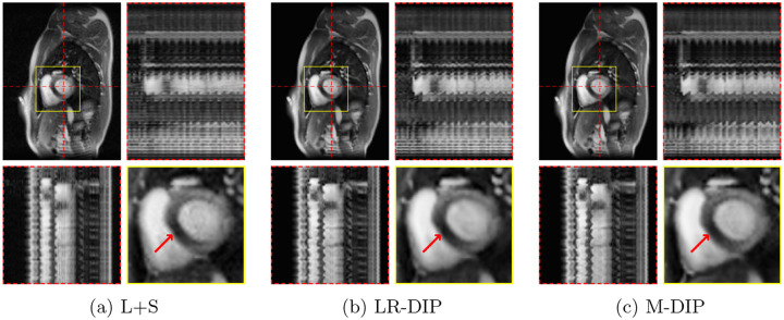
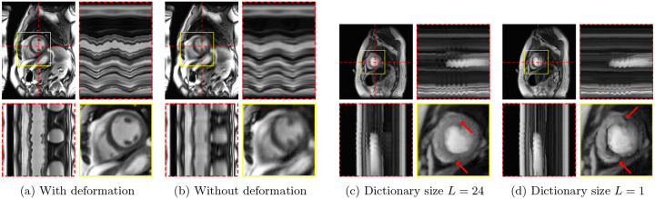
Methods: Multi-Dynamic Deep Image Prior (M-DIP), a novel unsupervised reconstruction framework for accelerated real-time cardiac MRI, is introduced. To capture contrast or content variation, M-DIP first employs a spatial dictionary to synthesize a time-dependent intermediate image. Then, this intermediate image is further refined using time-dependent deformation fields that model cardiac and respiratory motion. Unlike prior DIP-based methods, M-DIP simultaneously captures physiological motion and frame-to-frame content variations, making it applicable to a wide range of dynamic applications.

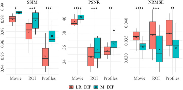
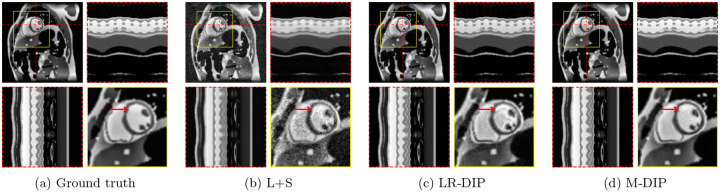
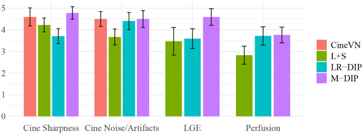
Results: We validate M-DIP using simulated MRXCAT cine phantom data as well as free-breathing real-time cine, single-shot late gadolinium enhancement (LGE), and first-pass perfusion data from clinical patients. Comparative analyses against state-of-the-art supervised and unsupervised approaches demonstrate M-DIP's performance and versatility. M-DIP achieved better image quality metrics on phantom data, higher reader scores on in-vivo cine and LGE data, and comparable scores on in-vivo perfusion data relative to another DIP-based approach.

Conclusion: M-DIP enables high-quality reconstructions of real-time free-breathing cardiac MRI without requiring external training data. Its ability to model physiological motion and content variations makes it a promising approach for various dynamic imaging applications.


Conflict of interest statement

MV is an employee of Siemens Healthineers AG. FK receives research funding from Siemens Healthineers AG, receives patent royalties for AI for MR image reconstruction from Siemens Healthineers AG, holds stock options from Subtle Medical Inc. and serves as scientific advisor to Imaginostics Inc.

Figures

Figure 1:
Figure 1:
Overview of M-DIP. (A) Trainable dynamic code vectors of dimensionality K, (B) trainable static code vectors of dimensionality N×c, (C) the network 𝒢ψ that generates a different deformation field for each t, (D) the network 𝒢ζ that generates a different set of weights for each t, (E) the network 𝒢θ that generates L time-invariant spatial dictionary elements, (F) x- and y-components of the deformation field generated by 𝒢ψ at t=τ, (G) weights generated by 𝒢ζ at t=τ, (H) spatial dictionary s(1:L) generated by 𝒢θ, (I) linear combination of the dictionary elements with time-specific weights at t=τ, (J) intermediate image at t=τ, (K) warping operation that applies deformation to the intermediate image, and (L) the output frame at t=τ.
Figure 2:
Figure 2:
M-DIP and LR-DIP results of the phantom study with results of Student’s t-tests. * indicates p0.05, ** indicates p0.01,*** indicates p0.001, and **** indicates p0.0001.
Figure 3:
Figure 3:
Exemplary MRXCAT phantom reconstructions. Each sub-figure illustrates an end-diastolic frame, temporal profiles, and a close-up of the heart. Red arrows highlight an artifact present in LR-DIP that is suppressed in M-DIP.
Figure 4:
Figure 4:
Bar plots with the scoring results for the in-vivo studies.
Figure 5:
Figure 5:
Exemplary in-vivo real-time cine reconstructions. Each sub-figure illustrates an end-diastolic frame, temporal profiles, and a close-up of the heart. Red arrows show an area where differences in image sharpness are particularly apparent.
Figure 6:
Figure 6:
Exemplary in-vivo free-breathing single-shot LGE reconstructions. Each sub-figure illustrates one frame, temporal profiles, and a close-up of the heart. Red arrows show an enhanced area that is better visible in M-DIP compared to LR-DIP.
Figure 7:
Figure 7:
Exemplary in-vivo first-pass perfusion reconstructions. Each sub-figure illustrates one frame, temporal profiles, and a close-up of the heart. Red arrows show a septal perfusion defect clearly visible in all three reconstructions.
Figure 8:
Figure 8:
M-DIP reconstructions of a real-time cine series with the deformation field generator 𝒢ψ (a) activated and (b) deactivated, and M-DIP reconstructions of a perfusion dataset with spatial dictionaries of (c) size L=24 and (d) size L=1. The red arrows point to the artifact observed in the perfusion case when L=1.

Similar articles

References

    1. Contijoch F., Rasche V., Seiberlich N., and Peters D., “The future of CMR: All-in-one vs. real-time CMR (Part 2),” J Cardiovasc Magn Reson, vol. 26, no. 1, p. 100 998, 2024, doi: 10.1016/j.jocmr.2024.100998. - DOI - PMC - PubMed
    1. Lustig M., Donoho D., Santos J., and Pauly J., “Compressed sensing MRI,” IEEE Signal Process Mag, vol. 25, no. 2, pp. 72–82, 2008, doi: 10.1109/MSP.2007.914728. - DOI
    1. Yang G., Yu S., Dong H., et al. , “DAGAN: Deep de-aliasing generative adversarial networks for fast compressed sensing MRI reconstruction,” IEEE Trans Med Imaging, vol. 37, no. 6, pp. 1310–1321, 2018, doi: 10.1109/TMI.2017.2785879. - DOI - PubMed
    1. Jalal A., Arvinte M., Daras G., Price E., Dimakis A., and Tamir J., “Robust compressed sensing MRI with deep generative priors,” ser. Proc Neural Inf Process Syst (NeurIPS), vol. 34, Virtual, 2021, pp. 14 938–14 954.
    1. Knoll F., Murrell T., Sriram A., et al. , “Advancing machine learning for MR image reconstruction with an open competition: Overview of the 2019 fastMRI challenge,” Magn Reson Med, vol. 84, no. 6, pp. 3054–3070, 2020, doi: 10.1002/mrm.28338. - DOI - PMC - PubMed

Publication types

LinkOut - more resources