. 2024 Nov 26;15(12):6999-7012.
doi: 10.1364/BOE.540754. eCollection 2024 Dec 1.

Silver mean sequence in extended depth of focus intraocular lenses: a comparative study of kinoform and stepwise designs

Affiliations

Silver mean sequence in extended depth of focus intraocular lenses: a comparative study of kinoform and stepwise designs

Anabel Martínez-Espert et al. Biomed Opt Express. .

Abstract

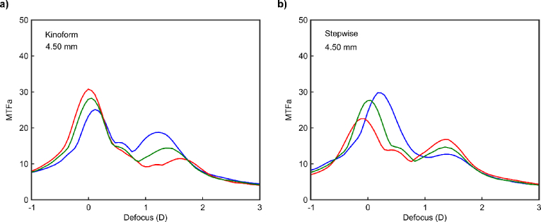
In this work, we present two new multifocal intraocular lens (MIOL) designs, both based on the silver mean kinoform diffractive lens. We demonstrate that a single aperiodic diffractive profile can be used to create two different MIOLs: one with a kinoform structure and the other with a stepwise profile. Quantitative assessment of the designs was carried out using the through focus modulation transfer function and the area under the modulation transfer function for the prediction of their visual performance. Our results show that both designs exhibit nearly identical optical performance at the design wavelength (λ = 550 nm), though their intrinsic longitudinal chromatic aberration differs significantly. Given that diffractive extended depth of focus (EDoF) intraocular lenses are prone to image degradation due to dysphotopic phenomena, we also compared the halos generated by these two designs and found notable differences in their behavior. Furthermore, under photopic conditions, the proposed lens designs demonstrated the potential to achieve visual acuity values of 0.2 logMAR or better across a vergence range from approximately 0 to 2 D. Finally, to qualitatively assess the behavior of the MIOLs, an objective experimental evaluation was conducted using an adaptive optics visual simulator in a model eye. Experimental results align with the quantitative assessment of the proposed designs.


Conflict of interest statement

The authors declare that there are no conflicts of interest related to this article.

Figures

Fig. 1.
Fig. 1.
Phase profile of a Kinoform Silver Mean Lens of order S = 4 (black line) represented in the normalized square radial coordinate and the phase profile with variations (blue line).
Fig. 2.
Fig. 2.
Axial Irradiance for a 3 mm pupil for different values of α. The dotted blue line indicates the selected factor for our designs α=1.2. The blue arrows indicate the positions of the four foci of the KSML corresponding to first diffraction order, the green arrow indicates the zero-order diffraction (far focus).
Fig. 3.
Fig. 3.
Diffractive profiles of the Silver MIOL designs: kinoform and stepwise (in blue). Green and red lines represent a pupillary radius of 3.0 mm and 4.5 mm in the lens plane. Dashed black lines mark the steps of the Silver Mean sequence. Heights are relative to the curvature of the base power (far vision) monofocal lens.
Fig. 4.
Fig. 4.
Monochromatic (λ=550 nm) TF-MTF at 50 lp/mm for a kinoform and stepwise profiles computed for pupil diameters of 3.0 mm (a); and 4.5 mm (b).
Fig. 5.
Fig. 5.
Monochromatic area under the Modulation Transfer Function (MTFa) curves obtained for wavelengths 450 nm (blue line), 550 nm (green line), 650 nm (red line) with the kinoform (a) and stepwise designs (b) using 4.5 mm pupil diameter.
Fig. 6.
Fig. 6.
MTFa curves for 3.0 mm (a) and 4.5 mm (b) pupils obtained for polychromatic light (V(λ)) for the kinoform, stepwise, and monofocal IOL (dotted line).
Fig. 7.
Fig. 7.
Polychromatic MTFa values for the distance (solid lines) and intermediate (dotted lines) foci obtained for pupils varying from 2.5 mm to 6.0 mm for the kinoform (red lines) and stepwise designs (black lines).
Fig. 8.
Fig. 8.
Simulated halos obtained with polychromatic light and a 4.5 mm pupil diameter: The retinal image was obtained by convolving the point-spread function with a 2 arcmin pinhole. The numerical values correspond to the diameter that encircles 50% of the intensity at the retina.
Fig. 9.
Fig. 9.
Calculated Visual Acuity (VA) defocus curves obtained from polychromatic MTFa values using Eq. (1), for 3.0 mm (a) and 4.5 mm (b) pupils.
Fig. 10.
Fig. 10.
a) Experimental Through-the-focus images obtained with the VAO system. The tumbling E optotype corresponds to visual acuities of 0.4 logMAR, 0.2 logMAR, and 0.0 logMAR. The three designs evaluated were: a monofocal lens (upper row), the bifocal design included in the VAO (middle row), and Silver kinoform MIOL (bottom row). b) Simulated images (obtained from the point-spread function computed with Zemax) of an optotype with letters whose sizes correspond to the same visual acuities at vergences 0.00, 0.75 and 1.50 D (outlined in red in the S row). All images were obtained using a 4.5 mm pupil.

References

    1. Wolffsohn J. S., Davies L. N., Sheppard A. L., “New insights in presbyopia: impact of correction strategies,” BMJ Open Ophthalmol 8(1), e001122 (2023).10.1136/bmjophth-2022-001122 - DOI - PMC - PubMed
    1. Rampat R., Gatinel D., “Multifocal and extended depth-of-focus intraocular lenses in 2020,” Ophthalmology 128(11), e164–e185 (2021).10.1016/j.ophtha.2020.09.026 - DOI - PubMed
    1. Alió J. L., Plaza-Puche A. B., Alió del Barrio J. L., et al. , “Clinical outcomes with a diffractive trifocal intraocular lens,” Eur J Ophthalmol 28(4), 419–424 (2018).10.1177/1120672118762231 - DOI - PubMed
    1. Bilbao-Calabuig R., Llover-Rausell A., Ortega-Usobiaga J., et al. , “Visual outcomes following bilateral lmplantation of two diffractive trifocal intraocular lenses in 10 084 eyes,” Am J Ophthalmol 179, 55–66 (2017).10.1016/j.ajo.2017.04.013 - DOI - PubMed
    1. Alfonso J. F., Fernández-Vega Cueto L., Belda-Salmerón L., et al. , “Visual function after implantation of a diffractive aspheric trifocal intraocular lens,” Eur J Ophthalmol 26(5), 405–411 (2016).10.5301/ejo.5000741 - DOI - PubMed

LinkOut - more resources