. 2025 Feb 5;16(2):e0327424.
doi: 10.1128/mbio.03274-24. Epub 2024 Dec 16.

Inhibition and evasion of neutrophil microbicidal responses by Legionella longbeachae

Affiliations

Inhibition and evasion of neutrophil microbicidal responses by Legionella longbeachae

Hannah E Hanford et al. mBio. .

Abstract

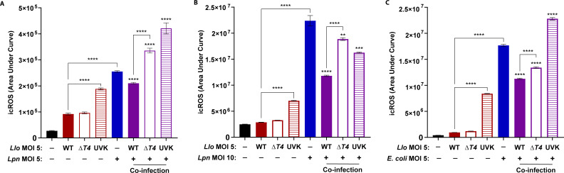
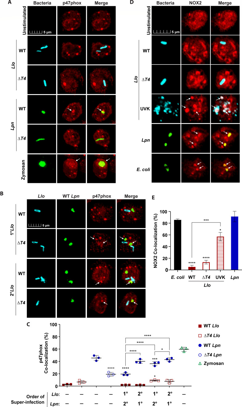
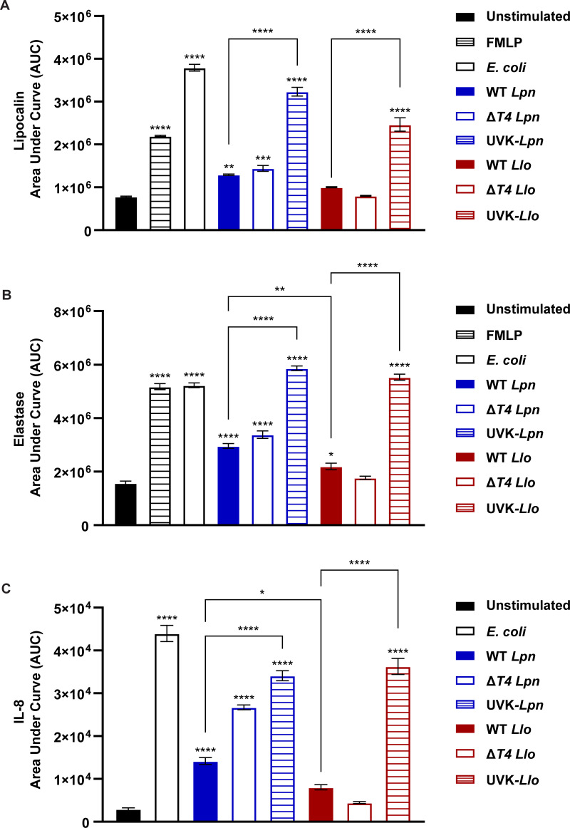
Legionella species evade degradation and proliferate within alveolar macrophages as an essential step for the manifestation of disease. However, most intracellular bacterial pathogens are restricted in neutrophils, which are the first line of innate immune defense against invading pathogens. Bacterial degradation within neutrophils is mediated by the fusion of microbicidal granules to pathogen-containing phagosomes and the generation of reactive oxygen species (ROS) by the phagocyte NADPH oxidase complex. Here, we show that human neutrophils fail to trigger microbicidal processes and, consequently, fail to restrict L. longbeachae. In addition, neutrophils infected with L. longbeachae fail to undergo a robust pro-inflammatory response, such as degranulation and IL-8 production. Here, we identify three strategies employed by L. longbeachae for evading restriction by neutrophils and inhibiting the neutrophil microbicidal response to other bacteria co-inhabiting in the same cell. First, L. longbeachae excludes the cytosolic and membrane-bound subunits of the phagocyte NADPH oxidase complex from its phagosomal membrane independent of the type 4 secretion system (T4SS). Consequently, infected neutrophils fail to generate robust ROS in response to L. longbeachae. Second, L. longbeachae impedes the fusion of azurophilic granules to its phagosome and the phagosomes of bacteria co-inhabiting the same cell through T4SS-independent mechanisms. Third, L. longbeachae protects phagosomes of co-inhabiting bacteria from degradation by ROS through a trans-acting T4SS-dependent mechanism. Collectively, we conclude that L. longbeachae evades restriction by human neutrophils via T4SS-independent mechanisms and utilizes trans-acting T4SS-dependent mechanisms for inhibition of neutrophil ROS generation throughout the cell cytosol.

Importance: Legionella longbeachae is commonly found in soil environments where it interacts with a wide variety of protist hosts and microbial competitors. Upon transmission to humans, L. longbeachae invades and replicates within alveolar macrophages, leading to the manifestation of pneumonia. In addition to alveolar macrophages, neutrophils are abundant immune cells acting as the first line of defense against invading pathogens. While most intracellular bacterial species are killed and degraded by neutrophils, we show that L. longbeachae evades degradation. The pathogen impairs the major neutrophils' microbicidal processes, including the fusion of microbicidal granules to the pathogen-containing vacuole. By inhibiting of assembly of the phagocyte NADPH oxidase complex, the pathogen blocks neutrophils from generating microbicide reactive oxygen species. Overall, L. longbeachae employs unique virulence strategies to evade the major microbicidal processes of neutrophils.

Keywords: NADPH oxidase; ROS; azurphilic granules; granules; lysosomes.


Conflict of interest statement

The authors declare no conflict of interest.

Figures

Fig 1
Fig 1
Intracellular survival of L. longbeachae in neutrophils. (A) To determine bacterial survival, infected neutrophils were lysed at 5, 15, 60, and 120 min post-infection. Serial dilutions of lysates were plated on BCYE agar for quantification of CFUs. ± SD, n = 3. (B) Representative confocal images of neutrophils infected with WT L. longbeachae (Llo), ΔT4 Llo, WT L. pneumophila (Lpn), or ΔT4 Lpn at 15 min post-infection. Bacteria were labeled with anti-Llo (red) or anti-Lpn (green) antibodies. Cell nuclei were stained with DAPI (blue). (C) Data are shown as mean percent killed and degraded Llo (red) or Lpn (blue) ±SD, n = 3 (scatter plot dots). (D) Representative TEM images of neutrophils infected for 1 h. Large white arrowheads indicate Llo or Lpn. Small white arrowheads indicate neutrophil granules. The data shown are representative of three independent biological repeats.
Fig 2
Fig 2
Trans-acting suppression of neutrophil microbicidal response by L. longbeachae. Quantification of killed and degraded Llo and Lpn during solo-infection or when co-inhabiting the same cell. (A–C) Neutrophils were solo-infected with WT or ΔT4 bacterial strains for 15 min, or subjected to co-infections with Llo strains and WT Lpn at a multiplicity of infection (MOI) 1. Following a 15-min infection, bacterial morphology was examined by confocal microscopy in neutrophils solo-infected (A) or super-infected (B). (C) Data are shown as mean percent killed and degraded bacteria ± SD, n = 3 (scatter plot dots). (D–F) Neutrophils were solo-infected with WT or ΔT4 bacterial strains for 15 min or subjected to 15-min primary infections (1°) followed by super-infection (2°) for an additional 15 min at an MOI of 1. Bacterial morphology was examined by confocal microscopy in neutrophils solo-infected (D) or super-infected (E). Bacteria were labeled with anti-Llo (red) or anti-Lpn (green) antibodies. Cell nuclei were stained with DAPI (blue). (F) Data are shown as mean percent killed and degraded Llo (red) or Lpn (blue) ±SD, n = 3 (scatter plot dots). The data shown are representative of three independent biological replicates.
Fig 3
Fig 3
Inhibited fusion of azurophilic granules to the L. longbeachae-containing phagosome. To determine the fusion of azurophilic granules to bacterial phagosomes, neutrophils were labeled for elastase. Neutrophils were solo-infected with WT or ΔT4 bacterial strains for 15 min or subjected to 15-min primary infections (1°) followed by super-infection (2°) for an additional 15 minutes. Representative confocal images of azurophilic granule fusion to phagosomes in neutrophils solo-infected (A) or super-infected (B). Bacteria were labeled with anti-Llo antibody (cyan) or anti-Lpn antibody (green). Zymosan A BioParticles Alexa Fluor 594 are shown in green. Azurophilic granules were labeled with anti-elastase antibodies (red). White arrows indicate positive co-localization of elastase with pathogen-containing phagosomes. (C) Data are shown as mean percent co-localization of elastase to Llo phagosomes (red) or Lpn phagosomes (blue) during infections. Zymosan was included as a positive control (green) ± SD, n = 3 (scatter plot dots). The data shown are representative of three independent biological repeats.
Fig 4
Fig 4
Fusion of specific granules to the L. longbeachae-containing phagosome. To determine the fusion of specific granules to bacterial phagosomes, neutrophils were labeled for lactoferrin (A–C) and NGAL (D–F). Neutrophils were solo-infected with WT or ΔT4 bacterial strains for 15 min or subjected to 15 min primary infections (1°) followed by super-infection (2°) for an additional 15 minutes. Representative confocal images of specific granule fusion to phagosomes during solo-infections (A and D) or super-infections (B and E). Bacteria were labeled with anti-Llo antibody (cyan) or anti-Lpn antibody (green). Zymosan A BioParticles Alexa Fluor 594 are shown in green. Specific granules were labeled with anti-lactoferrin or anti-NGAL antibodies (red). White arrows indicate positive co-localization of lactoferrin or NGAL with pathogen-containing phagosomes. (C and F) Data are shown as mean percent co-localization of lactoferrin or NGAL to Llo phagosomes (red) or Lpn phagosomes (blue) during infections. Zymosan was included as a positive control (green) ± SD, n = 3 (scatter plot dots). The data shown are representative of three independent biological repeats.
Fig 5
Fig 5
Failure of neutrophils to elicit robust ROS production in response to L. longbeachae. Neutrophils were infected with WT or ΔT4 Legionella strains alone, UV-killed (UVK) Llo alone, or co-infected as indicated on the x-axis. The kinetic of ROS production was determined up to 1 h. Neutrophils were solo-infected or co-infected with Legionella strains at a 1:1 infection ratio of Llo to Lpn with an MOI of 5 (A) or 1:2 infection ratio of Llo to Lpn (B) with an MOI of 5 and 10, respectively ±SD, n = 3. (C) Neutrophils were solo-infected or co-infected with E. coli and Llo strains at a 1:1 infection ratio with an MOI of 5. Data are shown as mean area under curve for kinetic ROS production during 1 h post-infection for unstimulated (black), Llo-infected (red), Lpn- or E. coli-infected (blue), and co-infected cells (purple) ±SD, n = 3. The data shown are representative of three independent biological repeats.
Fig 6
Fig 6
Failure of neutrophils to assemble the NADPH oxidase complex in response to L. longbeachae. To determine the assembly of the phagocyte NADPH oxidase complex at bacterial phagosomes, neutrophils were labeled with antibodies for cytosolic subunits and membrane-bound components of the NADPH oxidase complex. Neutrophils were solo-infected with WT or ΔT4 bacterial strains for 15 min or subjected to 15 min primary infections (1°) followed by super-infection (2°) for an additional 15 minutes. Representative confocal images of cytosolic p47phox localization to phagosomes in neutrophils solo-infected (A) or super-infected (B). Bacteria were labeled with anti-Llo antibody (cyan) or anti-Lpn antibody (green). Zymosan A BioParticles Alexa Fluor 594 are shown in green. p47phox was labeled with anti-p47phox antibody (red). White arrows indicate positive co-localization of p47phox with pathogen-containing phagosomes. (C) Data are shown as mean percent co-localization of p47phox to Llo phagosomes (red) or Lpn phagosomes (blue) during infections. Zymosan was included as a positive control (green). ±SD, n = 3 (scatter plot dots). (D) Representative confocal images of the membrane-bound catalytic core gp91phox/NOX2 (NOX2) subunit co-localizing with phagosomal membranes in neutrophils solo-infected with WT or ΔT4 Llo strains, UV-killed (UVK) Llo, WT Lpn, or m-cherry E. coli. White arrows indicate positive co-localization of gp91phox/NOX2 with pathogen-containing phagosomes. Bacteria were labeled with anti-Llo antibody (cyan) or anti-Lpn antibody (green). The gp91phox/NOX2 was labeled with anti-gp91phox/NOX2 antibody (red). White arrows indicate positive co-localization of p47phox with pathogen-containing phagosomes. (E) Data are shown as mean percent co-localization of NOX2 to Llo phagosomes (red) or Lpn phagosomes (blue) during infections ± SD, n = 3. The data shown are representative of three independent biological repeats.
Fig 7
Fig 7
Failure of robust degranulation and IL-8 production by neutrophils in response to L. longbeachae. Neutrophils were infected with WT, ΔT4, or UV-killed (UVK) Legionella strains for up to 20 h at an MOI of 5. Cells were stimulated with fMLP or infected with E. coli at an MOI of 5 as additional positive controls. Aliquots of cell supernatants were collected at 0.5, 1, 4, 6, 12, and 20 h post-infection to quantify degranulation of specific granules using lipocalin as a marker (A), azurophilic granules using elastase as a marker (B), or IL-8 production (C) by ELISA. Data are shown as mean area under curve for kinetic degranulation and cytokine production during 20 h post-infection for unstimulated (black), fMLP-stimulated (black and white bars), E. coli-infected (black outline), Lpn-infected (blue), and Llo-infected (red) cells ± SEM, n = 3. The data shown are representative of three independent biological repeats.
Fig 8
Fig 8
Three strategies employed by L. longbeachae for evasion and inhibition of microbicidal activities in neutrophils. Model of T4SS-independent and T4SS-dependent inhibition of neutrophil microbicidal activities by L. longbeachae via (1) T4SS-independent evasion of NADPH oxidase assembly at the L. longbeachae phagosome for inhibition of ROS production, (2) T4SS-independent inhibition of fusion of azurophilic granules to pathogen-containing phagosomes during solo-infection or dual-infections, and (3) T4SS-dependent inhibition of NADPH oxidase complex formation to intracellular phagosomes for impeding ROS generation during dual infections.

Similar articles

References

    1. Cazalet C, Gomez-Valero L, Rusniok C, Lomma M, Dervins-Ravault D, Newton HJ, Sansom FM, Jarraud S, Zidane N, Ma L, Bouchier C, Etienne J, Hartland EL, Buchrieser C. 2010. Analysis of the Legionella longbeachae genome and transcriptome uncovers unique strategies to cause Legionnaires’ disease. PLoS Genet 6:e1000851. doi:10.1371/journal.pgen.1000851 - DOI - PMC - PubMed
    1. Dolinsky S, Haneburger I, Cichy A, Hannemann M, Itzen A, Hilbi H. 2014. The Legionella longbeachae Icm/Dot substrate SidC selectively binds phosphatidylinositol 4-phosphate with nanomolar affinity and promotes pathogen vacuole-endoplasmic reticulum interactions. Infect Immun 82:4021–4033. doi:10.1128/IAI.01685-14 - DOI - PMC - PubMed
    1. Zhan X-Y, Yang J-L, Sun H, Zhou X, Qian Y-C, Huang K, Leng Y, Huang B, He Y. 2022. Presence of viable, clinically relevant Legionella bacteria in environmental water and soil sources of China. Microbiol Spectr 10:e0114021. doi:10.1128/spectrum.01140-21 - DOI - PMC - PubMed
    1. Mondino S, Schmidt S, Buchrieser C. 2020. Molecular mimicry: a paradigm of host-microbe coevolution illustrated by Legionella. mBio 11:e01201-20. doi:10.1128/mBio.01201-20 - DOI - PMC - PubMed
    1. Slow S, Anderson T, Murdoch DR, Bloomfield S, Winter D, Biggs PJ. 2022. Extensive epigenetic modification with large-scale chromosomal and plasmid recombination characterise the Legionella longbeachae serogroup 1 genome. Sci Rep 12:5810. doi:10.1038/s41598-022-09721-9 - DOI - PMC - PubMed

LinkOut - more resources