. 2025 Feb;66(2):430-443.
doi: 10.1111/epi.18182. Epub 2024 Dec 16.

TSPO-PET in pre-surgical evaluations: Correlation of neuroinflammation and SEEG epileptogenicity mapping in drug-resistant focal epilepsy

Affiliations

TSPO-PET in pre-surgical evaluations: Correlation of neuroinflammation and SEEG epileptogenicity mapping in drug-resistant focal epilepsy

Jennifer Kilmer et al. Epilepsia. 2025 Feb.

Abstract

Objectives: Resective surgery in drug-resistant focal epilepsy (DRFE) requires extensive evaluation to localize the epileptogenic zone (EZ). When non-invasive phase 1 assessments (electroencephalography, EEG; magnetic resonance imaging, MRI; and 18F-Fluorodeoxyglucose-positron emission tomography, [18F]FDG-PET) are inconclusive for EZ localization, invasive investigations such as stereo-EEG (SEEG) are necessary. Epileptogenicity maps (Ems) visualize the EZ using SEEG-identified ictal high-frequency oscillations (iHFOs). PET imaging with radioligands targeting the18-kDa translocator protein (TSPO), a marker of glial activation, may aid EZ localization. This study investigates the correlation between TSPO-PET imaging and SEEG iHFOs in DRFE to determine the utility of TSPO-PET in pre-surgical assessments, especially in complex or non-lesional cases.

Methods: Patients with DRFE and inconclusive phase 1 assessments were recruited from Bicêtre Hospital (AP-HP) for a prospective study (Eudract 2017-003381-27). They underwent SEEG and [18F]DPA-714 (N,N-diethyl-2-(2-(4-(2-(fluoro-18F)ethoxy)phenyl)-5,7-dimethylpyrazolo[1,5-a]pyrimidin-3-yl)acetamide) (TSPO radioligand) PET imaging. Statistical parametric mapping (SPM) techniques analyzed significant [18F]DPA-714-PET uptake (TSPO-map) and generated epileptogenicity maps (EM-map). Correlation analyses at regional and voxel-of-interest (VOI) levels assessed the relationship between TSPO-map and EM-map.

Results: We were able to obtain and analyze both maps in 12 of 17 patients recruited. A significant positive correlation between EM-map and TSPO-map in focal epilepsies was found regionally (r = .81, p < .00004) and at the VOI level (r = .79, p < .00003). Temporal, insular, parietal, and occipital regions showed particularly strong correspondence. In frontal epilepsies, TSPO-map was more focal than EM-map, suggesting increased specificity for SEEG planning. This study also demonstrated the benefit of the TSPO-map in identifying multiple foci in multifocal epilepsies, with or without lesions.

Significance: These findings suggest that neuroinflammation may be a molecular substrate of the EZ in non-lesional focal epilepsy. Identifying the EZ inpatients with complex DRFE and inconclusive MRI/[18F]FDG-PET imaging is essential to improve resective surgery outcomes. Combining TSPO-PET imaging with SEEG recordings may help bridge this gap.

Keywords: TSPO‐PET; drug‐resistant epilepsy; epilepsy surgery; epileptogenicity maps; neuroinflammation; statistical parametric mapping.


Conflict of interest statement

None of the authors have any conflicts of interest to disclose.

Figures
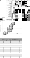
FIGURE 1
FIGURE 1
Correlation of parametric maps (index of epileptogenicity and [18F]DPA‐714 uptake) in DRFE patients by VOIs and cortical brain regions. Correlation of VOIs and grouped cortical regions with both significant EI and [18F]DPA‐714 radiotracer uptake in patients with DRFE. Demonstrated with a VOI matrix representing the areas of significant EI (dark gray), DPA uptake (gray), or both (light gray) (A); graphical representation of the regional correlations between EI and DPA uptake in each subject (B); and table of results (C). (A) The matrix defines the significant activity in each segmented VOI. VOIs with no SEEG electrodes implanted were not considered during final analysis and left blank; those with no significant SEEG/PET activity recorded are in black. The black‐outlined VOIs represent the patient's EZ, as identified with phase 1 and 2 pre‐surgical evaluations. VOIs in 14 of 16 EZs were found to have both significant electrical activity and inflammatory markers. Statistical significance was defined based on each individual map's p‐value and corresponding z‐score; for example, a combined z‐score >1.96 was considered significant for parametric maps created with p < .05 (two‐tailed). (B) The area of each bubble signifies the percent correlation of regions with both EI and DPA uptake as compared to the total number of regions with significant EI (limiting factor). Each number represents a different patient, with patients having up to three EMs (SEEGs) compared to their DPA‐PET maps. Three patients had bilateral SEEG exploration (P4, P10, P15). Bubbles with internal numbers signify overlapping patient data or (number of seizures). (C) Considering all ictal SEEGs from this population (n = 19), there is not a statistically significant correlation (r = .26; p = .26) between regional EI and DPA uptake; however, when those with no focal onset of seizures measured with SEEG (P15*, P17**) are removed from calculations there is a strong statistically significant positive correlation between maps (.81; p < .00005). There is a significant positive correlation between EM and DPA maps at the VOI level (r = .6; p < .005), which increases when the non‐focal seizures are removed (r = .79; p < .00003). *Dispersed network/no focus; **No focal onset. DRFE, drug‐resistant focal epilepsy; EI, epileptogenicity index; EM, epileptogenicity map; EZ, epileptogenic zone; No., number of; NS, not significant; VOI, voxel‐of‐interest.
FIGURE 2
FIGURE 2
Magnetic resonance imaging (MRI) and stereo‐electroencephalography (SEEG) results and corresponding parametric maps (index of epileptogenicity and [18F]DPA‐714 uptake) of two different seizure types in the same patient. Comparison of T1‐weighted MRI, SEEG‐defined iHFOs, SEEG‐based EMs, and [18F]DPA‐714 PET parametric maps from seizure types with MRI‐positive (lesional) and MRI‐negative (non‐lesional) EZs in patient 7. T1‐weighted MRI studies demonstrating lesional EZ (right occipital dysplasia; red arrow) and non‐lesional EZ (MRI‐negative temporal seizure type) in patient 7 (P7). Results of SEEG studies recorded during different seizure types, demonstrating increased number of ictal iHFOs in both lesional and non‐lesional types. Parametric maps overlaid on MRI and visualized in FreeSurfer (Freeview). In both lesional and non‐lesional EZs, there is an increased index of epileptogenicity and increase in [18F]DPA‐714 tracer uptake, highlighting the utility of TSPO‐PET imaging in cases where standard imaging is inadequate. EZ, epileptogenic zone; iHFO, ictal high‐frequency oscillations; TSPO, translocator protein.

References

    1. Aronica E, Crino PB. Inflammation in epilepsy: clinical observations. Epilepsia. 2011;52:26–32. 10.1111/j.1528-1167.2011.03033.x - DOI - PubMed
    1. Cosenza‐Nashat M, Zhao M‐L, Suh H‐S, et al. Expression of the translocator protein of 18 kDa by microglia, macrophages and astrocytes based on immunohistochemical localization in abnormal human brain. Neuropathology Appl Neurobio. 2009;35:306–328. 10.1111/j.1365-2990.2008.01006.x - DOI - PMC - PubMed
    1. Kreisl WC, Kim M‐J, Coughlin JM, Henter ID, Owen DR, Innis RB. PET imaging of neuroinflammation in neurological disorders. Lancet Neurol. 2020;19:940–950. 10.1016/S1474-4422(20)30346-X - DOI - PMC - PubMed
    1. Sauvageau A, Desjardins P, Lozeva V, Rose C, Hazell AS, Bouthillier A, et al. Increased expression of “peripheral‐type” benzodiazepine receptors in human temporal lobe epilepsy: implications for PET imaging of hippocampal sclerosis. Metab Brain Dis. 2002;17:3–11. 10.1023/A:1014044128845 - DOI - PubMed
    1. Johnson EW, De Lanerolle NC, Kim JH, et al. “Central” and “peripheral” benzodiazepine receptors: opposite changes in human epileptogenic tissue. Neurology. 1992;42:811. 10.1212/WNL.42.4.811 - DOI - PubMed

MeSH terms