. 2025 Feb;12(6):e2411739.
doi: 10.1002/advs.202411739. Epub 2024 Dec 16.

Ca2+- and cGAMP-Contained Semiconducting Polymer Nanomessengers for Radiodynamic-Activated Calcium Overload and Immunotherapy

Affiliations

Ca2+- and cGAMP-Contained Semiconducting Polymer Nanomessengers for Radiodynamic-Activated Calcium Overload and Immunotherapy

Danling Cheng et al. Adv Sci (Weinh). 2025 Feb.

Abstract

Various second messengers exert some vital actions in biological systems, including cancer therapy, but the therapeutic efficacy is often need to be improved. A semiconducting polymer nanomessenger (TCa/SPN/a) consisting of two second messengers, calcium ion (Ca2+) and cyclic guanosine monophosphate-adenosine monophosphate (cGAMP) for metastatic breast cancer therapy, is reported here. Such a TCa/SPN/a is constructed to exhibit X-ray response for the activatable delivery of mitochondria-targeting Ca compound and cGAMP as stimulator of interferon genes (STING) agonist. With X-ray irradiation, TCa/SPN/a could generate singlet oxygen (1O2) via radiodynamic effect for ablating solid tumors and improving the tumor immunogenicity by inducing immunogenic cell death (ICD). Furthermore, the released mitochondria-targeting Ca compounds show a high binging effect on mitochondria and cause reactive oxygen species (ROS) generation and mitochondria damage via calcium overload, while cGAMP boosts immunological effect through activating STING pathway. In this way, TCa/SPN/a enables a radiodynamic-activated calcium overload and immunotherapy to obviously inhibit the growths of bilateral tumors and also abolish tumor metastasis in metastatic breast cancer mouse models. This article should demonstrate the first smart dual-functional nanotherapeutic containing two second messengers for precise and specific cancer therapy.

Keywords: calcium overload; immunotherapy; radiodynamic therapy; second messengers; smart nanotherapeutic.


Conflict of interest statement

The authors declare no conflict of interest.

Figures

Figure 1
Figure 1
The X‐ray responsive second messenger release strategy by TCa/SPN/a for RDT‐combined tumor therapy. a) The schematic of fabrication processes of TCa/SPN/a. b) The schematic of X‐ray responsive second messenger release and RDT‐combined tumor therapy using TCa/SPN/a.
Figure 2
Figure 2
Preparation and characterization of SPN, SPN/a, TCa/SPN, and TCa/SPN/a. a) Appearance morphology images of SPN, SPN/a, TCa/SPN and TCa/SPN/a. b) Absorbance property of cGAMP, PFODBT, SPN, SPN/a, TCa/SPN and TCa/SPN/a. c) Zeta potentials of SPN, SPN/a, TCa/SPN and TCa/SPN/a (n = 3). d) Profiles of size distribution of SPN, SPN/a, TCa/SPN and TCa/SPN/a. e) Fluorescence spectra of SPN, SPN/a, TCa/SPN and TCa/SPN/a. f) Fluorescence intensity changes of SOSG probe (F/F 0) of aqueous solutions containing SPN, SPN/a, TCa/SPN and TCa/SPN/a at different radiation doses (n = 3). g) The release efficiency of cGAMP for SPN/a and TCa/SPN/a under X‐ray irradiation of different doses (n = 3). Data are presented with mean ± SD.
Figure 3
Figure 3
In vitro therapeutic efficacy and DNA damage studies. a) Cell viability analysis of SPN‐, SPN/a‐, TCa/SPN‐ and TCa/SPN/a‐treated 4T1 cells (n = 5). b) Cell viability analysis of 4T1 cancer cells after SPN, SPN/a, TCa/SPN and TCa/SPN/a incubations with or without X‐ray irradiation (n = 5). c) Fluorescence images of 4T1 cells after ROS generation. d) Quantitative analysis of the fluorescence intensity of the generated ROS (n = 5). e) The uptake efficacy of 4T1 cells in SPN, SPN/a, TCa/SPN and TCa/SPN/a groups. f) Quantitative analysis of the fluorescence signals of 4T1 cell images with nanoparticle uptake (n = 5). g) DNA damage analysis for 4T1 cells in SPN, SPN/a, TCa/SPN and TCa/SPN/a groups with or without X‐ray irradiation. Data are presented with mean ± SD, (**) p < 0.01, (***) p < 0.001, unpaired two‐tailed Student's t tests.
Figure 4
Figure 4
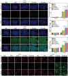
ICD effect and mitochondrial damage analysis in vitro. a) The detection of CRT for treated 4T1 cells via immunofluorescence staining analysis. b) Quantitative analysis of CRT staining images (n = 3). c) HMGB1 level analysis for treated 4T1 cells (n = 3). d) Extracellular ATP level analysis for the treated 4T1 cells (n = 3). e) CLSM images of JC‐1 aggregates (red) and JC‐1 monomer (green) in 4T1 cells after SPN, SPN/a, TCa/SPN and TCa/SPN/a incubation with or without X‐ray irradiation. Data are presented with mean ± SD, (**) p < 0.01, (***) p < 0.001, unpaired two‐tailed Student's t tests.
Figure 5
Figure 5
Evaluation of tumor accumulation and ICD effect in vivo. a) The accumulation analysis of SPN, SPN/a, TCa/SPN and TCa/SPN/a in tumors sites via IVIS spectrum imaging system (the tumor areas were represented by white dotted circles). b) Fluorescence intensity analysis of the tumor sites at certain post‐injection time point (n = 3). c) Fluorescence intensity of tumor tissues and organs (n = 3). d) CLSM images of the ROS generation in tumor site after treatments (n = 3). e) Determination of ATP release levels in tumors (n = 3). f) Immunofluorescence images of the tumors for evaluating the expression of CRT after treatments. g) Immunofluorescence images of the tumors for evaluating the expression of HMGB1 after treatments. Data are presented with mean ± SD, (**) p < 0.01, (***) p < 0.001, unpaired two‐tailed Student's t tests.
Figure 6
Figure 6
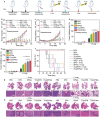
Antitumor and anti‐metastasis analysis. a) Strategies of bilateral tumor model construction, nanoparticle injection, X‐ray irradiation of primary tumors, and data analysis. b) The volume curves of primary tumors in SPN‐, SPN/a‐, TCa/SPN‐ and TCa/SPN/a‐treated mice with or without X‐ray irradiation (n = 6). c) The volume curves of distant tumors (n = 6). d) Analysis of tumor inhibition rates for primary tumors (n = 6). e) Analysis of tumor inhibition rates for distant tumors (n = 6). f) The survival curves of treated mice (n = 5). g) H&E staining images of isolated lungs in each group. h) H&E staining images of isolated livers in different treatment groups. Data are presented with mean ± SD, (*) p < 0.05, (**) p < 0.01, (***) p < 0.001, unpaired two‐tailed Student's t tests.
Figure 7
Figure 7
Evaluation of immunotherapy effect. a) The ratios of CD4+ and CD8+ T cells in CD3+ cells in primary tumor sites via flow cytometry analysis. The proportion analysis of b) CD4+ T cells and c) CD8+ T cells (n = 5). d) The ratios of CD4+ and CD8+ T cells in CD3+ cells in distant tumor sites via flow cytometry analysis. The proportion analysis of e) CD4+ T cells and f) CD8+ T cells (n = 5). g) The levels of Treg cells in primary tumor sites via flow cytometry analysis. h) The proportion analysis of Treg cells in primary tumors (n = 5). i) The levels of Treg cells in distant tumor sites via flow cytometry analysis. j) The proportion analysis of Treg cells in distant tumors (n = 5). Data are presented with mean ± SD, (**) p < 0.01, (***) p < 0.001, unpaired two‐tailed Student's t tests.

Similar articles

Cited by

References

    1. a) Oe Y., Wang X., Patriarchi T., Konno A., Ozawa K., Yahagi K., Hirai H., Tsuboi T., Kitaguchi T., Tian L., Nat. Commun. 2020, 11, 471; - PMC - PubMed
    2. b) Slavik K. M., Morehouse B. R., Ragucci A. E., Zhou W., Ai X., Chen Y., Li L., Wei Z., Bähre H., König M., Nature 2021, 597, 109; - PMC - PubMed
    3. c) Radhakrishnan A., Rohatgi R., Siebold C., Nat. Chem. Biol. 2020, 16, 1303. - PMC - PubMed
    1. a) Zheng P., Ding J., Asian J. Pharm. Sci. 2022, 17, 1; - PMC - PubMed
    2. b) Cui C., Merritt R., Fu L., Pan Z., Acta Pharm. Sin. B 2017, 7, 3; - PMC - PubMed
    3. c) Prise K. M., O'sullivan J. M., Nat. Rev. Cancer 2009, 9, 351; - PMC - PubMed
    4. d) Kang Y., Xu L., Dong J., Huang Y., Yuan X., Li R., Chen L., Wang Z., Ji X., Coord. Chem. Rev. 2023, 481, 215050.
    1. a) Xu L., Tong G., Song Q., Zhu C., Zhang H., Shi J., Zhang Z., ACS Nano 2018, 12, 6806; - PubMed
    2. b) Dong Z., Feng L., Hao Y., Li Q., Chen M., Yang Z., Zhao H., Liu Z., Chem 2020, 6, 1391;
    3. c) Zheng P., Ding B., Shi R., Jiang Z., Xu W., Li G., Ding J., Chen X., Adv. Mater. 2021, 33, 2007426. - PubMed
    1. a) Shae D., Becker K. W., Christov P., Yun D. S., Lytton‐Jean A. K., Sevimli S., Ascano M., Kelley M., Johnson D. B., Balko J. M., Nat. Nanotechnol. 2019, 14, 269; - PMC - PubMed
    2. b) Wu J., Sun L., Chen X., Du F., Shi H., Chen C., Chen Z. J., Science 2013, 339, 826; - PMC - PubMed
    3. c) Chin E. N., Yu C., Vartabedian V. F., Jia Y., Kumar M., Gamo A. M., Vernier W., Ali S. H., Kissai M., Lazar D. C., Science 2020, 369, 993; - PubMed
    4. d) Zhan M., Yu X., Zhao W., Peng Y., Peng S., Li J., Lu L., J. Nanobiotechnol. 2022, 20, 23. - PMC - PubMed
    1. a) Koshy S. T., Cheung A. S., Gu L., Graveline A. R., Mooney D. J., Adv. Biosyst. 2017, 1, 1600013; - PMC - PubMed
    2. b) Chattopadhyay S., Liu Y.‐H., Fang Z.‐S., Lin C.‐L., Lin J.‐C., Yao B.‐Y., Hu C.‐M. J., Nano Lett. 2020, 20, 2246; - PubMed
    3. c) Gong W., Zhao T., Yuan H., Yang G., Wang W., Li X., Ju H., Nano Today 2024, 58, 102465.

LinkOut - more resources