

Diffusion imaging shows microstructural alterations in untreated, non-lesional patients already after a first unprovoked seizure
- PMID: 39679966
- PMCID: PMC11908665
- DOI: 10.1111/epi.18213
Diffusion imaging shows microstructural alterations in untreated, non-lesional patients already after a first unprovoked seizure
Abstract
Objective: Patients with newly diagnosed epilepsy exhibit brain white matter (WM) abnormalities, but the temporal dynamics of these are unknown. The literature suggests these alterations might be present before diagnosis. This study investigates WM microstructural integrity using diffusion imaging in non-lesional (NL), interictal epileptiform discharge (IED)-free, unmedicated patients who experienced a first unprovoked seizure compared to healthy controls. Furthermore, we evaluated whether the patients who developed epilepsy within a 1-year follow-up had a divergent pattern of WM alterations in contrast to those who did not. We also evaluated patients with established epilepsy.
Methods: We performed a diffusion imaging analysis in a cohort of 82 subjects. Twenty patients recently experienced a first unprovoked seizure (first-seizure group), 32 patients had chronic epilepsy (chronic-epilepsy group), and 30 healthy controls. The first-seizure patients were later classified into patients who developed epilepsy (early-epilepsy) and those who did not (single-seizure). Fractional anisotropy (FA), mean diffusivity (MD), and radial diffusivity (RD) were calculated. Group differences were analyzed using tract-based spatial statistics and permutation analysis of linear models.
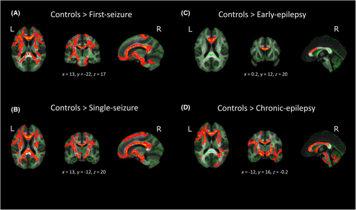
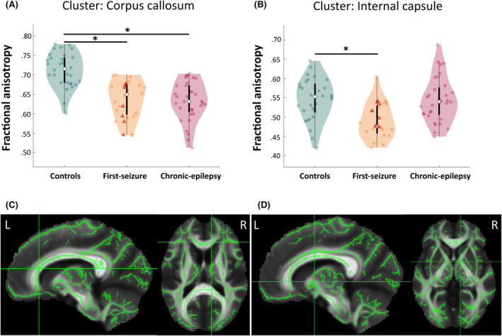
Results: Compared to controls, first-seizure patients showed decreased FA (p < .05, d = 1.3) in the corpus callosum and forceps minor, among other tracts. Similar changes were found in the single-seizure group (p < .05, d = 1.3), whereas the early-epilepsy patients showed decreases only in the corpus callosum (p < .05, d = 2.4). We confirmed that patients with chronic epilepsy have widespread FA decreases (p < .05, d = 1).
Significance: We provide evidence that, as early as after the first unprovoked seizure, patients considered NL by conventional methods harbor marked microstructural abnormalities detectable with diffusion magnetic resonance imaging (MRI). These findings suggest that WM alterations are present very early in the epileptogenic process even before the diagnosis can currently be made. These results have important implications for better understanding the epileptogenic process and preexisting structural difference in patients after a first seizure.
Keywords: TBSS; diffusion imaging; early epilepsy; imaging biomarkers; unprovoked seizures.
© 2024 The Author(s). Epilepsia published by Wiley Periodicals LLC on behalf of International League Against Epilepsy.
Conflict of interest statement
N.K.F. received honoraria from Arvelle/Angelini, Jazz Pharma, Bial, and Eisai, as well as research support from Jazz Pharma, all unrelated to this work. E.H.U.R. received honoraria from Jazz Pharma, Eisai, and Springer, all unrelated to this work. The remaining authors declare no conflicts of interest.
Figures
References
MeSH terms
Grants and funding
LinkOut - more resources
Full Text Sources
Medical
Miscellaneous
