. 2025 Jan 1;152(1):dev202955.
doi: 10.1242/dev.202955. Epub 2025 Jan 7.

CFI-1 functions unilaterally to restrict gap junction formation in C. elegans

Affiliations

CFI-1 functions unilaterally to restrict gap junction formation in C. elegans

Zan Wu et al. Development. .

Abstract

Electrical coupling is vital to neural communication, facilitating synchronized activity among neurons. Despite its significance, the precise mechanisms governing the establishment of gap junction connections between specific neurons remain elusive. Here, we identified that the PVC interneuron in Caenorhabditis elegans forms gap junction connections with the PVR interneuron. The transcriptional regulator CFI-1 (ARID3) is specifically expressed in the PVC but not PVR interneuron. Reducing cfi-1 expression in the PVC interneuron leads to enhanced gap junction formation in the PVR neuron, while ectopic expression of cfi-1 in the PVR neuron restores the proper level of gap junction connections in the PVC neuron, along with the normal touch response. These findings unveil the pivotal role of CFI-1 in bidirectionally regulating the formation of gap junctions within a specific neuronal pair, shedding light on the intricate molecular mechanisms governing neuronal connectivity in vivo.

Keywords: C. elegans; CFI-1/ARID3; Gap junction; Neural circuitry; Transcription factor.


Conflict of interest statement

Competing interests The authors declare no competing or financial interests.

Figures
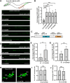
Fig. 1.
Fig. 1.
cfi-1 inhibits gap junction formation in Punc-53-expressing neurons. (A) Schematic (top) and the distribution of Punc-53::UNC-9::GFP puncta (green) in wild-type (wt), cfi-1(xd424), cfi-1(ot786), cfi-1(ky650), cfi-1(ky651), and cfi-1(xd424/ot786) mutant animals. (B) The relative change of UNC-9::GFP puncta number in various genotypes. One-way ANOVA with Tukey's multiple comparisons test was performed. ***P<0.001. n=20 for each genotype. (C) Domain distribution of CFI-1. The mutation sites in different alleles are labeled. (D) The distribution of Punc-53::UNC-7::GFP puncta (green) in wild-type (wt) and cfi-1(xd424) mutant animals. (E) The relative change of UNC-7::GFP puncta number in wild-type (wt) and cfi-1(xd424) animals. Two-tailed unpaired Student's t-test was performed. **P<0.01. n=12 for each genotype. (F) The distribution Punc-53::INX-13::GFP puncta (green) in wild-type (wt) and cfi-1(xd424) mutant animals. (G) The relative change of INX-13::GFP puncta number in wild-type (wt) and cfi-1(xd424) animals. Two-tailed unpaired Student's t-test was performed. ***P<0.001. n≥11 for each genotype. (H) The neurons labeled by Punc-53-driven GFP in wild-type (wt) and cfi-1(xd424) animals. (I) Quantification of the number of Punc-53::GFP-expressing cells in wild-type (wt) and cfi-1(ot786) animals. (J) Quantification of the GFP fluorescence intensity of Punc-53::GFP-expressing cells in wild-type (wt) and cfi-1(ot786) animals. Two-tailed unpaired Student's t-test. NS, not significant. n≥20. Data are mean±s.d. Scale bars: 25 µm.
Fig. 2.
Fig. 2.
cfi-1 could function within or outside Punc-53-expressing neurons. (A) The ectopic UNC-9::GFP puncta (green) phenotype in cfi-1 mutants could be rescued by Punc-53 or Pcfi-1 promoter-driven CFI-1. (B) The relative change in UNC-9::GFP puncta number in various genotypes. One-way ANOVA with Tukey's multiple comparisons test was performed. Data are mean±s.d. ***P<0.001. (C,D) Co-localization analysis of CFI-1::GFP knock-in (green) and Pcfi-1 driven CFI-1::mCherry (red) in the ventral cord (C) and tail region (D). (E) CFI-1::GFP knock-in (green) signal does not merge with Punc-53 driven mCherry (red) in the tail region. In D and E, dotted line indicates cell bodies of cells expressing CFI-1. (F) The cfi-1-expressing PVC and/or LUA neurons may form gap junctions with Punc-53-expressing neurons. Scale bars: 25 µm (A,C); 10 µm (D,E).
Fig. 3.
Fig. 3.
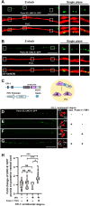
cfi-1 functions in PVC neurons. (A,B) Gap junctions formed by Punc-53-expressing neurons (green, labeled by Punc-53::UNC-9::GFP) are situated on PVC neurons (red) in wild-type (A) and cfi-1(xd424) mutant (B) animals. Z-stacks encompassing the PVC neurite and UNC-9::GFP were captured. Enlarged images from a single plane within the boxed regions are shown on the right. (C) Schematic of CFI-1 AID in PVC neurons. (D-G) Expression of CFI-1::wrmScarlet::degron (red) and UNC-9::GFP puncta (green) driven by Punc-53 with or without Pnmr-1::TIR1 or IAA. (H) The relative change in UNC-9::GFP puncta number in various treatments. One-way ANOVA with Tukey's multiple comparisons test was performed. Data are mean±s.d. ***P<0.001; *P<0.05. NS, not significant. Scale bars: 25 µm (main panels); 5 µm (D-G, enlarged panels).
Fig. 4.
Fig. 4.
PVC and PVR are gap junction partner cells. (A-C) The Punc-53::UNC-9::GFP puncta (green) are not present on PVQ (red) (A), PVW (red) (B) or PVP (red) (C). (D) The Punc-53::UNC-9::GFP puncta (green) are present on PVR (red). (E) The Pnmr-1::UNC-9::GFP puncta (green) (from PVC) are situated on PVR (red) neurons. (F) The Pflp-10::UNC-9::GFP puncta (green) (from PVR) are situated on PVC (red) neurons. Z-stacks encompassing the neurite and UNC-9::GFP were captured. Enlarged images from a single plane within the boxed regions are shown on the right in D-F. (G) Quantification of the percentage of UNC-9::GFP puncta on various types of neurons. (H) The Pnmr-1::UNC-9::GFP puncta (green) (from PVC) and Punc-53::UNC-7::wrmScarlet (red) (from PVR) are adjacent to each other. Z-stacks capturing the UNC-9::GFP and UNC-7::wrmScarlet signals were acquired. Enlarged images from a single plane within the boxed regions are displayed below. (I,J) The ectopic Punc-53::UNC-9::GFP puncta (green) observed in cfi-1(xd424) mutant animals are formed by PVR (red) (I), but not by PVQ (red), PVW (red) or PVP (red) (J). Z-stacks encompassing the PVR neurite and UNC-9::GFP were captured. In I, enlarged images from a single plane within the boxed region are shown on the right. (K) Quantification of the percentage of UNC-9::GFP puncta on various types of neurons in cfi-1(xd424) mutant animals. (L) Schematic of gap junction connections between PVC and PVR neurons in wild type (wt) and cfi-1(xd424) mutants. Data are mean±s.d. Scale bars: 25 µm.
Fig. 5.
Fig. 5.
cfi-1 affects gap junction formation between PVC and PVR neurons. (A) Schematic of endogenous gap junction labeling by NATF approach. (B-E) The UNC-9::split-GFP (green) puncta formed by PVR neurons in wild-type (wt) (B), cfi-1(xd424) (C), cfi-1(xd424);PVR CFI-1 (D), or cfi-1(xd424);PVC CFI-1 (E) animals. (F) The endogenous gap junctions (labeled by UNC-9::split-GFP) formed by PVR neurons are situated on PVC (red) neurons in both wild-type and cfi-1(xd424) mutant animals. (G) The ectopic gap junctions (labeled by UNC-9::split-GFP) formed by PVR neurons are indeed found on PVR (red) neurons in cfi-1(xd424) mutant animals. (H) The 3D reconstruction of endogenous gap junctions (labeled by UNC-9::split-GFP) formed by PVR neurons on PVC (red) neurons in cfi-1(xd424) mutant animals. (I) Quantification of the number of endogenous gap junctions formed by PVR neuron in various genotypes. One-way ANOVA with Tukey's multiple comparisons test was performed. n≥21 for each genotype. ***P<0.001. (J) The anterior touch response in wild-type and cfi-1(xd424) mutant animals. (K) The posterior touch response in various genotypes. Data are mean±s.d. n≥18. Scale bars: 25 µm (B-E); 2 µm (H).

Similar articles

Cited by

References

    1. Abascal, F. and Zardoya, R. (2013). Evolutionary analyses of gap junction protein families. Biochim. Biophys. Acta. 1828, 4-14. 10.1016/j.bbamem.2012.02.007 - DOI - PubMed
    1. Alcami, P. and Pereda, A. E. (2019). Beyond plasticity: the dynamic impact of electrical synapses on neural circuits. Nat. Rev. Neurosci. 20, 253-271. 10.1038/s41583-019-0133-5 - DOI - PubMed
    1. An, G., Miner, C. A., Nixon, J. C., Kincade, P. W., Bryant, J., Tucker, P. W. and Webb, C. F. (2010). Loss of Bright/ARID3a function promotes developmental plasticity. Stem Cells 28, 1560-1567. 10.1002/stem.491 - DOI - PMC - PubMed
    1. Barnes, T. M. and Hekimi, S. (1997). The Caenorhabditis elegans avermectin resistance and anesthetic response gene unc-9 encodes a member of a protein family implicated in electrical coupling of excitable cells. J. Neurochem. 69, 2251-2260. 10.1046/j.1471-4159.1997.69062251.x - DOI - PubMed
    1. Barrios, A., Ghosh, R., Fang, C., Emmons, S. W. and Barr, M. M. (2012). PDF-1 neuropeptide signaling modulates a neural circuit for mate-searching behavior in C. elegans. Nat. Neurosci. 15, 1675-1682. 10.1038/nn.3253 - DOI - PMC - PubMed

MeSH terms

Substances