. 2025 Mar 3;224(3):e202408092.
doi: 10.1083/jcb.202408092. Epub 2024 Dec 16.

Identification of the Polo-like kinase substrate required for homologous synapsis

Affiliations

Identification of the Polo-like kinase substrate required for homologous synapsis

Ariel L Gold et al. J Cell Biol. .

Abstract

The synaptonemal complex (SC) is a zipper-like protein structure that aligns homologous chromosome pairs and regulates recombination during meiosis. Despite its conserved appearance and function, how synapsis occurs between chromosome axes remains elusive. Here, we demonstrate that Polo-like kinases (PLKs) phosphorylate a single conserved residue in the disordered C-terminal tails of two paralogous SC subunits, SYP-5 and SYP-6, to establish an electrostatic interface between the SC central region and chromosome axes in C. elegans. While SYP-5/6 phosphorylation is dispensable for the ability of SC proteins to self-assemble, local phosphorylation by PLKs at the pairing center is crucial for SC elongation between homologous chromosome axes. Additionally, SYP-5/6 phosphorylation is essential for asymmetric SC disassembly and proper PLK-2 localization after crossover designation, which drives chromosome remodeling required for homolog separation during meiosis I. This work identifies a key regulatory mechanism by which localized PLK activity mediates the SC-axis interaction through phosphorylation of SYP-5/6, coupling synapsis initiation to homolog pairing.


Conflict of interest statement

Disclosures: The authors declare no competing interests exist.

Figures
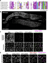
Figure 1.
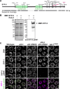
Figure 1.
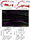
Phosphorylation of SYP-5 S541 during meiotic prophase. (A) Amino acid alignment of SYP-5/6 orthologs from Caenorhabditis species using the T-Coffee algorithm. Amino acids are colored using the Clustal X color scheme: hydrophobic (blue), positive (red), negative (purple), polar (green), cysteine (pink), glycine (orange), proline (yellow), aromatic (cyan), and unconserved (white). The black line above the sequence alignment indicates the phosphopeptide used to generate phospho-specific antibodies and the arrow indicates the PLK phosphorylation sites characterized in this study. (B) Composite projection images of a full-length gonad dissected from a worm strain expressing GFP::COSA-1, 3xFlag::SYP-5, and HA::SYP-6 are shown for DNA (top) and SYP-5 pS541 (bottom). Scale bar, 50 µm. (C) Zoomed-in images of leptotene/zygotene (upper panels) and pachytene nuclei (lower panels) as indicated by white boxes in B. 3xFlag::SYP-5 (magenta) and SYP-5 pS541 (green) staining are shown. Scale bar, 10 µm. (D) Zoomed-in images of late pachytene nuclei as indicated by the white box in B. DNA, 3xFlag::SYP-5 (magenta), SYP-5 pS541 (green), and GFP::COSA-1 (red) staining are shown. Scale bar, 5 µm.
Figure S1.
Figure S1.
Purification and characterization of the SYP-5 pS541 antibody. (A) Immune serum against the SYP-5 phosphopeptide (pS541) was passed through affinity columns coupled to non-phospho (SYP-5 S541) and phosphopeptides (SYP-5 pS541). The unbound (flow-through) and bound (elution) fractions were used in dot blots of serially diluted peptides. (B) Immunofluorescence images of pachytene nuclei from wild type, syp-5S541A, and syp-6T685A mutants showing DNA, 3xFlag::SYP-5 or HA::SYP-6 (magenta), and SYP-5 pS541 (green) staining. Scale bar, 5 µm. Source data are available for this figure: SourceData FS1.
Figure 2.
Figure 2.
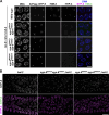
Phosphorylation of SYP-5 S541 requires both PLK-1 and PLK-2, but not the PLK-docking site on SYP-1. (A) Schematic showing in vitro PLK-2 phosphorylation sites on SYP-5, with sequence coverage from mass spectrometry analysis indicated by gray blocks. Conserved residues that conform to the PLK consensus motif are highlighted in magenta. (B) In vitro kinase assays using recombinant 6His-MBP-SYP-5 incubated with or without PLK-2. Coomassie staining of the purified proteins (left) and western blot for SYP-5 pS541 (right) are shown. 6His-MBP-SYP-5 is indicated by an arrow. (C) Immunofluorescence images of pachytene nuclei from wild type, plk-2(tm1395), plk-1(RNAi), plk-2(tm1395); plk-1(RNAi), and syp-1T452A animals, stained for SYP-1 (magenta), and SYP-5 pS541 (green). Scale bar, 5 µm. Source data are available for this figure: SourceData F2.
Figure S2.
Figure S2.
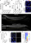
Prolonged CHK-2 activity and abnormal crossover designation in syp-5 S541A syp-6 T685A mutants. (A and B) Graphs showing the percent viable and male self-progeny from wild type (n = 1,921), syp-5S541A (n = 2,617), syp-6T685A (n = 3,022), and syp-5S541Asyp-6T685A mutants (n = 1,526) hermaphrodites. **, P < 0.01; ***, P < 0.001; ****, P < 0.0001 by unpaired t test. (C) Immunofluorescence images from wild type and syp-5S541Asyp-6T685A mutants stained for DNA (blue) and HIM-8 (red). Scale bar, 5 µm. (D) Dissected gonads from wild type and syp-5S541Asyp-6T685A mutants were stained for DNA and phospho-HIM-8/ZIMs (CHK-2 substrate). Composite projection immunofluorescence images are shown. The CHK-2 active zone is marked by yellow dotted lines. Scale bar, 50 µm. (E) Graph showing the CHK-2 active zone in wild type and syp-5S541Asyp-6T685A mutants, shown as a percent gonad length of the pHIM-8/ZIM-positive region relative to the region from meiotic entry to pachytene exit. The numbers of gonads scored are also indicated. **, P < 0.05 by unpaired t test. (F) Immunofluorescence images of pachytene nuclei from wild type and syp-5S541Asyp-6T685A animals stained for DNA (blue) and GFP::COSA-1 (green). Scale bar, 5 µm. (G) Graph showing the quantification of GFP::COSA-1 foci per nucleus. The numbers of nuclei scored are indicated on the top.
Figure 3.
Figure 3.
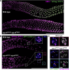
Phosphorylation of SYP-5 S541 and SYP-6 T685 is critical for proper SC assembly and disassembly. (A) Composite immunofluorescence images of full-length gonads from wild type and syp-5S541Asyp-6T685A mutant animals stained for HIM-3 (magenta) and 3xFlag::SYP-5 (green). Scale bar, 50 µm. (B) Composite immunofluorescence images of late pachytene and diplotene region from wild type and syp-5S541Asyp-6T685A animals showing 3xFlag::SYP-5 (magenta) and GFP::COSA-1 (green) staining. Scale bar, 50 µm. Insets show zoomed-in views of the boxed regions with DNA staining (blue). Scale bar, 1 µm. (C) Zoomed-in views of the boxed regions in diplotene as indicated by colored boxes in B. DNA (blue), SYP-5 (magenta), and GFP::COSA-1 (green) staining are shown. Scale bar, 1 µm.
Figure S3.
Figure S3.
Phospho-mimetic mutations at SYP-5 S541 and SYP-6 T685 cause defects in SC assembly. (A and B) Graph showing the percent viable and male self-progeny from the wild type (n = 1,684) and syp-5S541Dsyp-6T685D mutants (n = 1,431) hermaphrodites. ****, P < 0.0001 by unpaired t test. (C) Full-length gonads dissected from wild type and syp-5S541Dsyp-6T685D animals were stained for HIM-3 (magenta) and 3xFlag::SYP-5 (green). Composite projection images are shown. Scale bar, 50 µm. (D) Composite immunofluorescence images of dissected gonads from wild type and syp-5S541Dsyp-6T685D animals with DNA and phospho-HIM-8/ZIMs (CHK-2 substrate) staining are shown. Yellow dotted lines mark the CHK-2 active zone. Scale bar, 50 µm. (E) Graph showing the CHK-2 active zone in the wild type and syp-5S541Asyp-6T685A mutants, shown as a percent gonad length of the pHIM-8/ZIM-positive region relative to the region from meiotic entry to pachytene exit. The numbers of gonads scored are also indicated. **, P = 0.0015 by unpaired t test.
Figure S4.
Figure S4.
Additional characterization of SYP-5/6 phosphorylation and SC disassembly defects in syp-5/6 phosphomutants. (A) Immunofluorescence images of diplotene nuclei from wild type and syp-5S541Dsyp-6T685D mutant animals stained for HIM-3 (magenta) and SYP-5 (green). Scale bar, 25 µm. The inset shows the zoomed-in view of the boxed regions. Scale bar, 1 µm. (B) Immunofluorescence images of diplotene nuclei from cosa-1 and syp-5S541Dsyp-6T685D; cosa-1 mutants stained for HTP-3 (magenta) and SYP-5 (green). Scale bar, 25 µm. The inset shows the zoomed-in view of the boxed regions. Scale bar, 1 µm. (C) Immunofluorescence images of diplotene nuclei from cosa-1(tm3298) mutants. HTP-3 and SYP-5 pS541 staining are shown. Scale bar, 25 µm.
Figure 4.
Figure 4.
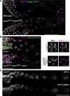
Phosphorylation of SYP-5/6 is required for proper PLK-2 localization and chromosome remodeling in late meiotic prophase. (A) Composite immunofluorescence images of wild type and syp-5S541Asyp-6T685A animals showing DNA (blue) and PLK-2::mRuby (green) staining. Scale bar, 25 µm. Insets show zoomed-in views of the boxed regions with 3xFlag::SYP-5 (magenta) staining. Scale bar, 1 µm. (B) Immunofluorescence images of diakinesis nuclei from wild type and syp-5S541Asyp-6T685A animals showing HTP-3, 3xFlag::SYP-5 (green), and HTP-1/2 (magenta). Scale bar, 20 µm. Insets show representative bivalents from indicated genotypes showing HTP-3, HTP-1/2 (magenta), and SYP-5 (green) staining. Scale bar, 1 µm. (C) Immunofluorescence images of diakinesis nuclei from wild type and syp-5S541Asyp-6T685A animals showing DNA, histone H3 pT3 (green), and HTP-3 (magenta) staining. Scale bar, 5 µm. Insets show zoomed-in views of the boxed regions. Scale bar, 1 µm.
Figure 5.
Figure 5.
Conserved tryptophan residues in SYP-5/6 C-termini are essential for proper SC assembly. (A and B) Electrostatic potential of the C-terminal tails of SYP-5 (aa 536–547) (A) and SYP-6 (aa 682–691) (B) using AlphaFold models (AF-Q9N3E8-F1 and AF-A0A7R9SUK8-F1). Conserved tryptophan residues (SYP-5 W543 and SYP-6 W687) are labeled in green. (C) Composite immunofluorescence images of dissected gonads from wild type, syp-5W543Asyp-6W687A, and syp-5S541A W543Asyp-6T685A W687A animals showing HIM-3 (magenta) and 3xFlag::SYP-5 (green). Scale bar, 50 µm. (D and E) Graphs showing the percent viable and male self-progeny from wild type (n = 1,921), syp-5S541Asyp-6T685A (n = 1,526), syp-5W543Asyp-6W687A (n = 1,962), and syp-5S541A, W543Asyp-6T685A, W687A hermaphrodites (n = 1,156). ***, P < 0.001; ****, P < 0.0001 by unpaired t test.
Figure S5.
Figure S5.
Characterization of SC disassembly and SYP-5 phosphorylation in syp-5 W543A syp-6 W687A mutants. (A) Immunofluorescence images of diplotene nuclei from wild type and syp-5W543Asyp-6W687A mutant animals stained for 3xFlag::SYP-5 (green) and HTP-3 (magenta). Scale bar, 25 µm. (B) Immunofluorescence images of pachytene nuclei from wild type and syp-5W543Asyp-6W687A animals showing DNA, 3Flag::SYP-5 (magenta), and SYP-5 pS541 (green) staining. Scale bar, 5 µm. (C) Western blot of worm lysates from the indicated genotypes probed against the Flag tag. HIM-3 was used as a loading control. (D) Graph showing the quantification of 3xFlag::SYP-5 signals normalized to the loading control, HIM-3. Changes in the normalized protein expression were analyzed by unpaired t test (n = 3). Source data are available for this figure: SourceData FS5.
Figure 6.
Figure 6.
Phosphorylation of SYP-5/6 C-termini mediate interactions between the SC central region and meiotic HORMA domain proteins. (A) Immunofluorescence images of pachytene nuclei from indicated genotypes lacking meiotic kleisin subunits (rec-8; coh-3 coh-4). DNA (blue), 3xFlag::SYP-5 (magenta), HIM-3 (green), and HTP-3 staining are shown. Scale bar, 5 µm. (B) Composite immunofluorescence images of early pachytene nuclei dissected from indicated genotypes showing SYP-5 (green) and HTP-3 (magenta) staining. Scale bar, 10 µm.
Figure 7.
Figure 7.
Model for PLK-mediated regulation of SC assembly and disassembly via phosphorylation of SYP-5/6 C-termini in C. elegans. (A) PLK-dependent phosphorylation of the C-termini of SYP-5/6 occurs just prior to meiotic entry. (B) Localized phosphorylation of SYP-5/6 by PLKs at the pairing centers links homolog pairing with the initiation of synapsis. (C) Phosphorylation of the SYP-5/6 C-termini establishes an electrostatic interface between chromosome axes and the central region of the SC, facilitated by cation–anion and π interactions. The structural model of phosphorylated SYP-5 was generated using AlphaFold3. (D) Phosphorylation of SYP-5/6 by PLKs reinforces the SC short-arm identity, ensuring accurate segregation of holocentric chromosomes during meiosis I.

Update of

References

    1. Agarwal, S., and Roeder G.S.. 2000. Zip3 provides a link between recombination enzymes and synaptonemal complex proteins. Cell. 102:245–255. 10.1016/S0092-8674(00)00029-5 - DOI - PubMed
    1. Altendorfer, E., Láscarez-Lagunas L.I., Nadarajan S., Mathieson I., and Colaiácovo M.P.. 2020. Crossover position drives chromosome remodeling for accurate meiotic chromosome segregation. Curr. Biol. 30:1329–1338.e7. 10.1016/j.cub.2020.01.079 - DOI - PMC - PubMed
    1. Blundon, J.M., Cesar B.I., Bae J.W., Čavka I., Haversat J., Ries J., Köhler S., and Kim Y.. 2024. Skp1 proteins are structural components of the synaptonemal complex in C. elegans. Sci. Adv. 10:eadl4876. 10.1126/sciadv.adl4876 - DOI - PMC - PubMed
    1. Börner, G.V., Barot A., and Kleckner N.. 2008. Yeast Pch2 promotes domainal axis organization, timely recombination progression, and arrest of defective recombinosomes during meiosis. Proc. Natl. Acad. Sci. USA. 105:3327–3332. 10.1073/pnas.0711864105 - DOI - PMC - PubMed
    1. Brandt, J.N., Hussey K.A., and Kim Y.. 2020. Spatial and temporal control of targeting Polo-like kinase during meiotic prophase. J. Cell Biol. 219:e202006094. 10.1083/jcb.202006094 - DOI - PMC - PubMed

MeSH terms

Substances

LinkOut - more resources