

Whole exome sequencing reveals heparan sulfate proteoglycan 2 (HSPG2) as a potential causative gene for kidney stone disease in a Thai family
- PMID: 39680213
- PMCID: PMC11649748
- DOI: 10.1007/s00240-024-01674-0
Whole exome sequencing reveals heparan sulfate proteoglycan 2 (HSPG2) as a potential causative gene for kidney stone disease in a Thai family
Abstract
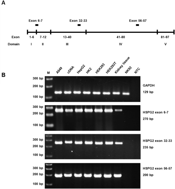
Kidney stone disease (KSD) is a prevalent and complex condition, with an incidence of 85 cases per 100,000 individuals in Thailand. Notably, over 40% of cases are concentrated in the northeastern region, indicating a potential genetic influence, which is supported by genetic mutations reported in several families by our research group. Despite this, the genetic basis of KSD remains largely unknown for many Thai families. This study aimed to identify the genetic mutation responsible for KSD in a specific Thai family, the UBRS131 family, which includes four affected individuals. Whole exome sequencing was performed, and variant filtering using the VarCards2 program identified 10 potentially causative mutations across 9 genes. These mutations were subjected to segregation analysis among family members and screened in 180 control and 179 case samples using real-time PCR-HRM or PCR-RFLP techniques. Prioritization of these variants using GeneDistiller identified the p.Asp775Glu mutation in the heparan sulfate proteoglycan 2 (HSPG2) gene as the likely causative mutation for KSD in this family. The Asp775 residue is highly conserved across vertebrates, and structural analysis suggests that the Glu775 substitution may disrupt the formation of two crucial hydrogen bonds, potentially altering the mutant protein's configuration. Immunohistochemistry confirmed the presence of perlecan (HSPG2 protein) in the proximal tubules in nephrons. These findings highlight the significant role of the HSPG2 gene in familial KSD within this study family.
Keywords: HSPG2; Heparan sulfate proteoglycan 2; Kidney stone disease; Whole exome sequencing.
© 2024. The Author(s).
Conflict of interest statement
Declarations. Conflict of interest: The authors declare no competing interests.
Figures


Similar articles
-
Exome sequencing identifies a rare HSPG2 variant associated with familial idiopathic scoliosis.G3 (Bethesda). 2014 Dec 12;5(2):167-74. doi: 10.1534/g3.114.015669. G3 (Bethesda). 2014. PMID: 25504735 Free PMC article.
-
Exome sequencing identified rare variants in genes HSPG2 and ATP2B4 in a family segregating developmental dysplasia of the hip.BMC Med Genet. 2017 Mar 21;18(1):34. doi: 10.1186/s12881-017-0393-8. BMC Med Genet. 2017. PMID: 28327142 Free PMC article.
-
Novel HSPG2 mutations causing Schwartz‑Jampel syndrome type 1 in a Chinese family: A case report.Mol Med Rep. 2018 Aug;18(2):1761-1765. doi: 10.3892/mmr.2018.9143. Epub 2018 Jun 6. Mol Med Rep. 2018. PMID: 29901129
-
Characteristics and Yield of Modern Approaches for the Diagnosis of Genetic Causes of Kidney Stone Disease.Genes (Basel). 2024 Nov 14;15(11):1470. doi: 10.3390/genes15111470. Genes (Basel). 2024. PMID: 39596670 Free PMC article. Review.
-
Border patrol: insights into the unique role of perlecan/heparan sulfate proteoglycan 2 at cell and tissue borders.Matrix Biol. 2014 Feb;34:64-79. doi: 10.1016/j.matbio.2013.08.004. Epub 2013 Aug 31. Matrix Biol. 2014. PMID: 24001398 Free PMC article. Review.
References
-
- Resnick M, Pridgen DB, Goodman HO (1968) Genetic predisposition to formation of calcium oxalate renal calculi. N Engl J Med 278(24):1313–1318. 10.1056/nejm196806132782403 - PubMed
-
- Daga A, Majmundar AJ, Braun DA, Gee HY, Lawson JA, Shril S, Jobst-Schwan T, Vivante A, Schapiro D, Tan W, Warejko JK, Widmeier E, Nelson CP, Fathy HM, Gucev Z, Soliman NA, Hashmi S, Halbritter J, Halty M, Kari JA, El-Desoky S, Ferguson MA, Somers MJG, Traum AZ, Stein DR, Daouk GH, Rodig NM, Katz A, Hanna C, Schwaderer AL, Sayer JA, Wassner AJ, Mane S, Lifton RP, Milosevic D, Tasic V, Baum MA, Hildebrandt F (2018) Whole exome sequencing frequently detects a monogenic cause in early onset nephrolithiasis and nephrocalcinosis. Kidney Int 93(1):204–213. 10.1016/j.kint.2017.06.025 - PMC - PubMed
-
- Nettuwakul C, Praditsap O, Sawasdee N, Pungsrinont T, Sritippayawan S, Ahsan N, Yenchitsomanus PT, Rungroj N (2022) Genetic heterogeneity of kidney stone disease in northeastern Thai patients. Genomics Genet 15(1):1–15
-
- Jabalameli MR, Fitzpatrick FM, Colombo R, Howles SA, Leggatt G, Walker V, Wiberg A, Kunji ERS, Ennis S (2021) Exome sequencing identifies a disease variant of the mitochondrial ATP-Mg/Pi carrier SLC25A25 in two families with kidney stones. Mol Genet Genomic Med 9(12):e1749. 10.1002/mgg3.1749 - PMC - PubMed
MeSH terms
Substances
Grants and funding
LinkOut - more resources
Full Text Sources
