Case Reports
. 2024 Dec 16:25:e945519.
doi: 10.12659/AJCR.945519.

Perihilar Cholangiocarcinoma Originating in Peribiliary Glands: Insights from a Case without Precancerous Lesions

Affiliations
Case Reports

Perihilar Cholangiocarcinoma Originating in Peribiliary Glands: Insights from a Case without Precancerous Lesions

Yukihiro Shirota et al. Am J Case Rep. .

Abstract

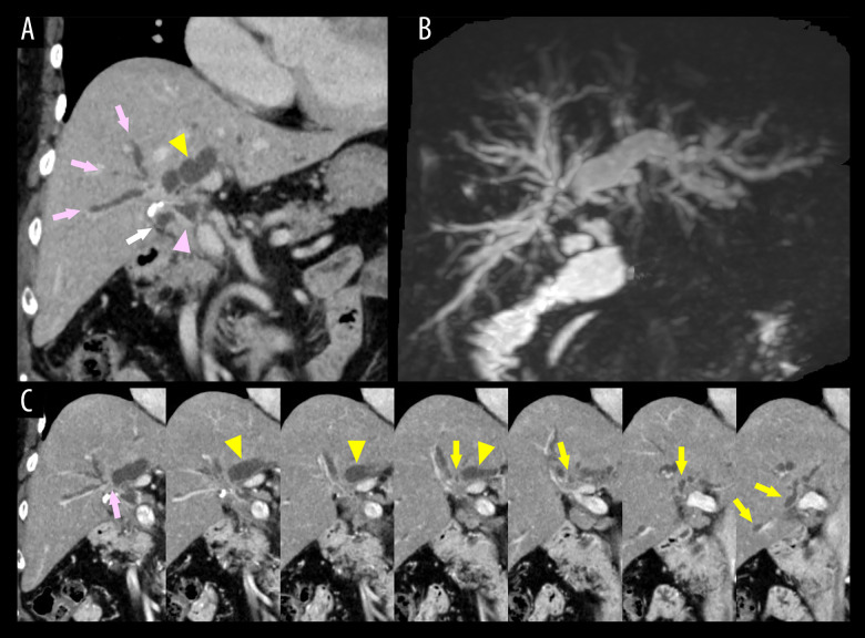
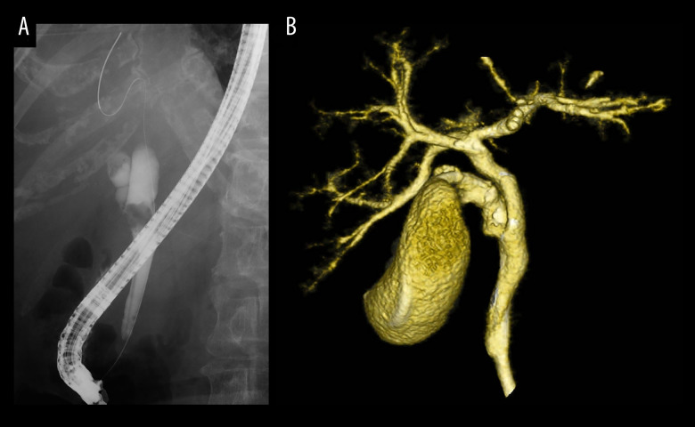
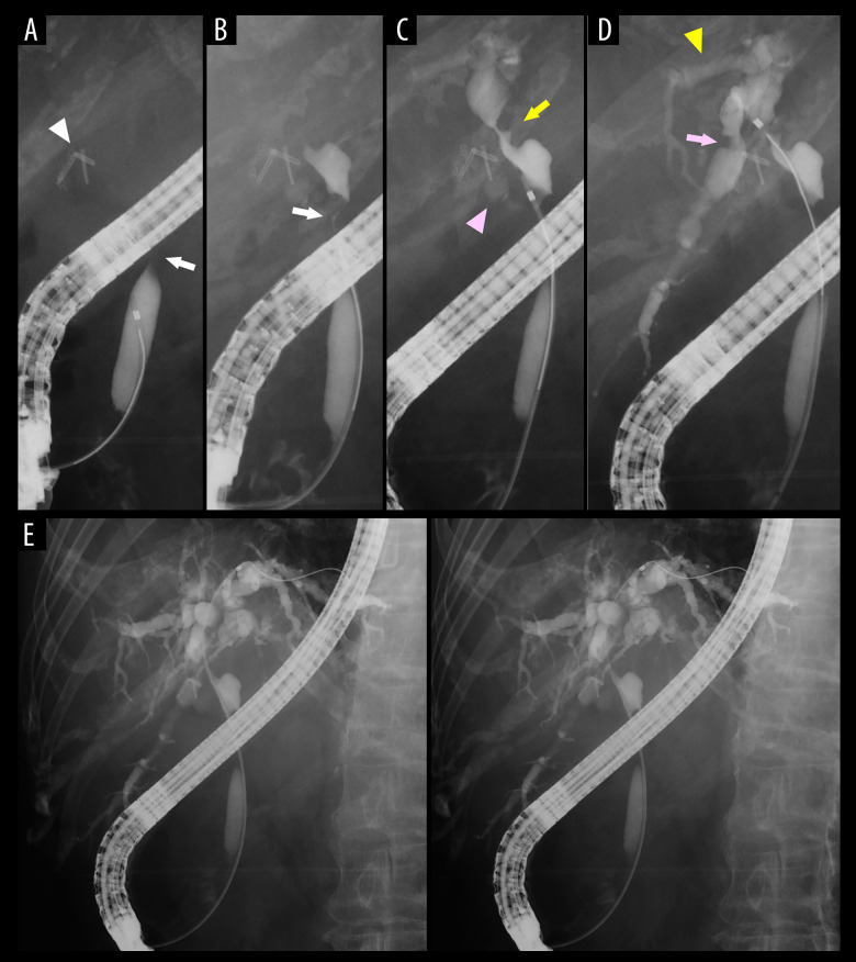
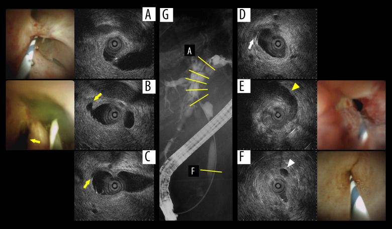
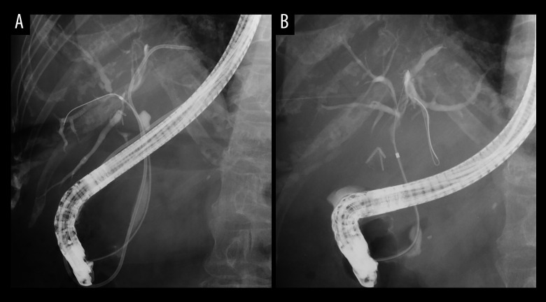
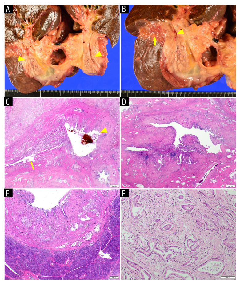
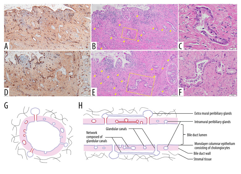
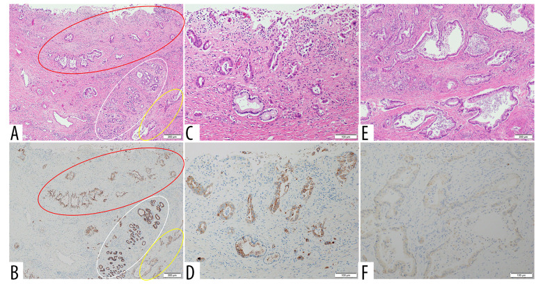
BACKGROUND Recent studies have shown that peribiliary glands may be the potential cell origin of cholangiocarcinoma, and that precancerous lesions such as biliary intraepithelial neoplasms and intraductal papillary neoplasms of the bile duct may arise from these peribiliary glands. However, whether and how these precancerous lesions progress to cholangiocarcinoma is controversial. CASE REPORT Herein, an autopsy case of perihilar cholangiocarcinoma, exclusively periductal-infiltrating, is reported. Since repeated transpapillary biopsies and cytology showed no carcinoma cells, the patient was treated for sclerosing cholangitis until death. The findings at cholelithiasis treatment 1 year earlier had not aroused suspicion of the presence of precancerous lesions. The changes in the spread of bile duct stenoses on cholangiography and the unique findings at autopsy, namely (i) the distribution of cancer growing locally within the peribiliary gland compartment without invading the bile duct mucosa and (ii) the existence of in situ-like carcinoma cells replacing the epithelium of the peribiliary glands throughout the extrahepatic bile duct, suggested that cholangiocarcinoma arose from the peribiliary glands in the hilum without a detectable precancerous lesion and then spread to the lower end of the common bile duct via the peribiliary gland network. CONCLUSIONS This case report may help further our understanding of the natural history of cholangiocarcinoma and provide clues about cholangiocarcinogenesis and progression. In addition, histological and cytological diagnosis could be theoretically difficult by sampling tissue from the bile duct lumen in cholangiocarcinoma, as in this case.


Conflict of interest statement

Conflict of interest: None declared

Figures

Figure 1.
Figure 1.
Radiological findings of bile duct at onset. Representative coronal reformatted image section of contrast-enhanced CT (A) in the portal phase shows upstream dilatation of the left hepatic duct (yellow arrow head) and local dilatation of the common hepatic duct with wall thickening (pink arrow head). Mild dilated right anterior sectoral duct branches (pink arrow) and a remnant cystic duct (white arrow) are also shown. Continuous coronal sections of CT from anterior to posterior (B) show stenoses of bifurcations of the right posterior (yellow arrow) and anterior (pink arrow) sectoral ducts from the left hepatic duct (yellow arrow head) with wall thickening. Magnetic resonance cholangiopancreatography shows these stenoses and dilatation stereoscopically (C).
Figure 2.
Figure 2.
Radiological findings at cholelithiasis treatment 1 year earlier. Endoscopic retrograde cholangiopancreatography (ERCP) at electrohydraulic lithotripsy (A) and volume rendering image of drip infusion cholecystocholangiography (DIC-CT) before cholecystectomy (B) about 1 year earlier, showing no abnormality of the bile duct, such as stenosis. The ERCP shows stones at the orifice of and in the cystic duct. At DIC-CT, a transpapillary gallbladder stent is placed. The DIC-CT shows the anomaly of branching at the hilar bile duct: the right posterior sectoral duct and right anterior sectoral duct individually join the left hepatic duct in this order from upstream to form the common hepatic duct.
Figure 3.
Figure 3.
Endoscopic retrograde cholangiopancreatography (ERCP) findings 1 week after onset. ERCP images from anterior in chronological order of contrast agent spreading showing multiple, non-continuous, smooth stenoses at the upper common bile duct (A and B, white arrow), common hepatic duct (C, yellow arrow), and right posterior sectoral duct root (D, pink arrow). A lightly stained remnant cystic duct (C, pink arrow head) and right anterior sectoral duct (D, yellow arrow head) are also shown. Clips on the cystic duct at laparoscopic cholecystectomy about 1 year earlier are shown (A, white arrow head). ERCP stereo image from anterior when the contrast agent has spread sufficiently (E).
Figure 4.
Figure 4.
Direct cholangioscopy and intraductal ultrasonography (IDUS) images from left hepatic duct branches to the lower portion of the common bile duct. The images, in which the cholangioscopy and IDUS images are arranged side by side (A, B, E, and F), are images of almost the same bile duct site. Each site of these images is indicated by a yellow line on the endoscopic retrograde cholangiopancreatography (ERCP) image (G), from (A) to (F), from upstream to downstream. The IDUS image at left hepatic duct branches shows no wall thickening (A). The images at the levels of the bifurcation of the right posterior sectoral duct from the left hepatic duct (B and C, yellow arrow), of the bifurcation of the anterior sectoral duct from the common hepatic duct (D, white arrow), and of the lower common bile duct (F, white arrow head shows pancreatic duct) show relatively uniform and diffuse wall thickening continuously, extending beyond the sites of stenosis indicated by ERCP. The IDUS images at the 2 sites of stenosis of the common hepatic duct (E, yellow arrow head shows the right hepatic artery) and of the upper common bile duct show wall thickening higher than that at the other sites, but its outer boundary is almost preserved. Direct cholangioscopic images show whitish smooth surface mucosa not only at the level at which wall thickening is not detected (A), but also at the levels at which wall thickening is detected by IDUS (B and F). At the small parts of the tightest stenoses of the common hepatic duct and the upper common bile duct are shown relatively well-ordered blood vessel distension (E).
Figure 5.
Figure 5.
The changes of endoscopic retrograde cholangiopancreatography (ERCP) findings during the clinical course. ERCP performed 3 weeks after endoscopic nasal biliary drainage (ENBD; 1 month after onset) shows that the stenoses extend to the anterior and posterior sectoral duct roots more clearly (A). A guide wire and a cannula have been inserted to the right anterior sectoral duct. An ENBD tube has been inserted to the left hepatic duct branch. The right posterior sectoral duct is branched from the left hepatic duct upstream of the bifurcation of the right anterior sectoral duct. ERCP performed after treatment by ENBD for stent dysfunction (7 months after onset) shows that the stenosis extends from the left hepatic duct and right anterior branch to the lower end of the common bile duct continuously (B). The right posterior branch is not visualized. A guide wire has been inserted to the left hepatic duct branch. A cannula has been inserted to the common bile duct.
Figure 6.
Figure 6.
Autopsy bile duct findings. The common bile duct (A, yellow arrow head) shows an irregularly thickened wall and partially severely constricted lumen, but with a smooth surface. Around the common hepatic duct (B, yellow arrow head) and the duodenal bulb (B, yellow arrow) severe adhesion is shown. The hematoxylin and eosin-stained specimens in the low-power fields show periductal fibrous thickening continuously at the bifurcation of the right posterior or anterior sectoral duct (C, yellow arrow: it was difficult to distinguish between the right posterior and anterior sectoral ducts on liver excisions) from the left hepatic duct (C, yellow arrow head), at the upper common bile duct (D), and at the lower common bile duct (E). The specimen in the mid-power field shows well-differentiated adenocarcinoma with desmoplastic stroma in a periductal infiltrating pattern (F).
Figure 7.
Figure 7.
Microscopic findings of the peribiliary glands (PBGs). Two representative sections of the bile duct wall with few invasive cancer components (A–C and D–F) show the bile duct lumen and intramural PBGs using serial sections (A corresponds to B, and D to E). Immunohistochemical study using anti-laminin antibody delineating the basement membrane of the PBGs and glandular canals (A and D). Vessels are also delineated more clearly. The hematoxylin and eosin-stained specimens in the mid-power field show the relationship of the intramural PBG and the adenocarcinoma (B and E). The specimens in the high-power field (C and F, magnified image of the yellow square in B and E, respectively) show the in situ-like carcinoma cells that extend, replacing the epithelium of the PBG. The epithelium of most of the PBGs and glandular canals is replaced by carcinoma cells (B and E, yellow arrow head), whereas some intact PBGs are seen (white circle). The carcinoma cells in the bile duct lumen are hardly seen in these specimens. Drawings schematically show a cross-section (G) and a longitudinal section (H) of a segment of the extrahepatic bile duct containing both intramural and extramural PBGs. The observed distribution of the carcinoma cells in this case is shown by the red lines.
Figure 8.
Figure 8.
Immunohistochemical study using anti-E-cadherin antibody. The hematoxylin and eosin-stained specimen of bile duct wall in the mid-power field (A) shows both the in situ-like carcinoma cells that extend, replacing the epithelium of the peribiliary gland (PBG; red and yellow circles) and intact PBGs (white circle). Immunohistochemical study using anti-E-cadherin antibody of this specimen using serial section (B) shows that the epithelial cells of the PBG are positive for E-cadherin in an apicolateral pattern (white circle). In contrast, the expression of E-cadherin decreases in carcinoma cells (red and yellow circles), and the decreasing trend is stronger in the deeper region (yellow circle) than in the shallow region (red circle). The hematoxylin and eosin-stained specimens in the high-power field of the intramural (C) and the invasive portion of outside the bile duct wall (E) show carcinoma cells in which the expression of E-cadherin is decreased in the intramural region (D) and is lost in the invasive portion (F) on the immunohistochemical study using anti-E-cadherin antibody.

Similar articles

References

    1. Lanzoni G, Cardinale V, Carpino G. The hepatic, biliary, and pancreatic network of stem/progenitor cell niches in humans: A new reference frame for disease and regeneration. Hepatology. 2016;64(1):277–86. - PubMed
    1. Moeini A, Haber PK, Sia D. Cell of origin in biliary tract cancers and clinical implications. JHEP Rep. 2021;3(2):100226. - PMC - PubMed
    1. Banales JM, Marin JJG, Lamarca A, et al. Cholangiocarcinoma 2020: the next horizon in mechanisms and management. Nat Rev Gastroenterol Hepatol. 2020;17(9):557–88. - PMC - PubMed
    1. Umemura A, Suto T, Sasaki A, et al. Pure laparoscopic left hemihepatectomy for hepatic peribiliary cysts with biliary intraepithelial neoplasia. Case Rep Surg. 2016;2016:7236427. - PMC - PubMed
    1. Miyata T, Uesaka K, Nakanuma Y. Cystic and papillary neoplasm at the hepatic hilum possibly originating in the peribiliary glands. Case Rep Pathol. 2016;2016:9130754. - PMC - PubMed

Publication types