. 2024 Dec 24;121(52):e2413883121.
doi: 10.1073/pnas.2413883121. Epub 2024 Dec 16.

The distinctive mechanical and structural signatures of residual force enhancement in myofibers

Affiliations

The distinctive mechanical and structural signatures of residual force enhancement in myofibers

Anthony L Hessel et al. Proc Natl Acad Sci U S A. .

Abstract

In muscle, titin proteins connect myofilaments together and are thought to be critical for contraction, especially during residual force enhancement (RFE) when steady-state force is elevated after an active stretch. We investigated titin's function during contraction using small-angle X-ray diffraction to track structural changes before and after 50% titin cleavage and in the RFE-deficient, mdm titin mutant. We report that the RFE state is structurally distinct from pure isometric contractions, with increased thick filament strain and decreased lattice spacing, most likely caused by elevated titin-based forces. Furthermore, no RFE structural state was detected in mdm muscle. We posit that decreased lattice spacing, increased thick filament stiffness, and increased non-cross-bridge forces are the major contributors to RFE. We conclude that titin directly contributes to RFE.

Keywords: X-ray diffraction; elasticity; force transmission; mouse; ultrastructure.


Conflict of interest statement

Competing interests statement:T.C.I. provides consulting and collaborative research studies to Edgewise Therapeutics inc. and A.L.H./M.K. are owners of Accelerated Muscle Biotechnologies LLC, but such work is unrelated to the content of this article. All other authors declare that they have no competing interests.

Figures
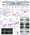
Fig. 1.
Fig. 1.
Mechanical properties of TC fiber bundles before and after 50% TC during IsoS, IsoL, and IsoR conditions. (A) Schematic of skeletal half-sarcomere with relevant structural periodicities, I-band titin segments, and TEVP cleavage site indicated. (B and C) Tension traces of fibers during mechanical experiments before (B) and after (C) 50% TC, normalized to the total tension during ISOL. Active tension is total tension during contractions, minus the passive tension at those lengths. (D) Passive stiffness after TC (n = 12-31), normalized to its paired pre-stiffness value at IsoS. (E) Tension before (blue) and after (red) TC for passive, active, and total conditions (n = 23-28). Tension is normalized by individual to the active tension during IsoShort. (F) Transmission electron micrographs of TC sarcomeres after full mechanical protocols with sham (−TEVP) or treatment (+TEVP) conditions. (Scale bar, 1 µm.) Expanded image available in SI Appendix, Fig. S2. (G) RFE in fibers before and after titin treatment (n = 23-27). RFE is calculated as active tension during ISOR—active tension during ISOL and normalized to active tension during ISOS. (H) Representative X-ray diffraction pattern of skeletal psoas fibers during IsoS before TC, with labeled reflections indicating relevant periodic structures that are referenced in (A). An X-ray pattern from a sample after TEV protease treatment is presented in SI Appendix, Fig. S3. Statistics: ANOVA design with random effect of individual, followed by Tukey HSD multiple comparison procedure on significant main effects (P < 0.05). Data displayed as connecting letters: Different letters are significantly different (Tukey HSD P < 0.05). Data throughout reported as mean ± SEM. Full statistical details in SI Appendix, Table S1.
Fig. 2.
Fig. 2.
Sarcomeric structural parameters of TC fibers before and after 50% TC. D1,0 (n = 25-27) (A), I1,1/I1,0 (n = 25-27) (B), σA (n = 24-26) (C), σD (n = 23-25) (D) SM3 (n = 24-26) (E), IM3 (n = 24-26) (F), SgActin (n =12-16) (G), ST3 (n =19-24) (H), and SM6 (n = 20-25) (I) were recorded before (blue) and after (red) 50% TC, at three conditions: IsoS, IsoL, and IsoR. Statistics: ANOVA design with random effect of individual, followed by Tukey HSD multiple comparison procedure on significant main effects (P < 0.05). Data displayed as connecting letters: Different letters are significantly different (Tukey HSD P < 0.05). Full statistical details in SI Appendix, Tables S2 and S3. Intrasample pre–post differences in SI Appendix, Fig. S5.
Fig. 3.
Fig. 3.
Mechanism of RFE. Configuration of sarcomeric proteins during (A) IsoS, (B) IsoL, and (C) IsoR that can account for their distinctive mechanical and structural signatures. Filament extension is dramatized for visualization purposes and is smaller in real sarcomeres. In passive muscle, low-level titin-thin filament interactions occur in such a way that passive stretch is enough to detach-reattach and/or drag titin along the thin filament so that titin-based free length and extension are still like if they were not attached at all. During contraction, the titin-thin filament interaction becomes stronger, so that during an eccentric contraction, titin extension occurs above that in passive, producing elevated titin-based force and explaining the mechanical and structural signatures in IsoR. Increased titin-based force contributes to RFE, which also leads to smaller lattice spacing and increased thick filament stiffness, improving force production and force transmission, respectively.

Update of

References

    1. Dickinson M. H., et al. , How animals move: An integrative view. Science 288, 100–106 (2000). - PubMed
    1. Nishikawa K. C., Muscle function from organisms to molecules. Integr. Comp. Biol. 58, 194–206 (2018). - PubMed
    1. Dick T. J. M., Biewener A. A., Wakeling J. M., Comparison of human gastrocnemius forces predicted by Hill-type muscle models and estimated from ultrasound images. J. Exp. Biol. 220, 1643–1653 (2017). - PMC - PubMed
    1. Seiberl W., Power G. A., Herzog W., Hahn D., The stretch-shortening cycle (SSC) revisited: Residual force enhancement contributes to increased performance during fast SSCs of human m. adductor pollicis. Physiol. Rep. 3, e12401 (2015). - PMC - PubMed
    1. Lindstedt S. L., Skeletal muscle tissue in movement and health: Positives and negatives. J. Exp. Biol. 219, 183–188 (2016). - PubMed

LinkOut - more resources