Review
. 2025 Jan 22;125(2):933-971.
doi: 10.1021/acs.chemrev.4c00681. Epub 2024 Dec 16.

Engineering Phages to Fight Multidrug-Resistant Bacteria

Affiliations
Review

Engineering Phages to Fight Multidrug-Resistant Bacteria

Huan Peng et al. Chem Rev. .

Abstract

Facing the global "superbug" crisis due to the emergence and selection for antibiotic resistance, phages are among the most promising solutions. Fighting multidrug-resistant bacteria requires precise diagnosis of bacterial pathogens and specific cell-killing. Phages have several potential advantages over conventional antibacterial agents such as host specificity, self-amplification, easy production, low toxicity as well as biofilm degradation. However, the narrow host range, uncharacterized properties, as well as potential risks from exponential replication and evolution of natural phages, currently limit their applications. Engineering phages can not only enhance the host bacteria range and improve phage efficacy, but also confer new functions. This review first summarizes major phage engineering techniques including both chemical modification and genetic engineering. Subsequent sections discuss the applications of engineered phages for bacterial pathogen detection and ablation through interdisciplinary approaches of synthetic biology and nanotechnology. We discuss future directions and persistent challenges in the ongoing exploration of phage engineering for pathogen control.


Conflict of interest statement

The authors declare no competing financial interest.

Figures
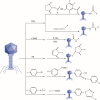
Figure 1
Figure 1
Representative chemical reactions for phage chemical modification.
Figure 2
Figure 2
Engineering phages with homologous recombination. Phage infects the host bacterium and injects the genome DNA into the host cell (A). The phage DNA recombines with the plasmid DNA which is partially (in green) homologous to the phage genome (B) and generates recombinant phage genomes (C). The genomes are packaged in recombinant phage particles (D) that are subsequently released (E).
Figure 3
Figure 3
Engineering phages with recombineering. Phage infects the host bacterium, injects the genome into the host cell and foreign DNA is transformed into the cell (A). The phage genome recombines with the foreign DNA which can partially (in green) homology to the phage genome (B) and generates recombinant phage genomes (C). The genomes are packaged in recombinant phage particles (D) that are subsequently released (E).
Figure 4
Figure 4
Engineering phages with BRED. Phage genome DNA is extracted (A) and coelectroporated into cells with recombineering DNA substrates (B). The recombination between the homologous parts (in black) yields recombinant genome DNA (C), which are packaged in recombinant phage particles (D) that are subsequently released (E).
Figure 5
Figure 5
Yeast-based assembly of phage genomes. Phage genome DNA is extracted (A) and coelectroporated into yeast cells with YAC molecules (B). The recombination between the homologous parts (in green) yields recombinant phage genome DNA (C), which is extracted (D) and in coelectroporated into bacterial cells (E). The resulting recombinant phage particles (F) are subsequently released (G).
Figure 6
Figure 6
Synthetic phage genome in vitro. The phage genome is extracted (A), subcloned into different pieces (B) and further manipulated with desired mutations (C, D). The recombinant DNA fragments are inserted into the phage genome (E, F) and electroporated into E. coli (G) or L-form bacterium (H), yielding recombinant phages (I).
Figure 7
Figure 7
Gene assembly in vitro. The synthetic oligonucleotides (A) are annealed, assembled, and ligated to get double strand phage genome DNA (A), which is circularized when appropriate (B) and electroporated into host cells (C). The phage genome DNA is packaged into phage particles (D) that is subsequently released (E).
Figure 8
Figure 8
Rebooting phage in a cell-free environment. In a cell-free expression system, the phage genome DNA is transcribed into mRNA (A) and translated into protein (B), and DNA can replicate (C,D). Subsequently, the phage genomes are packaged into phage particles (E).
Figure 9
Figure 9
Utilizing CRISPR-Cas systems for phage genome editing. During the crRNA biogenesis (A), the repeat-spacer array is transcribed into crRNA (brown, blue, and green rectangles). During the interference (B), the crNRAs combine with the Cas proteins to form effector complexes, breaking down the phage genome DNA (C). Through homogeneous recombination, the DNA fragments acquire mutations in the desired genes from a donor DNA construct to form recombinant phage genome DNA (D,E), which is packaged and released subsequently (F).
Figure 10
Figure 10
Representative CRISPR-Cas systems for phage genome editing, Type I-E from Escherichia coli, Type II-A from Streptococcus pyogenes, Type III-A from Staphylococcus epidermidis, Type V-A from Francisella novicida, Type VI-A from Leptotrichia shahii.
Figure 11
Figure 11
Schematic illustration of GOTraP to extend phage host range. (I) T7 phages without tail genes infects E. coli hosts carrying a plasmid with randomly mutated tail gene, antibiotic resistance gene, and packing signal gene. (II) The resulting phage library contains phage mutants with mutated tail protein. (III) Phage mutants are incubated with potential host strains and phages with compatible tail proteins recognizing the host bacteria can inject the plasmid with improved efficiency. (IV) Hosts requiring plasmid are selected on plates with antibiotics. The plasmids are extracted, mutated and transformed into the hosts. This process is repeated to optimize the tail gene and selected genes are sequenced to identify the desired mutations. Adapted with permission from ref (179). Copyright 2017 Elsevier.
Figure 12
Figure 12
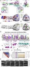
Structure-guided rational design of chimeric RBPs. (A–C) Crystal structure of the PSA tail spike. (A) Ribbon diagram of the Gp15CTD homotrimeric. (B,C) Molecular surface of the RBP complex (B) and the PSA RBP with host-range mutants (C). (D) Analysis of the stem-neck conversation and crystal structure. (E–G) Head domains analysis revealed limited sequence identity of SV 4a, SV 5, SV 6b, and SV 1/2 (E), and they were used to construct the stem- and neck-chimeric phages (F). Host ranges and infection efficiencies of the synthetic phage chimeras (G). Adapted with permission from ref (280). Copyright 2019 Elsevier.
Figure 13
Figure 13
Engineered phages expressing reporter genes for bacterial detection. (A,C,F) Schemes illustrating bacterial detection with engineered phage for bioluminescence (A), colorimetric (C), and electrochemical (F) detection. (B) Bioluminescence time course assays of bacteria infected with the reporter phage. Adapted with permission from ref (292). Copyright 2020 Meile et al. (D) Absorbance intensities vs detection time with T7lacZ phage for bacterial detection. Adapted with permission from ref (293). Copyright 2017 American Chemical Society. (E) Fluorescence images of mCherrybombϕ infections in the presence of p-nitrobenzoic acid for detection of M. smegmatis. Adapted with permission from ref (294). Copyright 2018 Rondón et al. (G) Dependence of peak current obtained from differential pulse voltammetry curve on varying E. coli concentrations for different incubation times. Adapted with permission from ref (295). Copyright 2017 American Chemical Society.
Figure 14
Figure 14
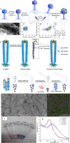
Bacterial detection with engineered phages and conjugated inorganic nanoparticles., (A) Scheme illustrating bacterial detection with engineered T7 phage and quantum dots (QD). (B) TEM images of T7 phage or phage bound to streptavidin-functionalized QDs targeted bacteria. (C–E) Using flow cytometry to detect phage–QD complexes. Plots of bacterial cells targeted by T7-myc (C) or T7-bio (D) phage after adding QDs. (E) Cell number vs fluorescence with the control and biotinylated phage. Adapted with permission from ref (328). Copyright 2006 the National Academy of Sciences of the USA. (F) Scheme illustrating bacterial detection with engineered M13 phage and gold nanoparticles (AuNPs). (G,H) TEM images of M13 phage before and after thiolation engineering. (I) Confocal microscopy image of engineered M13 phage capturing the host cell. Digital photos (J) and UV–vis spectra (K) exhibit the detection of bacteria with engineered M13 phage and AuNPs. Adapted with permission from ref (78). Copyright 2018 American Chemical Society.
Figure 15
Figure 15
Engineered phages to reduce bacterial virulence or increase susceptibility to antibiotics., (A) Schematic illustration of engineered ϕlexA3 phage enhancing ablation of E. coli EMG2 with antibiotics. (B) Bacterial cell ablation ability of no phage, wild-type phage ϕunmod, and modified phage ϕlexA3 with 1 μg/mL ofloxacin. Adapted with permission from ref (355). Copyright 2009 the National Academy of Sciences of the USA. (C,D) Reducing Shiga toxin (Stx2) production by genetically engineered a temperate phage. (C) Schematic illustration of using for in situ repression of the virulence factor in a mouse model. (D) Schematic illustration of using phage to induce enforced lysogeny of endogenous prophage stx2 repression. Adapted with permission from ref (357). Copyright 2020 Hsu et al.
Figure 16
Figure 16
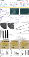
Engineered phages against bacterial biofilm. (A–G) Engineered DspB-T7 phage disperses biofilms. (A) Schematic illustration of removing biofilm with engineered T7 phages expressing DspB. (B,F) Time course of viable cell counts (B) and dose–response curves of mean cell densities (F) treated with T7control and T7DspB. (C,D) Scanning electronic microscopy images of T7DspB-treated biofilm (C) and untreated biofilm (D) after 20 h. (E,G) Time course of phage counts (E) and dose–response curves of phage number (G) after inoculating biofilm with T7DspB and the control. Adapted with permission from ref (358). Copyright 2007 the National Academy of Sciences of the USA. (H–J) Engineering of phages Y2::dpoL1-C inhibits biofilm formation. (H) Plaque of Y2::dpoL1-C (right) gives them a clearer appearance compared with that of Y2 (left). (I) Time course of E. amylovora CFBP 1430 infected by different phages. (J) Engineered Y2::dpoL1-C inhibited E. amylovora to colonize on flowers. Adapted with permission from ref (315). Copyright 2017 American Society for Microbiology. (K–P) Inhibiting biofilm development by engineered T7 phage expressing quorum-quenching enzyme. (K–M) Degradation of AHLs in the condition of buffer (K) or wild-type T7 (L) or engineered T7aiiA (M). (N–P) Engineered phage degrading biofilms. (N) Different bacterial species are plated in the presence/absence of wild-type or engineered T7 phage. (O) Analysis of the dosage of phages on the development of bacterial biofilm. (P) Analysis of effect of wild-type and engineered T7 phages on the formation of dual-species bacterial biofilm. Adapted with permission from ref (359). Copyright 2014 American Society for Microbiology.
Figure 17
Figure 17
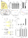
Engineered phages delivering Cas nuclease to kill bacteria specifically. (A–C) Delivering CRISPR system to specifically ablate S. aureus. (A) Schematic illustrating ΦNM1 phage delivering a specific phagemid to S. aureus to kill the bacterial cells. (B) Effects of phagemid targeting drug resistance gene or the control. (C) Results of eliminating USA300Φ in a bacterial community using the phagemid. Adapted with permission from ref (360). Copyright 2014 Springer Nature America, Inc. (D–F) Phages deliver RNA-guided nucleases (RGN) constructs as sequence-specific antimicrobials. (D) Scheme illustrating phage delivering RGN to influence the physiology of the hosts. (E) Ablation of wild-type or drug-resistant EMG2 bacteria with engineered phage or control. (F) In vivo experimental results in G. mellonella larvae model treated with buffer, EHEC, ΦRGNeae, and ΦRGNndm-1. Adapted with permission from ref (361). Copyright 2014 Springer Nature America, Inc. (G–I) Utilizing lytic phages to enhance the population of bacteria sensitive to antibiotics. (G) Scheme illustrating strategies to enhance bacterial population that are sensitive to antibiotics. (H) Enhancing the population E. coli resistant to phage. (I) Enhancing the population of E. coli sensitive to antibiotics. Surviving colonies from each culture were inoculated on plates having or lacking streptomycin or gentamicin. Adapted with permission from ref (362). Copyright 2015 the National Academy of Sciences of the USA.
Figure 18
Figure 18
Engineered phages delivering antimicrobial nanomaterials. (A–C) Chimeric M13 phages deliver gold nanorods to specifically kill bacterial pathogens.,, (A) Schematic illustration of constructing phanorod and killing bacterial pathogens specifically by irradiating the gold nanorods. (B,C) Cell viability assay of bacterial biofilm on mammalian cells, treated with phanorods, before (B) and after (row C) NIR irradiation. Adapted with permission from ref (71). Copyright 2020 the National Academy of Sciences of the USA. (D,E) Engineered phages deliver AIEgens for imaging, targeting and killing of bacterial pathogens. (D) Scheme illustrating preparing phage-AIE conjugates TVP-PAP. (E) In vivo experimental results of using TVP–PAP to treat wounds infected with MDR P. aeruginosa in a mice model. Adapted with permission from ref (154). Copyright 2020 American Chemical Society. (F–I) Engineered phages with PEI for intracellular pathogen inhibition. (F) Scheme illustration of capping phage head with PEI enables endosomal escape in cell to eliminate intracellular bacteria. (G–I) Evaluation of bacterial treatment with PEI@P in vivo. Photos of the major organs from mice treated with PEI@P and the control groups (G). Distribution of bacteria in the major organs (H,I). Adapted with permission from ref (363). Copyright 2022 The American Association for the Advancement of Science.

Similar articles

Cited by

References

    1. Ikuta K. S.; Swetschinski L. R.; Robles Aguilar G.; Sharara F.; Mestrovic T.; Gray A. P.; Davis Weaver N.; Wool E. E.; Han C.; Gershberg Hayoon A.; et al. Global mortality associated with 33 bacterial pathogens in 2019: a systematic analysis for the Global Burden of Disease Study 2019. Lancet 2022, 400, 2221–2248. 10.1016/S0140-6736(22)02185-7. - DOI - PMC - PubMed
    1. Butler M. S.; Henderson I. R.; Capon R. J.; Blaskovich M. A. T. Antibiotics in the clinical pipeline as of December 2022. J. Antibiot. 2023, 76, 431–473. 10.1038/s41429-023-00629-8. - DOI - PMC - PubMed
    1. Beyer P.; Paulin S. The Antibacterial Research and Development Pipeline Needs Urgent Solutions. ACS Infect. Dis. 2020, 6, 1289–1291. 10.1021/acsinfecdis.0c00044. - DOI
    1. Anderson M.; Panteli D.; van Kessel R.; Ljungqvist G.; Colombo F.; Mossialos E. Challenges and opportunities for incentivising antibiotic research and development in Europe. Lancet Reg. Health Eur. 2023, 33, 100705.10.1016/j.lanepe.2023.100705. - DOI - PMC - PubMed
    1. Mu A.; Lawley T. D. Bacteriophage discovery to advance biotechnology and biotherapeutics. Nat. Rev. Microbiol. 2023, 21, 279–279. 10.1038/s41579-023-00870-w. - DOI - PubMed

Substances