Microsurgical management of previously embolized intracranial aneurysms: A single center experience and literature review
- PMID: 39681331
- PMCID: PMC11984270
- DOI: 10.7461/jcen.2024.E2024.05.004
Microsurgical management of previously embolized intracranial aneurysms: A single center experience and literature review
Abstract
Background: Endovascular treatment of intracranial aneurysms (IAs) provides less invasiveness and lower morbidity than microsurgical clipping, albeit with a long-term recurrence rate estimated at 20%. We present our single-center experience and a literature review concerning surgical clipping of recurrent previously coiled aneurysms.
Methods: Retrospective analysis of nine (9) patients' data and final clinical/angiographic outcomes, who underwent surgical clipping of IAs in our center following initial endovascular treatment, over a 12-year period (2010-2022). Regarding the literature review, data were extracted from 48 studies including 969 patients with 976 aneurysms.
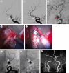
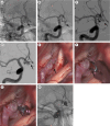
Results: 9 patients (5 males - 4 females) were included in the study with a mean age of 49 years. Subarachnoid hemorrhage was the initial presentation in 78% of patients. Aneurysms' most common location was the middle cerebral artery bifurcation (5/9) followed by the anterior communicating artery (3/9) and the internal carotid artery bifurcation (1/9). Indications for surgery were coil loosening, coil compaction, sac regrowth, and residual neck. Procedure-related morbidity and mortality were zero whereas complete aneurysm occlusion was achieved after surgical clipping in all cases (100%). All patients had minimal symptoms or were asymptomatic (mRS 0-1) at the final follow-up.
Conclusions: Surgical clipping seems a feasible and safe technique for selected cases of recurrent previously coiled intracranial aneurysms. A universally accepted recurrence classification system and a guideline template for the management of such cases are needed.
Keywords: Endovascular embolization; Intracranial aneurysm; Microsurgery; Microsurgical clippin; Recurrence.
Conflict of interest statement
The authors report no conflict of interest concerning the materials or methods used in this study or the findings specified in this paper.
Figures


Similar articles
-
Microsurgical clipping for recurrent aneurysms after initial endovascular coil embolization.World Neurosurg. 2015 Feb;83(2):211-8. doi: 10.1016/j.wneu.2014.08.013. Epub 2014 Aug 10. World Neurosurg. 2015. PMID: 25118057
-
Coil embolization for intracranial aneurysms: an evidence-based analysis.Ont Health Technol Assess Ser. 2006;6(1):1-114. Epub 2006 Jan 1. Ont Health Technol Assess Ser. 2006. PMID: 23074479 Free PMC article.
-
Microsurgical clipping of previously coiled intracranial aneurysms.Clin Neurol Neurosurg. 2013 Aug;115(8):1343-9. doi: 10.1016/j.clineuro.2012.12.030. Epub 2013 Jan 24. Clin Neurol Neurosurg. 2013. PMID: 23352715
-
Treatment of Recurrent Intracranial Aneurysms After Clipping: A Report of 23 Cases and a Review of the Literature.World Neurosurg. 2016 Aug;92:434-444. doi: 10.1016/j.wneu.2016.05.053. Epub 2016 May 27. World Neurosurg. 2016. PMID: 27241096 Review.
-
Microsurgical Treatment of Previously Coiled Giant Aneurysms: Experience with 6 Cases and Literature Review.World Neurosurg. 2023 Mar;171:e336-e348. doi: 10.1016/j.wneu.2022.12.016. Epub 2022 Dec 10. World Neurosurg. 2023. PMID: 36513298 Review.
References
-
- Aoun SG, Rahme RJ, El Ahmadieh TY, Bendok BR, Hunt Batjer H. Incorporation of extruded coils into the third nerve in association with third nerve palsy. Journal of Clinical Neuroscience. 2013 Sep;20(9):1299–302. - PubMed
-
- Asgari S, Doerfler A, Wanke I, Schoch B, Forsting M, Stolke D. Complementary management of partially occluded aneurysms by using surgical or endovascular therapy. J Neurosurg. 2002 Oct;97(4):843–50. - PubMed
-
- Boet R, Poon WS, Yu SC. The management of residual and recurrent intracranial aneurysms after previous endovascular or surgical treatment - a report of eighteen cases. Acta Neurochir (Wien) 2001 Nov;143(11):1093–101. - PubMed
-
- Campi A, Ramzi N, Molyneux AJ, Summers PE, Kerr RSC, Sneade M, et al. Retreatment of ruptured cerebral aneurysms in patients randomized by coiling or clipping in the international subarachnoid aneurysm trial (ISAT) Stroke. 2007 May;38(5):1538–44. - PubMed
-
- Chung J, Lim YC, Kim B, Lee D, Lee K-S, Shin YS. Early and late microsurgical clipping for initially coiled intracranial aneurysms. Neuroradiology. 2010 Dec;52(12):1143–51. - PubMed

