. 2024 Dec 16;14(1):30478.
doi: 10.1038/s41598-024-79572-z.

Renal phenotyping in a hypomorphic murine model of propionic aciduria reveals common pathomechanisms in organic acidurias

Affiliations

Renal phenotyping in a hypomorphic murine model of propionic aciduria reveals common pathomechanisms in organic acidurias

Anke Schumann et al. Sci Rep. .

Abstract

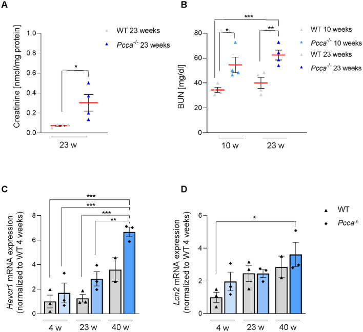
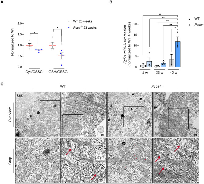
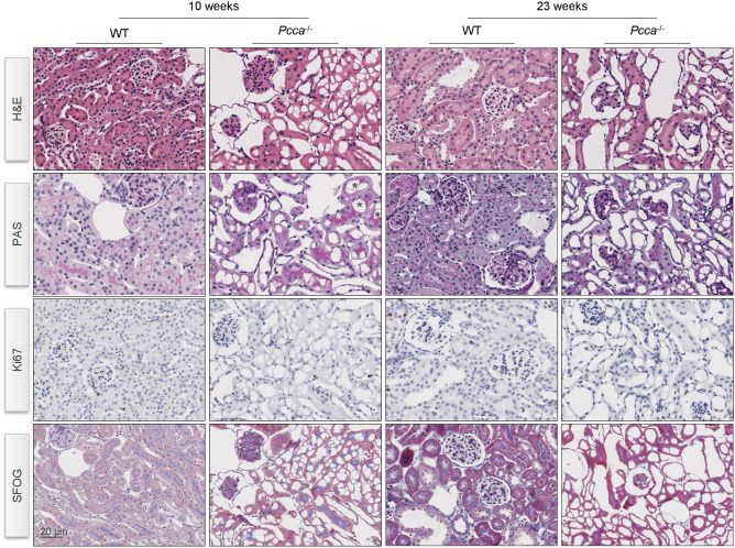
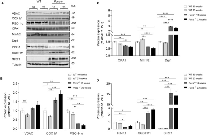
Mutations in the mitochondrial enzyme propionyl-CoA carboxylase (PCC) cause propionic aciduria (PA). Chronic kidney disease (CKD) is a known long-term complication. However, good metabolic control and standard therapy fail to prevent CKD. The pathophysiological mechanisms of CKD are unclear. We investigated the renal phenotype of a hypomorphic murine PA model (Pcca-/-(A138T)) to identify CKD-driving mechanisms. Pcca-/-(A138T) mice show elevated retention parameters and express markers of kidney damage progressing with time. Morphological assessment of the Pcca-/-(A138T) mouse kidneys indicated partial flattening of tubular epithelial cells and focal tubular-cystic dilation. We observed altered renal mitochondrial ultrastructure and mechanisms acting against oxidative stress were active. LC-MS/MS analysis confirmed disease-specific metabolic signatures and revealed disturbances in mitochondrial energy generation via the TCA cycle. Our investigations revealed altered mitochondrial networks shifted towards fission and a marked reduction of mitophagy. We observed a steep reduction of PGC-1-α, the key mediator modulating mitochondrial functions and a counter actor of mitochondrial fission. Our results suggest that impairment of mitochondrial homeostasis and quality control are involved in CKD development in PA. Therapeutic targeting of the identified pathways might help to ameliorate CKD in addition to the current treatment strategies.

Keywords: Chronic kidney disease; Mitochondrial dysfunction; Mitochondrial energy metabolism; Mitochondrial fission; Mitochondrial homeostasis; Mitochondrial quality control; Propionic aciduria.


Conflict of interest statement

Declarations. Competing interests: The authors declare no competing interests. Ethical approval: No material of human origin was used in this publication. Human and animal rights: All animal studies were approved by the Institutional Ethical Committee for Animal Experimentation (Universidad Autónoma de Madrid, references CEI 963-A026 and CEI-134-2830) and by the Regional Environment Department (Comunidad de Madrid, reference PROEX 194/19 and PROEX 204.5/24). The work with mice was performed in accordance with the ARRIVE guidelines (approval number PROEX 194-2019 and PROEX 204.5/24). Animal housing and maintenance protocols followed the local authority guidelines.

Figures

Fig. 1
Fig. 1
Kidney disease in Pcca-/-(A138T) mice. (A) LC–MS/MS quantification of creatinine concentrations in 23-week-old WT and Pcca-/-(A138T) mouse kidneys. Creatinine concentration in nmol/mg protein. n = 4 per group. (B) Blood urea nitrogen (BUN) concentration in the serum of 10- and 23-week-old WT and Pcca-/-(A138T) mice. BUN concentration in mg/dl. n = 4 per group. (C) Transcript levels of kidney injury molecule-1 (KIM-1, Havcr1) and lipocalin 2 (LCN2, Lcn2) obtained by RT-qPCR in kidney extracts of 4-, 23- and 40-week-old (w) WT and Pcca-/-(A138T) mice. n = 3 per group, n = 2 for WT 40-week-old mice.
Fig. 2
Fig. 2
Altered mitochondrial morphology and function in Pcca-/-(A138T) mouse kidneys. (A) Quantitative LC–MS/MS analysis ratio of reduced (cysteine) to oxidized (CSSC) of cysteine and reduced (GSH) to oxidized (GSSG) levels of glutathione. Data normalized to WT. n = 4 per group. (B) Transcript levels of fibroblast growth factor 2 (FGF21, Fgf21) obtained by RT-qPCR in kidney extracts of 4-, 23- and 40-week-old (w) WT and Pcca-/-(A138T) mice. n = 3 per group, n = 2 for WT 40-week-old mice. (C) Representative transmission electron microscopy of proximal (PT) and distal tubular (DT) segments of 40-week-old WT and Pcca-/-(A138T) mouse kidneys (n = 2). Scale bar: 2 μm. n = 2 per group.
Fig. 3
Fig. 3
Partial flattening and focal tubular-cystic dilation in tubular cells of Pcca-/-(A138T) mice. Immunostaining in kidneys of WT and Pcca-/-(A138T) mice at 10 and 23 weeks of age addressing tissue structure and proliferation. H&E: hematoxylin and eosin staining; PAS: Periodic acid-Schiff staining; Ki67: Marker of proliferation Ki67; SFOG-staining: acid-fuchsin, orange-G, and aniline-blue staining. Representative images of n = 3 mice/group. Scale bar: 20 μm.
Fig. 4
Fig. 4
Protein expression profile of 10- and 23-week-old Pcca-/-(A138T) mice. (A) Immuno-blot analysis for mitochondrial homeostasis: voltage-dependent anion channel (VDAC1), cytochrome c oxidase IV (COX IV), peroxisome proliferator-activated receptor-gamma coactivator-1α (PGC-1-α); mitochondrial dynamics: optic atrophy-1 (OPA1), mitofusin1/2 (Mfn1/2), dynamin-related protein 1 (Drp1); mitochondrial quality control: sequestosome 1 (SQSTM1), PTEN-induced kinase 1 (PINK1), sirtuin1 (SIRT1). Tubulin was used as loading control. (B) Quantification of proteins for mitochondrial homeostasis. Data normalized to WT. n = 3 per group. (C) Quantification of proteins for mitochondrial dynamics. Data normalized to WT. n = 3 per group. (D) Quantification of proteins for mitochondrial quality control. Data normalized to WT. n = 3 per group.

References

    1. Forny, P. et al. Guidelines for the diagnosis and management of methylmalonic acidaemia and propionic acidaemia: First revision. J. Inherit. Metab. Dis.44, 566–592. 10.1002/jimd.12370 (2021). - PMC - PubMed
    1. Quintero, J. et al. The role of liver transplantation in propionic Acidemia. Liver Transpl.24, 1736–1745. 10.1002/lt.25344 (2018). - PubMed
    1. Charbit-Henrion, F. et al. Early and late complications after liver transplantation for propionic Acidemia in children: a two centers study. Am. J. Transplant.15, 786–791. 10.1111/ajt.13027 (2015). - PubMed
    1. Shchelochkov, O. A. et al. Chronic kidney disease in propionic Acidemia. Genet. Med.21, 2830–2835. 10.1038/s41436-019-0593-z (2019). - PMC - PubMed
    1. Vernon, H. J., Bagnasco, S., Hamosh, A. & Sperati, C. J. Chronic kidney disease in an adult with propionic acidemia. JIMD Rep.12, 5–10. 10.1007/8904_2013_237 (2014). - PMC - PubMed

MeSH terms

Substances

LinkOut - more resources