. 2025 Mar;39(3):555-567.
doi: 10.1038/s41375-024-02493-3. Epub 2024 Dec 16.

Co-targeting of the thymic stromal lymphopoietin receptor to decrease immunotherapeutic resistance in CRLF2-rearranged Ph-like and Down syndrome acute lymphoblastic leukemia

Affiliations

Co-targeting of the thymic stromal lymphopoietin receptor to decrease immunotherapeutic resistance in CRLF2-rearranged Ph-like and Down syndrome acute lymphoblastic leukemia

Tommaso Balestra et al. Leukemia. 2025 Mar.

Abstract

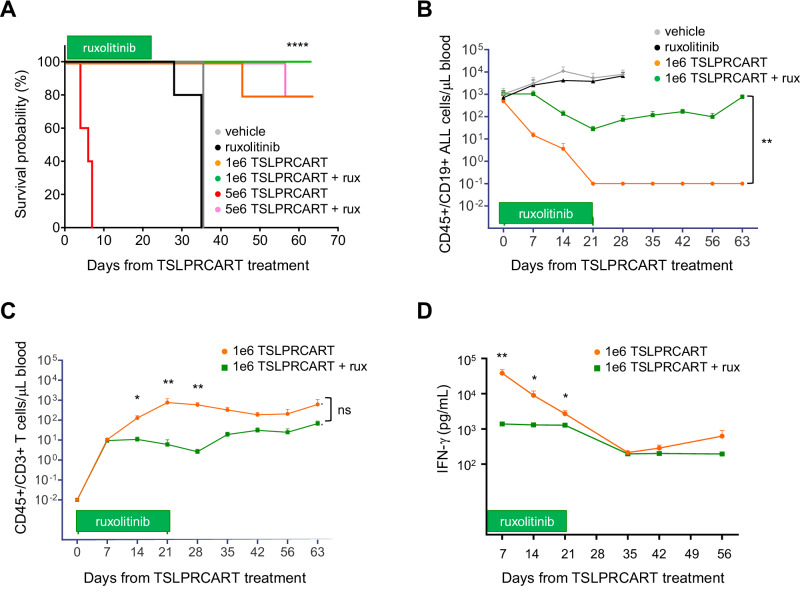
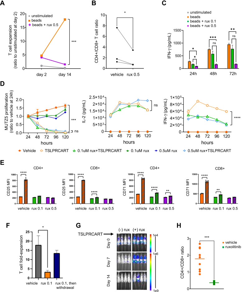
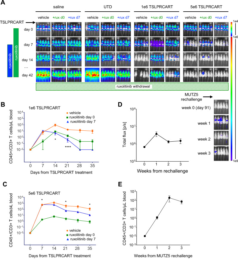
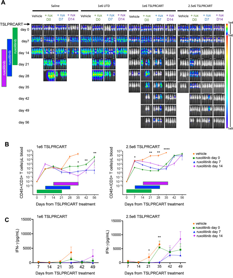
CRLF2 rearrangements occur in >50% of Ph-like and Down syndrome (DS)-associated B-acute lymphoblastic leukemia (ALL) and induce constitutive kinase signaling targetable by the JAK1/2 inhibitor ruxolitinib under current clinical investigation. While chimeric antigen receptor T cell (CART) immunotherapies have achieved remarkable remission rates in children with relapsed/refractory B-ALL, ~50% of CD19CART-treated patients relapse again, many with CD19 antigen loss. We previously reported preclinical activity of thymic stromal lymphopoietin receptor-targeted cellular immunotherapy (TSLPRCART) against CRLF2-overexpressing ALL as an alternative approach. In this study, we posited that combinatorial TSLPRCART and ruxolitinib would have superior activity and first validated potent TSLPRCART-induced inhibition of leukemia proliferation in vitro in CRLF2-rearranged ALL cell lines and in vivo in Ph-like and DS-ALL patient-derived xenograft (PDX) models. However, simultaneous TSLPRCART/ruxolitinib or CD19CART/ruxolitinib treatment during initial CART expansion diminished T cell proliferation, blunted cytokine production, and/or facilitated leukemia relapse, which was abrogated by time-sequenced/delayed ruxolitinib co-exposure. Importantly, ruxolitinib co-administration prevented fatal TSLPRCART cytokine-associated toxicity in ALL PDX mice. Upon ruxolitinib withdrawal, TSLPRCART functionality recovered in vivo with clearance of subsequent ALL rechallenge. These translational studies demonstrate an effective two-pronged therapeutic strategy that mitigates acute CART-induced hyperinflammation and provides potential anti-leukemia 'maintenance' relapse prevention for CRLF2-rearranged Ph-like and DS-ALL.


Conflict of interest statement

Competing interests: AB is a current employee of Carisma Therapeutics. SLR is a current employee of Parexel International. KRR has received preclinical research support from Incyte Corporation. TJF was a prior part-time employee of and consultant for Sana Biotechnology. TJF is an inventor on patent US11834509B2 (‘Thymic stromal lymphopoietin receptor-specific chimeric antigen receptors and methods using same’). SKT receives clinical research funding from Incyte Corporation for conduction of the Children’s Oncology Group AALL1521 phase 2 clinical trial (NCT02723994). The remaining authors declare no competing interests. Ethics approval and consent to participate: Viably cryopreserved primary pediatric, adolescent, and young adult ALL specimens used to create PDX models for these studies were obtained from leukemia biorepositories of the Children’s Oncology Group, Children’s Hospital of Philadelphia (CHOP), or Texas Children’s Hospital under institutional review board (IRB)-approved research protocols following obtainment of informed consent in accordance with the Declaration of Helsinki. Use of coded leukemia specimens without identifying patient health information in these studies was deemed non-human subjects research and exempt from further review by the CHOP IRB and ethics committee. All animal studies were conducted under an Institutional Animal Care and Use Committee-approved protocol at CHOP in accordance with all guidelines and regulations.

Figures

Fig. 1
Fig. 1. Simultaneous JAK inhibition ameliorates in vivo CAR T cell-induced mortality and suppresses TSLPRCART activity in ruxolitinib-insensitive CRLF2-rearranged Ph-like ALL.
A Kaplan-Meier survival analysis of IGH::CRLF2 Ph-like ALL patient-derived xenograft (PDX) mice (JH331 model). Cohorts of 5 mice were randomized and treated intravenously (IV) with 5e6 TSLPRCART or vehicle (saline) control with or without simultaneous exposure to ruxolitinib 2 g/kg rodent chow administered continuously (ad libitum) days 0 through 21 (horizontal green bar). B Human CD45+/CD19+B-ALL cells and (C) human CD45+/CD3+CAR T cells were quantified weekly via flow cytometric analysis of sampled peripheral blood from JH331 mice treated with vehicle, ruxolitinib monotherapy, or lower-dose 1e6 TSLPRCART with or without simultaneous ruxolitinib treatment from days 0 to day 21 (green bar). Lower TSLPRCART numbers are initially detected at early ruxolitinib co-administration timepoints, then normalize in subsequent weeks after ruxolitinib withdrawal. D IFN-γ levels in plasma from JH331 mice treated with 1e6 TSLPRCART in (B) and (C) are lower in mice treated with simultaneous ruxolitinib. Depicted data represent mean ± standard error of the mean (SEM). Statistical analyses were performed with Kaplan-Meier survival analysis with log-rank (Mantel-Cox) for comparison, 2-way ANOVA with Šidák correction, or unpaired t-tests at relevant time points. ns not significant, *p < 0.05, **p < 0.01, ****p < 0.0001.
Fig. 2
Fig. 2. Ruxolitinib co-administration inhibits T cell expansion and cytokine production and preferentially affects CD4 + T cells.
A Normal T cells from healthy donors (n = 3 donors) were cultured with CD3/CD28 beads with or without ruxolitinib (0.5 µM) in vitro for 2 weeks, with unstimulated T cells cultured without CD3/CD28 beads as a control. T cell expansion was assessed via Cell Titer Glo viability assays. Depicted data represent the mean of 3 independent T cell donors plated in technical triplicates ±SEM. B The ratio of CD4/CD8+ T cells was measured by flow cytometric immunophenotyping of normal healthy donor T cells exposed to 0.5 μM ruxolitinib for 72 h. C IFN-γ in culture supernatant of CD3/CD28 bead activated T cells treated with or without ruxolitinib at the indicated concentrations was quantified by ELISA at the indicated timepoints. Depicted data represent the mean of 3 independent T cell donors plated in technical triplicates ±SEM. D Luciferase-transduced MUTZ5 (a TSLPR+ human CRLF2-rearranged ALL cell line) cells were co-incubated in vitro with TSLPRCART at 1:15 effector-to-target (E:T) ratio and either vehicle or ruxolitinib at 0.1 and 0.5 μM concentrations. ALL cytotoxicity via luciferase reporter assays (left) and IL-2 (middle) and IFN-γ (right) production via ELISA were measured at the indicated time points. Depicted data represent the mean of technical triplicates ± SEM. E TSLPRCART were incubated in the absence (solid bars) or presence (striped bars) of MUTZ5 B-ALL cells in 1:1 E:T ratio for 24 h with or without ruxolitinib at the indicated concentrations. Induction of CD25 (left two panels) or CD71 (right two panels) surface expression on CD4+ and CD8+ T cells was evaluated by flow cytometry analysis. Quantification of median fluorescent intensity (MFI) for CD4+ and CD8+ T cell subsets with technical triplicates for each condition is displayed ±SEM. Ruxolitinib blunted upregulation of cell surface T cell activation markers CD25 and CD71 on both CD4+ and CD8+ TSLPRCART when co-incubated with MUTZ5 cells. F TSLPRCART were co-incubated 1:1 with MUTZ5 cells with or without ruxolitinib at the indicated concentrations. Every 3–4 days, TSLPRCART were sampled for enumeration by quantitative flow cytometry analysis to determine expansion from prior plating, and the remaining cells were re-stimulated 1:1 with MUTZ5 and fresh ruxolitinib-containing media. On day 7, a subset of ruxolitinib-exposed TSLPRCART were replated in the absence of ruxolitinib (withdrawal condition) for 4 days, and this ruxolitinib-induced decrease in in vitro TSLPRCART expansion was observed to be reversible upon drug removal. Depicted data represent the mean of technical triplicates ±SEM. G Ruxolitinib co-administration also inhibited anti-leukemia activity of TSLPRCART in vivo in a bioluminescent IGH::CRLF2/JAK2R683-mutant Ph-like ALL PDX model (ALL121). Luciferase-expressing ALL121 cells were injected (1e6) IV into NSG mice. Once engraftment was confirmed by bioluminescent imaging, cohorts of 4–8 mice were treated with 1e6 TSLPRCART IV and simultaneously exposed to ruxolitinib-infused chow (rux) or control rodent chow. H At day 14 of the experiment in (G), human CD4+ and CD8+ T cells in murine peripheral blood were quantified by flow cytometry and demonstrate significantly decreased CD4:CD8 ratio. Depicted data represent mean ±SEM. After data normality assessment, statistical analyses were performed for (A) and (C) with two-way ANOVA with Tukey post-test for multiple comparisons, for (B) with a paired t-test, for (D) with one-way ANOVA and Dunnett post-test for multiple comparisons using the TSLPRCART condition as the comparator, for (E) and (F) with one-way ANOVA and Tukey post-test for multiple comparisons, and for (H) with an unpaired t-test. ns not significant, *p < 0.05, **p < 0.01, ***p < 0.001, ****p < 0.0001.
Fig. 3
Fig. 3. Ruxolitinib-induced inhibition of TSLPRCART is improved with delayed co-administration and is reversible.
A Luciferase-transduced MUTZ5 cells were injected IV into NSG mice. Once engraftment was documented by bioluminescent imaging (BLI), cohorts of 5 mice were randomized to IV treatment on day 0 with saline, 1e6 untransduced T cells (UTD), or lower-dose (1e6) or higher-dose (5e6) TSLPRCART. Ruxolitinib chow (rux) ad libitum was administered simultaneously at day 0 (green) or day 7 after T cell treatments (blue) and continued to day 42. Leukemia burden was measured weekly by BLI. Human CD3+/CD45+ T cells were quantified weekly by flow cytometry analysis of peripheral blood of mice treated with B lower-dose or C higher-dose TSLPRCART. Statistical analysis was performed by two-way ANOVA with Tukey post-test for multiple comparisons with differences indicated at relevant timepoints. D Ruxolitinib was then removed at day 42 (striped green bar) for relevant cohorts, and mice continued to be followed by BLI to monitor potential re-emergence of leukemia. After 49 additional days without ruxolitinib exposure (day 91), mice were injected IV with 1e7 luciferase-expressing TSLPR + MUTZ5 cells to simulate relapse (week 0 antigen rechallenge) and followed by BLI. D Summary BLI radiance data following MUTZ5 rechallenge are displayed graphically for the 5e6 TSLPRCART/original ruxolitinib day 7 cohort shown in the lower right aspect of (A) with (E) enumeration of human CD3+/CD45+ T cells in murine peripheral blood at these same time points by quantitative flow cytometry. TSLPRCART with prior day 7 delayed-ruxolitinib exposure expanded robustly following MUTZ5 rechallenge, and low or undetectable leukemia burden was maintained. Statistical analyses were performed for (B) and (C) with one-way ANOVA and Tukey post-test for multiple comparisons. *p < 0.05, ****p < 0.0001.
Fig. 4
Fig. 4. Delayed JAK inhibitor co-treatment improves in vivo TSLPRCART activity against a ruxolitinib-sensitive CRLF2-rearranged Ph-like ALL PDX model.
A Luciferase-transduced IGH::CRLF2/JAK2R683G-mutant ALL121 PDX model cells (1e6) were injected IV in NSG mice. Once engraftment was documented by BLI, cohorts of 5 mice were randomized to IV treatment with saline, 1e6 untransduced T cells (UTD), or lower-dose (1e6) or higher-dose (2.5e6) TSLPRCART. Ruxolitinib (rux) chow ad libitum was administered simultaneously at day 0 (green), day 7 (blue), or day 14 (purple) after T cell treatments and continued for 21 days in each cohort until days 21, 28, or 35, respectively. Leukemia burden was measured weekly by BLI. B Human CD3+/CD45+ T cells were quantified weekly by flow cytometry analysis of peripheral blood of mice treated with lower-dose (left panel) or higher-dose (right panel) TSLPRCART. C IFN-γ was measured by ELISA in plasma prepared from weekly peripheral venous blood from mice treated with lower-dose (left panel) or higher-dose (right panel) TSLPRCART. Time-sequenced ruxolitinib co-administration at day 14 (at initial peak of detected CAR T cell expansion) improved long-term leukemia clearance and PDX model ‘remission’ in both lower-dose and higher-dose TSLPRCART cohorts. Statistical analyses were performed for (B) and (C) with one-way ANOVA and Tukey post-test for multiple comparisons for surviving cohorts at day 56. *p < 0.05, **p < 0.01, ***p < 0.001, ****p < 0.0001.
Fig. 5
Fig. 5. ‘Maintenance’ therapy with ruxolitinib following TSLPRCART-induced ALL clearance prevents in vivo leukemia relapse.
A Luciferase-transduced IGH::CRLF2/JAK2R683G-mutant ALL121 PDX model cells (1e6) were injected IV in NSG mice. Once engraftment was documented by BLI, all mice (n = 10) were treated IV with 2.5e6 TSLPRCART and followed by weekly BLI measurements. After documentation of TSLPRCART-induced leukemia clearance, mice were rechallenged IV with 1e7 ALL121 cells, and cohorts of 5 mice were randomized at day 21 to continued receipt of control chow (orange) or new administration of ruxolitinib chow (green) ad libitum for 2 weeks (horizontal green bar). Mice co-treated with ruxolitinib at ALL121 rechallenge remained in remission, while control chow-fed mice experienced leukemia progression, as assessed by BLI. B Weekly flow cytometric quantification of human CD3+/CD45+ T cells in murine peripheral blood showed no reduction of TSLPRCART numbers in ruxolitinib- (green) versus control-treated (orange) mice or in (C) the CD4:CD8 ratio of T cells harvested from end-study murine spleens at day 36. D Ruxolitinib treatment significantly decreased surface expression of the exhaustion marker PD-1 in CD4+ and CD8+ T cell subsets is reported as median fluorescence intensity (MFI) measured by flow cytometric analyses. PD-1 expression on TSLPRCART is decreased with late ruxolitinib co-treatment. After data normality assessment, statistical analyses were performed for (A) and (B) by unpaired Mann-Whitney tests at each timepoint, (C) with an unpaired t-test, and (D) with one-way ANOVA and Šidák post-test for multiple comparisons between vehicle and ruxolitinib conditions for CD4+ and CD8+ subpopulations. ns not significant, *p < 0.05, **p < 0.01.
Fig. 6
Fig. 6. In vivo ruxolitinib and TSLPRCART sensitivity of CRLF2-rearranged Ph-like ALL is recapitulated in Down syndrome-associated ALL.
A NSG mice were engrafted with 5e5 DSALL47 (left) or 1e6 DSALL515 (right) PDX model cells. Once >1% CD10+/CD19+ human ALL cells were detectable in murine peripheral blood, cohorts of 5 mice were randomized to treatment with control (orange) or ruxolitinib (purple) chow ad libitum. Leukemia burden was monitored weekly by quantitative flow cytometric analysis of human CD10+/CD19+ALL cells in peripheral blood (top panels) and in end-study spleens (bottom panels), which was determined by rate of leukemia progression in control mice for each model. Significant reduction of DS-ALL cell numbers in peripheral blood and spleens was detected in both tested models with near-curative effect after 4 weeks of ruxolitinib treatment. B Flow cytometric quantification of TSLPR surface antigen density demonstrates similar ranges of expression in CRLF2-rearranged Ph-like and DS-ALL PDX models, suggesting similar potential for therapeutic activity of TSLPRCART. CRLF2 wild-type NALM-6 and CRLF2-rearranged MUTZ5 ALL cell lines were used as negative and positive controls, respectively. C NSG mice were engrafted with 1e6 TCHK150 ALL PDX model cells. Once >1% CD10+/CD19+ human ALL cells were detectable in murine peripheral blood, cohorts of 5 mice were randomized to IV treatment with saline, 2.5e6 UTD, or 2.5e6 TSLPRCART. Additional cohorts of TSLPRCART-treated mice (n = 5) were also randomized to simultaneous (day 0, green bar) or delayed (day 7, blue bar or day 14, purple bar) administration of ruxolitinib chow ad libitum. Animals were monitored weekly by quantitative flow cytometry analysis of human CD45+/CD10+/CD19+ ALL cells in murine peripheral blood and (D) end-study spleens. Delayed ruxolitinib co-treatment with TSLPRCART improved leukemia clearance compared to monotherapy or simultaneous co-treatment in this DS-ALL model. TSLPR surface expression remained unchanged by quantitative flow cytometric analysis in residual ALL cells in TSLPRCART-treated animals where applicable (data not shown). E Flow cytometric quantification of CD45+/CD3+ T cells in murine peripheral blood demonstrated no inhibition of TSLPRCART proliferation in vivo with day 14 ruxolitinib co-administration (lavender) compared to TSLPRCART monotherapy (light orange), whereas day 0 (light green) and day 7 (light blue) ruxolitinib exposure significantly impaired T cell numbers. F ELISA was performed to quantify human IFN-γ in murine plasma prepared from weekly peripheral venous blood. Significant dampening of IFN-γ production was detected with TSLPRCART and day 0 ruxolitinib co-treatment compared to TSLPRCART monotherapy (light green versus light orange). Statistical analyses were performed for (A) with unpaired t-tests and for (C), (D), (E), and (F) with 2-way ANOVA/mixed effects analysis and Dunnett post-test for multiple comparisons using the TSLPRCART condition as the comparator. ns not significant, *p < 0.05, **p < 0.01, ****p < 0.0001.

Similar articles

Cited by

References

    1. Den Boer ML, van Slegtenhorst M, De Menezes RX, Cheok MH, Buijs-Gladdines JG, Peters ST, et al. A subtype of childhood acute lymphoblastic leukaemia with poor treatment outcome: a genome-wide classification study. Lancet Oncol. 2009;10:125–34. - PMC - PubMed
    1. Roberts KG, Li Y, Payne-Turner D, Harvey RC, Yang YL, Pei D, et al. Targetable kinase-activating lesions in Ph-like acute lymphoblastic leukemia. N. Engl J Med. 2014;371:1005–15. - PMC - PubMed
    1. Tran TH, Tasian SK. How I Treat Philadelphia Chromosome-like Acute Lymphoblastic Leukemia in Children, Adolescents, and Young Adults. Blood. 2024 10.1182/blood.2023023153. - PubMed
    1. Roberts KG, Gu Z, Payne-Turner D, McCastlain K, Harvey RC, Chen IM, et al. High frequency and poor outcome of Philadelphia chromosome-like acute lymphoblastic leukemia in adults. J Clin Oncol. 2017;35:394–401. - PMC - PubMed
    1. Tasian SK, Dai YF, Devidas M, Roberts KG, Harvey RC, Chen IML, et al. Outcomes of patients with CRLF2-overexpressing acute lymphoblastic leukemia without Down syndrome: a report from the Children’s Oncology Group. Blood. 2020;136:45–46.

MeSH terms