. 2024 Dec;14(12):e70095.
doi: 10.1002/brb3.70095.

Baihui-Penetrating-Qubin Acupuncture Attenuates Neurological Deficits Through SIRT1/FOXO1 Reducing Oxidative Stress and Neuronal Apoptosis in Intracerebral Hemorrhage Rats

Affiliations

Baihui-Penetrating-Qubin Acupuncture Attenuates Neurological Deficits Through SIRT1/FOXO1 Reducing Oxidative Stress and Neuronal Apoptosis in Intracerebral Hemorrhage Rats

Shan-Shan Dong et al. Brain Behav. 2024 Dec.

Abstract

Background: Intracerebral hemorrhage (ICH) is a significant global disease with high mortality and disability. As of now, there is no effective therapy available. Oxidative stress and neuronal apoptosis play essential roles in ICH, determining neuronal survival. In our preliminary studies, we found that Baihui-penetrating-Qubin acupuncture could improve neurological deficits and neuropathological damage in the perihematomal area in ICH rats. The SIRT1/FOXO1 signaling pathway has been reported to mediate antioxidant and anti-neuronal apoptosis. This study aimed to investigate the effects of Baihui-penetrating-Qubin acupuncture on oxidative stress and neuronal apoptosis after ICH and the role of SIRT1/FOXO1 in acupuncture's neuroprotection.

Methods: ICH rat models were established by autologous tail blood (50 µL) infusion into the caudate nucleus. EX527, SIRT1-specific inhibitor was intraperitoneally administered 3 days before ICH. Baihui-penetrating-Qubin acupuncture treatment was performed once a day for 30 min after ICH. Neurological deficits were evaluated using the modified neurological severity score (mNSS). Brain edema was evaluated using brain water content. HE staining and Nissl staining were used to evaluate neuropathological damage in the perihematomal area. Terminal deoxynucleotidyl transferase dUTP nick end labeling was used to quantify neuronal apoptosis. Specific kits were used to detect the levels of SOD, CAT, GSH-Px in the brain. The oxidative DNA damage was evaluated using enzyme-linked immunosorbent assay to detect the level of 8-hydroxyguanosine (8-OHdG). Western blot was used to evaluate the expressions of SIRT1, Ac-FOXO1, FOXO1, Bcl-2, and Bax. Immunofluorescence staining was conducted to detect the cellular localization of SIRT1.

Results: Baihui-penetrating-Qubin acupuncture improved the neurological deficits and brain edema, reduced the pathological injury and neuronal degeneration in 3 days in the perihematomal area after ICH. Mechanistically, acupuncture reduced oxidative stress injury and neuronal apoptosis via activating SIRT1/FOXO1 pathway. The neuroprotective effects of acupuncture were abolished by injection of the SIRT1 inhibitor EX527.

Conclusions: Baihui-penetrating-Qubin acupuncture could reduce oxidative stress and neuronal apoptosis, at least in part, through the SIRT1/FOXO1 signaling pathway, improving neurological deficits and neuropathological damage after ICH. These findings suggest that Baihui-penetrating-Qubin acupuncture is an effective therapy for ICH, as well as targeting SIRT1 signaling to promote neuron survival could be a potential therapeutic strategy.

Keywords: SIRT1/FOXO1; acupuncture; brain injury; intracerebral hemorrhage (ICH); neuronal apoptosis; neuroprotection; oxidative stress.


Conflict of interest statement

The authors declare no conflicts of interest.

Figures
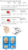
FIGURE 1
FIGURE 1
Description of experimental setup, Baihui‐penetrating‐Qubin acupuncture technique, and ICH rat models. (A) Experimental design and animal groups. Exp: experiment; ICH: intracerebral hemorrhage; WB: western blot; ELISA: enzyme‐linked immunosorbent assay; IF: immunofluorescence; mNSS: modified neurological severity score; BWC: brain water content; H&E: hematoxylin and eosin staining; TUNEL: terminal deoxynucleotidyl transferase‐ mediated dUTP nick end labeling; i.p.: intraperitoneal injection; mNSS: modified neurological severity score; 8‐OHdG: 8‐hydroxyguanosine. (B) The illustration of Baihui‐penetrating‐Qubin acupuncture treatment. (a) Location of GV20 and GB7 in human. (b) Schematic diagram of Baihui‐penetrating‐Qubin acupuncture in a rat. Baihui (GV20) is located at the intersection of the sagittal midline and the line between the two ears; Qubin (GB7) is located in the posterior 2/3 of the line connecting the right orbital margin and porus acusticus externus.(C)Location of the ICH hematoma. Representative images of coronal brain sections from the Sham and ICH groups, showing the site of blood transfusion within the caudate putamen region.
FIGURE 2
FIGURE 2
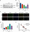
Time course of SIRT1 and 8‐OHdG expression in the perihematomal area after ICH. (A and B) Representative Western blot bands and quantitative analysis of SIRT1 expression levels in the perihematomal area after ICH. ICH: intracerebral hemorrhage; SIRT1: silent information regulator 1. (C) Representative micrographs of SIRT1 (red) /NeuN (green) co‐immunofluorescence staining in the perihematomal area after ICH (400×, n = 3). DAPI: diamidino phenylindole. (D) Time course of 8‐OHdG expression in the perihematomal area after ICH. Data are presented as the mean ± SD(n = 6); *< 0.05, **p < 0.01 vs Sham group. 8‐OHdG: 8‐hydroxyguanosine. (E) Correlation analysis of SIRT1 and 8‐OHdG expression levels in the perihematomal area after ICH.
FIGURE 3
FIGURE 3
Effects of Baihui‐penetrating‐Qubin acupuncture on the neurobehavioral deficits and neuropathological damage at 3 days after ICH. (A) The mNSS score after ICH (n = 6). mNSS: modified neurological severity score; ICH+Acu: intracerebral hemorrhage + Acupuncture.(B) Quantification of brain water content after ICH (n = 6). Data are presented as the mean ± SD; *p < 0.05, **p < 0.01 vs Sham group;# p < 0.05, ## p < 0.01 vs ICH group. (C and D) H&E staining and Nissl staining in the perihematomal area after ICH (200×, 400×, n = 3). The pictures of a, c, e represent the H&E and Nissl staining of tissue around the hematoma. The b, d, and f insets are magnifications of a, c, and e, respectively. Scale bars, 200×: 100 µm; 400×: 50 µm. mNSS: Modified neurological severity score; H&E: hematoxylin and eosin staining.
FIGURE 4
FIGURE 4
Effects of Baihui‐penetrating‐Qubin acupuncture on neuronal apoptosis and oxidative stress in the perihematomal area at 3 days after ICH. (A and B) Representative micrographs and quantitative analysis of TUNEL‐positive neurons in the perihematomal area after ICH (400×). TUNEL: terminal deoxynucleotidyl transferase‐mediated dUTP nick end labeling; DAPI: diamidino phenylindole; ICH+Acu: intracerebral hemorrhage.(C and D) Representative micrographs and quantitative analysis of 8‐OHdG (red)/NeuN (green) co‐immunofluorescence staining in the perihematomal area after ICH (400×). Scale bars: 50 µm. Data are presented as the mean ± SD (n = 6); *p < 0.05, **p < 0.01 vs Sham group; # p < 0.05, ## p < 0.01 vs ICH group. 8‐OHdG: 8‐hydroxyguanosine.
FIGURE 5
FIGURE 5
Effects of Baihui‐penetrating‐Qubin acupuncture on neurobehavioral deficits and neuropathological damage after ICH with pre‐injection of EX527. (A) The mNSS score after ICH (n = 6). (B) The brain water content after ICH (n = 6). Data are presented as the mean ± SD; *p < 0.05, **p < 0.01 VS. Sham group; # p < 0.05, ## p < 0.01 VS. ICH group; & p < 0.05, && p < 0.01 VS. ICH + Acu group. (C–D) H&E and Nissl staining in the perihematomal area after ICH (200×, 400×, n = 3). The pictures of a, c, e, and g represent the H&E and Nissl staining of tissue around the hematoma. The b, d, f, and h insets are magnifications of a, c, e, and g, respectively. Scale bars: 200×: 100 µm; 400×:50 µm. mNSS: Modified neurological severity score; H&E: hematoxylin and eosin staining.
FIGURE 6
FIGURE 6
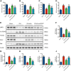
Effects of Baihui‐penetrating‐Qubin acupuncture on the SIRT1‐FOXO1 signaling pathway and expressions of downstream signaling molecules in the perihematomal area at 3 days after ICH. (A‐F) Representative Western blot bands and quantitative analysis of SIRT1, Ac‐FOXO1, FOXO1, Bcl‐2, and Bax in the perihematomal area after ICH. (G‐J) SOD, CAT, GSH‐Px, and MDA levels in the perihematomal area after ICH. Data are presented as the mean ± SD (n = 6); *p < 0.05, **p < 0.01 vs Sham group; # p < 0.05, ## p < 0.01 vs ICH group; & p < 0.05, && p < 0.01 vs ICH + Acupuncture group. CAT: catalase; GSH‐Px: glutathione peroxidase; ICH: intracerebral hemorrhage; MDA: malondialdehyde; SOD: superoxide dismutase.

References

    1. Bederson, J. B. , Pitts L. H., Tsuji M., Nishimura M. C., Davis R. L., and Bartkowski H.. 1986. “Rat Middle Cerebral Artery Occlusion: Evaluation of the Model and Development of a Neurologic Examination.” Stroke; A Journal of Cerebral Circulation 17, no. 3: 472–476. 10.1161/01.str.17.3.472. - DOI - PubMed
    1. Cai, Y. , Xu L., Xu H., and Fan X.. 2016. “SIRT1 and Neural Cell Fate Determination.” Molecular Neurobiology 53, no. 5: 2815–2825. 10.1007/s12035-015-9158-6. - DOI - PubMed
    1. Cantó, C. , Gerhart‐Hines Z., Feige J. N., et al. 2009. “AMPK Regulates Energy Expenditure by Modulating NAD+ Metabolism and SIRT1 Activity.” Nature 458, no. 7241: 1056–1060. 10.1038/nature07813. - DOI - PMC - PubMed
    1. Chen, X. , Pan Z., Fang Z., et al. 2018. “Omega‐3 Polyunsaturated Fatty Acid Attenuates Traumatic Brain Injury‐induced Neuronal Apoptosis by Inducing Autophagy Through the Upregulation of SIRT1‐mediated Deacetylation of Beclin‐1.” Journal of Neuroinflammation 15, no. 1: 310. 10.1186/s12974-018-1345-8. - DOI - PMC - PubMed
    1. Cordonnier, C. , Demchuk A., Ziai W., and Anderson C. S.. 2018. “Intracerebral Haemorrhage: Current Approaches to Acute Management.” Lancet 392, no. 10154: 1257–1268. 10.1016/s0140-6736(18)31878-6. - DOI - PubMed