. 2024 Dec 3;16(23):4052.
doi: 10.3390/cancers16234052.

Carcinoembryonic Antigen Expression in Human Tumors: A Tissue Microarray Study on 13,725 Tumors

Affiliations

Carcinoembryonic Antigen Expression in Human Tumors: A Tissue Microarray Study on 13,725 Tumors

Kristina Jansen et al. Cancers (Basel). .

Abstract

Background/objectives: Carcinoembryonic antigen (CEA) is a cell-surface glycoprotein serving as a drug target, diagnostic marker, and serum marker for cancer monitoring. However, prevalence data on CEA expression in cancer tissues vary considerably. This study was designed to determine CEA expression in normal and neoplastic tissues.

Methods: A tissue microarray containing 13,725 samples from 120 different tumor types, as well as 76 different normal tissue types, was analyzed by immunohistochemistry (IHC).

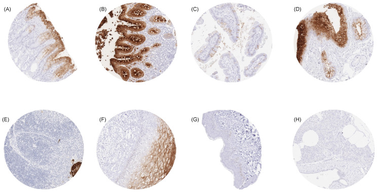
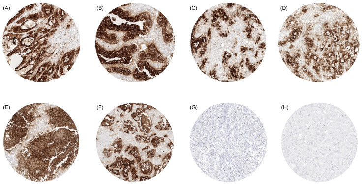
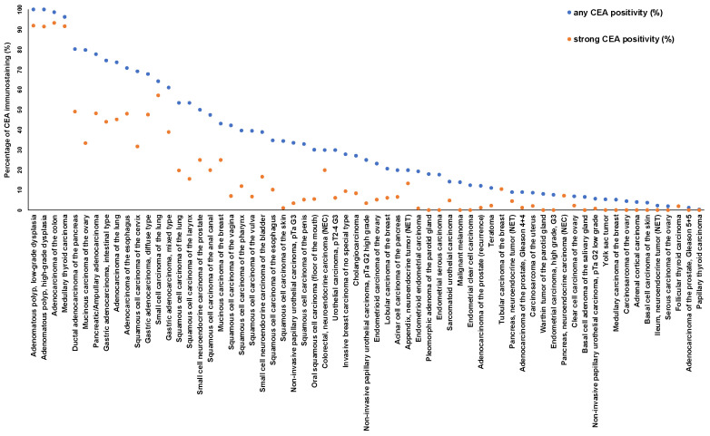
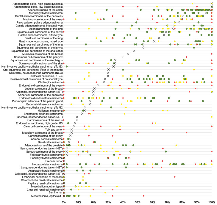
Results: CEA was detectable in 65 (54.2%) of 120 tumor categories, including 49 (40.8%) tumor types with at least one strongly positive case. CEA positivity was most common in colorectal adenomas (100%) and carcinomas (98.7%), other gastrointestinal adenocarcinomas (61.1-80.3%), medullary carcinomas of the thyroid (96.3%), pulmonary adenocarcinoma (73.7%), mucinous carcinomas of the ovary (79.8%) and the breast (43.2%), small-cell carcinomas of the lung (64.3%), and urinary bladder (38.9%). CEA overexpression was linked to high tumor grade and invasive growth (p < 0.0001 each) in urinary bladder cancer, and estrogen and HER2 receptor positivity (p ≤ 0.0158) in invasive breast cancer of no special type. In colorectal adenocarcinomas, reduced CEA expression was associated with mismatch repair deficiency (p < 0.0001).

Conclusions: The comprehensive list of CEA-positive human tumor types demonstrates that CEA is expressed in a broad range of epithelial neoplasms, many of which might benefit from CEA serum monitoring and anti-CEA therapies.

Keywords: CEA; diagnostic marker; human tumors; immunohistochemistry; tissue microarray.


Conflict of interest statement

The rabbit recombinant antibody, clone MSVA-465R was provided from MS Validated Antibodies GmbH (owned by a family member of GS). All other authors declare no conflicts of interest.

Figures

Figure 1
Figure 1
CEA immunostaining of normal tissues. A membranous and cytoplasmic CEA staining of variable intensity is seen in surface epithelial cells of the stomach (A), epithelial cells (predominantly at the surface) of the colon (B), goblet cells of the small intestine (C), respiratory epithelial cells (D), Hassal’s corpuscles of the thymus (E), and in superficial cell layers of the squamous epithelium of the cervix uteri (F). CEA staining is absent in tissues from the epidermis of the skin (G) and in the pancreas (H). Original magnifications 10×, spot size 600 μm.
Figure 2
Figure 2
CEA immunostaining in cancer. The panels show a strong CEA staining in an adenocarcinoma of the colon (A), an adenocarcinoma of the esophagus (B), a ductal adenocarcinoma of the pancreas (C), a cholangiocellular carcinoma of the liver (D), a small-cell neuroendocrine carcinoma of the lung (E), and an adenocarcinoma of the lung (F). CEA staining is lacking in a malignant mesothelioma of the pleura (H) and a hepatocellular carcinoma in the liver (G). Original magnifications 10×, spot size 600 μm.
Figure 3
Figure 3
Ranking order of CEA immunostaining in cancers. Both the percentage of positive cases (blue dots) and the percentage of strongly positive cases (orange dots) are shown.
Figure 4
Figure 4
CEA immunostaining and patient prognosis in invasive breast carcinoma of no special type.
Figure 5
Figure 5
Comparison of CEA expression with previous works in the literature. An “X” represents the proportion of CEA-positive cancers in the present study, dots indicate the frequencies reported in the literature for comparison: studies with ≤10 tumors analyzed are marked with red dots, studies with 11 to 25 tumors analyzed are marked with yellow dots, and green dots mark studies with >25 tumors analyzed.

Similar articles

Cited by

References

    1. Taheri M., Saragovi U., Fuks A., Makkerh J., Mort J., Stanners C.P. Self recognition in the Ig superfamily. Identification of precise subdomains in carcinoembryonic antigen required for intercellular adhesion. J. Biol. Chem. 2000;275:26935–26943. doi: 10.1016/S0021-9258(19)61463-8. - DOI - PubMed
    1. Nap M., Mollgard K., Burtin P., Fleuren G.J. Immunohistochemistry of carcino-embryonic antigen in the embryo, fetus and adult. Tumor Biol. 1988;9:145–153. doi: 10.1159/000217555. - DOI - PubMed
    1. Zhang Y., Yang J., Li H., Wu Y., Zhang H., Chen W. Tumor markers CA19-9, CA242 and CEA in the diagnosis of pancreatic cancer: A meta-analysis. Int. J. Clin. Exp. Med. 2015;8:11683–11691. - PMC - PubMed
    1. Grunnet M., Sorensen J.B. Carcinoembryonic antigen (CEA) as tumor marker in lung cancer. Lung Cancer. 2012;76:138–143. doi: 10.1016/j.lungcan.2011.11.012. - DOI - PubMed
    1. Fakih M.G., Padmanabhan A. CEA monitoring in colorectal cancer. What you should know. Oncology. 2006;20:579–587; discussion 588, 594, 596. - PubMed

LinkOut - more resources