Review
. 2024 Dec 7;16(23):4099.
doi: 10.3390/cancers16234099.

Role of Imaging in Multiple Myeloma: A Potential Opportunity for Quantitative Imaging and Radiomics?

Affiliations
Review

Role of Imaging in Multiple Myeloma: A Potential Opportunity for Quantitative Imaging and Radiomics?

Anna Michalska-Foryszewska et al. Cancers (Basel). .

Abstract

Multiple myeloma (MM) is the second most prevalent hematologic malignancy, particularly affecting the elderly. The disease often begins with a premalignant phase known as monoclonal gammopathy of undetermined significance (MGUS), solitary plasmacytoma (SP) and smoldering multiple myeloma (SMM). Multiple imaging modalities are employed throughout the disease continuum to assess bone lesions, prevent complications, detect intra- and extramedullary disease, and evaluate the risk of neurological complications. The implementation of advanced imaging analysis techniques, including artificial intelligence (AI) and radiomics, holds great promise for enhancing our understanding of MM. The integration of advanced image analysis techniques which extract features from magnetic resonance imaging (MRI), computed tomography (CT), or positron emission tomography (PET) images has the potential to enhance the diagnostic accuracy for MM. This innovative approach may lead to the identification of imaging biomarkers that can predict disease prognosis and treatment outcomes. Further research and standardized evaluations are needed to define the role of radiomics in everyday clinical practice for patients with MM.

Keywords: artificial intelligence; imaging; multiple myeloma; prognosis; radiomics.


Conflict of interest statement

The authors declare no conflict of interest.

Figures
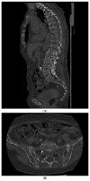
Figure 1
Figure 1
A 77-year-old woman with a diagnosis of multiple myeloma. (A)—Whole-body low-dose computed tomography demonstrates lytic lesions in (A) vertebral bodies and (B) sacral bone and iliac crest. Source: Radiological Diagnostics Center, The National Institute of Medicine of the Ministry of Interior and Administration, Warsaw, Poland.
Figure 2
Figure 2
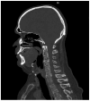
A 63-year-old man with lytic lesions on the cervical spine, shown in whole-body low-dose computed tomography. Source: Radiological Diagnostics Center, The National Institute of Medicine of the Ministry of Interior and Administration, Warsaw, Poland.
Figure 3
Figure 3
A 65-year old woman with multiple myeloma, presenting some lytic lesions in the bones of the skull on whole-body low-dose computed tomography. Source: Radiological Diagnostics Center, The National Institute of Medicine of the Ministry of Interior and Administration, Warsaw, Poland.
Figure 4
Figure 4
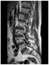
Sagittal T1-weighted image of a spine demonstrating a focal lesion in a 73-year-old man with multiple myeloma diagnosis. Source: Radiological Diagnostics Center, The National Institute of Medicine of the Ministry of Interior and Administration, Warsaw, Poland.
Figure 5
Figure 5
Coronal T1-weighted image of a spine demonstrating a “salt-and-pepper” pattern of marrow involvement in a 63-year-old woman with a multiple myeloma diagnosis. Source: Radiological Diagnostics Center, The National Institute of Medicine of the Ministry of Interior and Administration, Warsaw, Poland.

References

    1. Kumar S.K., Rajkumar S.V. The multiple myelomas—Current concepts in cytogenetic classification and therapy. Nat. Rev. Clin. Oncol. 2018;15:409–421. doi: 10.1038/s41571-018-0018-y. - DOI - PubMed
    1. Padala S.A., Barsouk A., Barsouk A., Rawla P., Vakiti A., Kolhe R., Kota V., Ajebo G.H. Epidemiology, Staging, and Management of Multiple Myeloma. Med. Sci. 2021;9:3. doi: 10.3390/medsci9010003. - DOI - PMC - PubMed
    1. Rajkumar S.V., Dimopoulos M.A., Palumbo A., Blade J., Merlini G., Mateos M.-V., Kumar S., Hillengass J., Kastritis E., Richardson P., et al. International Myeloma Working Group updated criteria for the diagnosis of multiple myeloma. Lancet Oncol. 2014;15:e538–e548. doi: 10.1016/S1470-2045(14)70442-5. - DOI - PubMed
    1. van de Donk N.W.C.J., Pawlyn C., Yong K.L. Multiple myeloma. Lancet. 2021;397:410–427. doi: 10.1016/S0140-6736(21)00135-5. - DOI - PubMed
    1. Wu F., Bernard S., Fayad L.M., Ilaslan H., Messiou C., Moulopoulos L.A., Mulligan M.E. Updates and Ongoing Challenges in Imaging of Multiple Myeloma: AJR Expert Panel Narrative Review. Am. J. Roentgenol. 2021;217:775–785. doi: 10.2214/AJR.21.25878. - DOI - PubMed

LinkOut - more resources