

Image Quality Assessment and Reliability Analysis of Artificial Intelligence-Based Tumor Classification of Stimulated Raman Histology of Tumor Biobank Samples
- PMID: 39682609
- PMCID: PMC11640452
- DOI: 10.3390/diagnostics14232701
Image Quality Assessment and Reliability Analysis of Artificial Intelligence-Based Tumor Classification of Stimulated Raman Histology of Tumor Biobank Samples
Abstract
Background: Stimulated Raman histology (SRH) is a label-free optical imaging method for rapid intraoperative analysis of fresh tissue samples. Analysis of SRH images using Convolutional Neural Networks (CNN) has shown promising results for predicting the main histopathological classes of neurooncological tumors. Due to the relatively low number of rare tumor representations in CNN training datasets, a valid prediction of rarer entities remains limited. To develop new reliable analysis tools, larger datasets and greater tumor variety are crucial. One way to accomplish this is through research biobanks storing frozen tumor tissue samples. However, there is currently no data available regarding the pertinency of previously frozen tissue samples for SRH analysis. The aim of this study was to assess image quality and perform a comparative reliability analysis of artificial intelligence-based tumor classification using SRH in fresh and frozen tissue samples.
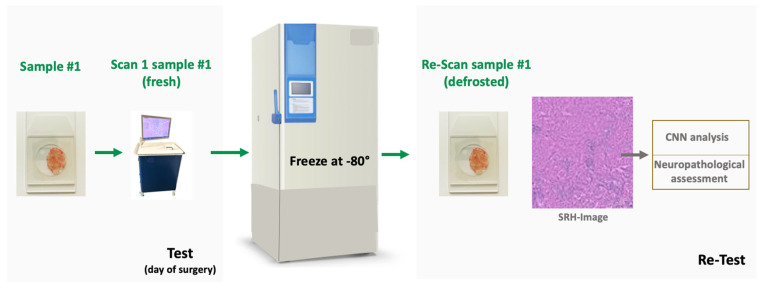
Methods: In a monocentric prospective study, tissue samples from 25 patients undergoing brain tumor resection were obtained. SRH was acquired in fresh and defrosted samples of the same specimen after varying storage durations at -80 °C. Image quality was rated by an experienced neuropathologist, and prediction of histopathological diagnosis was performed using two established CNNs.
Results: The image quality of SRH in fresh and defrosted tissue samples was high, with a mean image quality score of 1.96 (range 1-5) for both groups. CNN analysis showed high internal consistency for histo-(Cα 0.95) and molecular (Cα 0.83) pathological tumor classification. The results were confirmed using a dataset with samples from the local tumor biobank (Cα 0.91 and 0.53).
Conclusions: Our results showed that SRH appears comparably reliable in fresh and frozen tissue samples, enabling the integration of tumor biobank specimens to potentially improve the diagnostic range and reliability of CNN prediction tools.
Keywords: artificial intelligence; brain tumors; digital pathology; stimulated Raman histology.
Conflict of interest statement
The authors declare no conflicts of interest.
Figures


References
-
- Orringer D.A., Pandian B., Niknafs Y.S., Hollon T.C., Boyle J., Lewis S., Garrard M., Hervey-Jumper S.L., Garton H.J.L., Maher C.O., et al. Rapid Intraoperative Histology of Unprocessed Surgical Specimens via Fibre-Laser-Based Stimulated Raman Scattering Microscopy. Nat. Biomed. Eng. 2017;1:0027. doi: 10.1038/s41551-016-0027. - DOI - PMC - PubMed
-
- Movahed-Ezazi M., Nasir-Moin M., Fang C., Pizzillo I., Galbraith K., Drexler S., Krasnozhen-Ratush O.A., Shroff S., Zagzag D., William C., et al. Clinical Validation of Stimulated Raman Histology for Rapid Intraoperative Diagnosis of Central Nervous System Tumors. Mod. Pathol. 2023;36:100219. doi: 10.1016/j.modpat.2023.100219. - DOI - PMC - PubMed
-
- Eichberg D.G., Shah A.H., Di L., Semonche A.M., Jimsheleishvili G., Luther E.M., Sarkiss C.A., Levi A.D., Gultekin S.H., Komotar R.J., et al. Stimulated Raman Histology for Rapid and Accurate Intraoperative Diagnosis of CNS Tumors: Prospective Blinded Study. J. Neurosurg. 2019;134:137–143. doi: 10.3171/2019.9.JNS192075. - DOI - PubMed
-
- Einstein E.H., Ablyazova F., Rosenberg A., Harshan M., Wahl S., Har-El G., Constantino P.D., Ellis J.A., Boockvar J.A., Langer D.J., et al. Stimulated Raman Histology Facilitates Accurate Diagnosis in Neurosurgical Patients: A One-to-One Noninferiority Study. J. Neurooncol. 2022;159:369–375. doi: 10.1007/s11060-022-04071-y. - DOI - PubMed
Grants and funding
LinkOut - more resources
Full Text Sources