Review
. 2024 Nov 26;13(23):1958.
doi: 10.3390/cells13231958.

Human Cytochrome P450 Cancer-Related Metabolic Activities and Gene Polymorphisms: A Review

Affiliations
Review

Human Cytochrome P450 Cancer-Related Metabolic Activities and Gene Polymorphisms: A Review

Innokenty M Mokhosoev et al. Cells. .

Abstract

Background: Cytochromes P450 (CYPs) are heme-containing oxidoreductase enzymes with mono-oxygenase activity. Human CYPs catalyze the oxidation of a great variety of chemicals, including xenobiotics, steroid hormones, vitamins, bile acids, procarcinogens, and drugs.

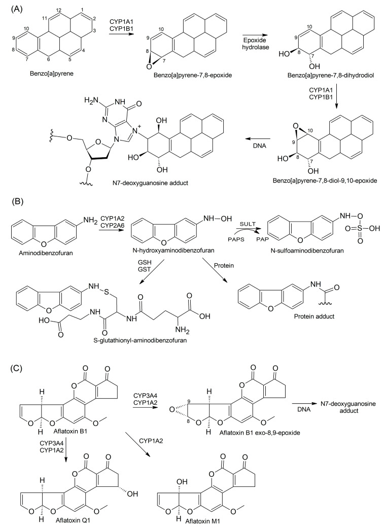
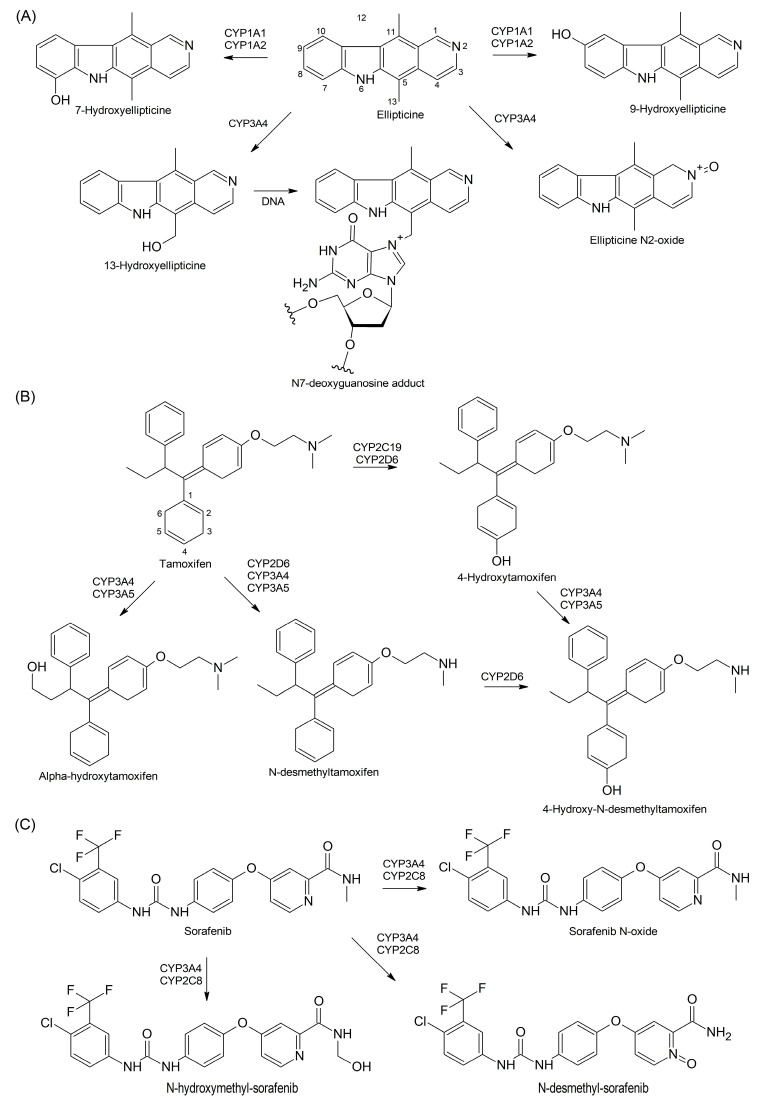
Findings: In our review article, we discuss recent data evidencing that the same CYP isoform can be involved in both bioactivation and detoxification reactions and convert the same substrate to different products. Conversely, different CYP isoforms can convert the same substrate, xenobiotic or procarcinogen, into either a more or less toxic product. These phenomena depend on the type of catalyzed reaction, substrate, tissue type, and biological species. Since the CYPs involved in bioactivation (CYP3A4, CYP1A1, CYP2D6, and CYP2C8) are primarily expressed in the liver, their metabolites can induce hepatotoxicity and hepatocarcinogenesis. Additionally, we discuss the role of drugs as CYP substrates, inducers, and inhibitors as well as the implication of nuclear receptors, efflux transporters, and drug-drug interactions in anticancer drug resistance. We highlight the molecular mechanisms underlying the development of hormone-sensitive cancers, including breast, ovarian, endometrial, and prostate cancers. Key players in these mechanisms are the 2,3- and 3,4-catechols of estrogens, which are formed by CYP1A1, CYP1A2, and CYP1B1. The catechols can also produce quinones, leading to the formation of toxic protein and DNA adducts that contribute to cancer progression. However, 2-hydroxy- and 4-hydroxy-estrogens and their O-methylated derivatives along with conjugated metabolites play cancer-protective roles. CYP17A1 and CYP11A1, which are involved in the biosynthesis of testosterone precursors, contribute to prostate cancer, whereas conversion of testosterone to 5α-dihydrotestosterone as well as sustained activation and mutation of the androgen receptor are implicated in metastatic castration-resistant prostate cancer (CRPC). CYP enzymatic activities are influenced by CYP gene polymorphisms, although a significant portion of them have no effects. However, CYP polymorphisms can determine poor, intermediate, rapid, and ultrarapid metabolizer genotypes, which can affect cancer and drug susceptibility. Despite limited statistically significant data, associations between CYP polymorphisms and cancer risk, tumor size, and metastatic status among various populations have been demonstrated.

Conclusions: The metabolic diversity and dual character of biological effects of CYPs underlie their implications in, preliminarily, hormone-sensitive cancers. Variations in CYP activities and CYP gene polymorphisms are implicated in the interindividual variability in cancer and drug susceptibility. The development of CYP inhibitors provides options for personalized anticancer therapy.

Keywords: CYPs; bioactivation vs. detoxification; drugs as substrates, inducers, and inhibitors; gene polymorphisms; hormone-dependent cancers; nuclear receptors and efflux transporters.


Conflict of interest statement

The authors declare no conflicts of interest.

Figures

Figure 1
Figure 1
Steroid hormone biosynthesis. The side chain of cholesterol is initially oxidized and cleaved by CYP11A1 with the participation of its redox partners, Fdx and FdR, and StAR to yield pregnenolone (P5). In the ∆4 pathway, P5 undergoes oxidation of the hydroxyl group at C3 by 3βHSD2 to yield progesterone, whereas, in the ∆5 pathway, CYP17A1 hydroxylates P5 at C17 to produce 17α-OHP5, which is further converted to DHEA and androstenedione, both C19 androgen and C18 estrogen precursors. CYP19A1 converts androstenedione to estrone and testosterone to 17β-estradiol whereas CYP11B1 and CYP11B2 are essential for corticosteroid biosynthesis.
Figure 2
Figure 2
The implication of CYPs in detoxification versus bioactivation of xenobiotics in the liver. (A) CYP3A4, CYP2D6, and CYP4F12 catalyze the bioactivation of C10 benzalkonium chloride (C10 BAC) via the ω-oxidation reactions, and their major products include ω-hydroxy-, (ω − 1)-hydroxy-, (ω, ω − 1)-diol-, (ω − 1)-ketone-, and ω-carboxylic acid metabolites. (B) Parathion is activated to diethyl 4-nitrophenyl phosphate (paraoxon) by CYP1A2 and CYP2B6; however, CYP2C19 favors inactivation via oxidative cleavage of parathion to p-nitrophenol and diethyl-phosphate. (C) CYP1A1 and CYP1A2 catalyze the activation of carbaryl to 5-hydroxycarbaryl whereas CYP1A2 and CYP3A4 activate carbaryl to 4-hydroxycarbaryl.
Figure 3
Figure 3
CYP-mediated bioactivation and inactivation of procarcinogens. (A) CYP1A1 and CYP1B1 catalyze the conversion of benzo[a]pyrene (B[a]P) to 7,8-epoxy-benzo[a]pyrene, that is further converted by liver epoxide hydrolase to B[a]P-7,8-dihydrodiol, which gives rise to other epoxy-derivatives with the capability to interact with DNA, yielding toxic adducts. (B) Metabolic activation of 3-aminodibenzofuran by CYP1A2 and CYP2A6 via the formation of N-hydroxylated derivative, which can either be conjugated with sulfuric acid and GSH or interact with proteins. (C) CYP3A4 and CYP1A2 are involved in the inactivation of aflatoxin B1 to its Q1 and M1 derivatives as well as in its activation to exo- and endo-8,9-epoxides that give rise to toxic DNA adducts.
Figure 4
Figure 4
CYP-catalyzed drug metabolism. (A) CYP1A1 and CYP1A2 catalyze the ellipticine detoxification by forming 7-hydroxy- and 9-hydroxyellipticine, whereas CYP3A4 catalyzes its bioactivation via its N-hydroxylation to ellipticine N2-oxide and the formation of 13-hydroxyellipticine that can produce DNA adducts. (B) Tamoxifen is metabolized to 4-hydroxytamoxifen by CYP2C19 and CYP2D6 and to N-desmethyltamoxifen by CYP2D6, CYP3A4, and CYP3A5. Both metabolites can be converted to 4-hydroxy-N-desmethyltamoxifen by CYP3A4/CYP3A5 and CYP2D6, respectively. CYP3A4/CYP3A5 are also active in converting tamoxifen to α-hydroxytamoxifen. (C) Anticancer drug sorafenib is metabolized by CYP3A4 and CYP2C8 to form N-hydroxymethyl, N-desmethyl metabolites, and sorafenib N-oxide.
Figure 5
Figure 5
Metabolism of estrogens and androgens by CYPs. (A) The conversions of 17β-estradiol by CYP1A1, CYP1A2, and CYP1B1 to 2,3-catechols and, further, to 2,3-quinones can be followed by either methylation by COMT, conjugation with glucuronic acid by UGT, or DNA adduct formation. (B) Testosterone can be either reversibly converted to androstenedione by 17βHSD or reduced by SRD5A1/2 isoenzymes to DHT, which can be, alternatively, produced from androstenedione by 17βHSD and SRD5A1/2 isoenzymes. Both testosterone and DHT are mostly conjugated with glucuronic acid and, to a lesser extent, sulfuric acid. (C) The metabolism of sunitinib by CYP1A2 and CYP3A4 via oxidative defluorination and the formation of quinoneimine. The presence of the CYP3A4 inducer rifampicin causes sunitinib N-dealkylation to form N-desethyl-sunitinib.

Similar articles

Cited by

References

    1. Bray F., Laversanne M., Weiderpass E., Soerjomataram I. The ever-increasing importance of cancer as a leading cause of premature death worldwide. Cancer. 2021;127:3029–3030. doi: 10.1002/cncr.33587. - DOI - PubMed
    1. Mattiuzzi C., Lippi G. Current cancer epidemiology. J. Epidemiol. Glob. Health. 2019;4:217–222. doi: 10.2991/jegh.k.191008.001. - DOI - PMC - PubMed
    1. Sung H., Ferlay J., Siegel R.L., Laversanne M., Soerjomataram I., Jemal A., Bray F. Global Cancer Statistics 2020: GLOBOCAN estimates of incidence and mortality worldwide for 36 cancers in 185 countries. CA Cancer J. Clin. 2021;71:209–249. doi: 10.3322/caac.21660. - DOI - PubMed
    1. Anderson N.M., Simon M.C. The tumor microenvironment. Curr. Biol. 2020;30:R921–R925. doi: 10.1016/j.cub.2020.06.081. - DOI - PMC - PubMed
    1. Neophytou C.M., Panagi M., Stylianopoulos T., Papageorgis P. The role of tumor microenvironment in cancer metastasis: Molecular mchanisms and therapeutic opportunities. Cancers. 2021;13:2053. doi: 10.3390/cancers13092053. - DOI - PMC - PubMed

MeSH terms

Substances

LinkOut - more resources