. 2024 Nov 26;13(23):1960.
doi: 10.3390/cells13231960.

Impairment of Intermediate Filament Expression Reveals Impact on Cell Functions Independent from Keratinocyte Transformation

Affiliations

Impairment of Intermediate Filament Expression Reveals Impact on Cell Functions Independent from Keratinocyte Transformation

Charlotte Klein et al. Cells. .

Abstract

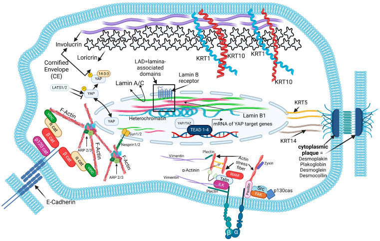
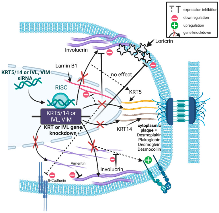
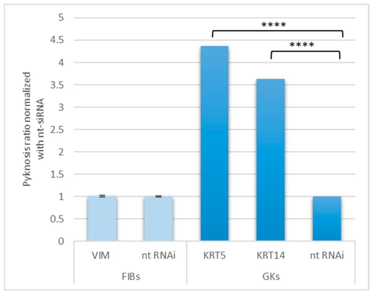
Although cytoplasmic intermediate filaments (cIFs) are essential for cell physiology, the molecular and cell functional consequences of cIF disturbances are poorly understood. Identifying defaults in cell function-controlled tissue homeostasis and understanding the interrelationship between specific cIFs and distinct cell functions remain key challenges. Using an RNAi-based mechanistic approach, we connected the impairment of cell-inherent cIFs with molecular and cell functional consequences, such as proliferation and differentiation. To investigate cIF disruption consequences in the oral epithelium, different cell transformation stages, originating from alcohol-treated oral gingival keratinocytes, were used. We found that impairment of keratin (KRT) KRT5, KRT14 and vimentin (VIM) affects proliferation and differentiation, and modulates the chromatin status. Furthermore, cIF impairment reduces the expression of nuclear integrity participant lamin B1 and the terminal keratinocyte differentiation marker involucrin (IVL). Conversely, impairment of IVL reduces cIF expression levels, functionally suggesting a regulatory interaction between cIFs and IVL. The findings demonstrate that the impairment of cIFs leads to imbalances in proliferation and differentiation, both of which are essential for tissue homeostasis. Thus, targeted impairment of cIFs appears promising to investigate the functional role of cIFs on cell-dependent tissue physiology at the molecular level and identifies putative interactions of cIFs with epithelial differentiation.

Keywords: apoptosis; chromatin; differentiation; intermediate filaments; involucrin; keratin; keratinocytes; proliferation; transformation; vimentin.


Conflict of interest statement

The authors declare no conflicts of interest.

Figures

Figure 1
Figure 1
Light microscopic images to visualize the different cell morphologies and degrees of confluence: (A) confluent gingival keratinocytes (GKs), (B) post-confluent GKs, (C) confluent epithelium-like phenotypes (EPIs), (D) post-confluent EPIs, (E) confluent fibroblast-like phenotypes (FIBs), (F) post-confluent FIBs. Bars correspond to 200 µm.
Figure 2
Figure 2
Model cell that visualizes the biomarkers of interest and their molecular context. In addition to the analyzed cIFs and involucrin (IVL), the following molecules were subject of the investigation: CDH1 (E-cadherin), LOR (loricrin), ITGB1 (integrin β1), ITGB3 (integrin β3), LMNB1 (lamin B1), ACTR2 (actin-related protein 2) and YAP1 (yes-associated protein). CDH1 is a constituent of adherens junctions, which connect neighboring epithelial cells to each other, thereby being indispensable for epithelial morphogenesis [48]. Intracellularly, it binds to the actin filament system via linker proteins of the catenin family [49]. In the context of epithelial keratinocyte differentiation in addition to IVL, LOR is a further marker of terminal differentiation and together with IVL participates in cornified envelope formation. LOR can form crosslinks with itself, thereby yielding different molecular weights [40]. Within the lamina-associated domains, the lamin B receptor tethers heterochromatin via LMNB1 to the inner nuclear membrane [50]. After shuttling to the nucleus, the co-transcriptional activator YAP (and its homologue TAZ, neglected in this study) binds to the TEAD family of transcription factors (TEA Domain Transcription Factor1-4), thereby inducing the expression of YAP target genes [51]. At sites of cell matrix interaction, the cIF VIM is involved in the formation of actin-bound FAs via interaction with the FAs constituent plectin [52,53]. Further constituents of FAs are ITGB1 and ITGB3, whereas ITGB3 appears to play a role in the recruitment of VIM and plectin to FAs [54]. Both of the basal cell-specific keratins, the cIFs KRT5 and KRT14, are connected with DP (Desmoplakin) [55], one of the constituents of the cytoplasmic plaque [56]. At sites of adherens junction-mediated cell-to-cell adhesion, CDH1 is linked to the actin cytoskeleton through members of the catenin family and vinculin (VCL) [57]. At sites of the nuclear envelope, ARP2/3 is involved in the formation of the actin cap, which interacts with the inner part of the nucleus through nesprin 1/2 interaction, the latter connected with the nuclear lamins nIFs via members of the Sun-family proteins Sun 1/2 (Sad1/UNC-84 domain-containing proteins) [58]. Created with BioRender.com.
Figure 3
Figure 3
Indirect immunofluorescence of biomarkers under study in GK, EPI and FIB phenotypes of 4-day-old cultures, within the subconfluent culture stage. (AC) Vimentin (VIM) expression in all phenotypes with decreasing intensity and changed cellular topology in FIBs (A), EPIs (B) and GKs (C). (DF) Cytokeratin 5 staining (KRT5) in FIBs, EPIs and GKs with increasing intensity and different cellular topology. (G) Involucrin (IVL) staining in FIBs, (H) EPIs and (I) GKs with changed cytoplasmic distribution. (JL) Loricrin (LOR) staining in FIBs was almost not detectable (J) and in EPIs (K) and GKs (L) with a faint cytoplasmic per-nuclear distribution. Cytokeratin 10 (KRT10) staining in FIBs (M) was again almost not detectable and in EPIs (N) and GKs (O) detectable with an inhomogeneous cytoplasmic distribution. (PR) Lamin B1 (LMNB1) staining in FIBs (P), EPIs (Q) and GKs (P) with nuclear localization. Biomarker under study with green fluorescence, red cytoskeleton staining in red and DAPI nuclear counterstain in blue. 60× magnification. Bars correspond to 100 µm.
Figure 4
Figure 4
The cartoon illustrates based on Figure 1 the used gene knockdowns/impairments and their respective effect on biomarkers under study. The figure shows in detail the RNAi-based intervention of KRT5/14, IVL and VIM, and their upstream and downstream effects in the cell. It shows either the up-regulation of the expression with a plus sign or the down-regulation with a minus sign of the corresponding genes. Created with BioRender.com.
Figure 5
Figure 5
Consequences of selectively evaluated impairments of KRT5, KRT14 and VIM on the proportion of pyknotic nuclei in FIBs and GKs. The total number of DAPI-stained cell nuclei was compared with the number of pyknotic cell nuclei, statistically analyzed using an unpaired t-test and compared with the nt-siRNA control (n = 3, mean value ± SEM, p < 0.0001 ****).

Similar articles

References

    1. Griswold M.G., Fullman N., Hawley C., Arian N., Zimsen S.R., Tymeson H.D., Venkateswaran V., Tapp A.D., Forouzanfar M.H., Salama J.S. Alcohol use and burden for 195 countries and territories, 1990–2016: A systematic analysis for the Global Burden of Disease Study 2016. Lancet. 2018;392:1015–1035. doi: 10.1016/S0140-6736(18)31310-2. - DOI - PMC - PubMed
    1. Im P.K., Yang L., Kartsonaki C., Chen Y., Guo Y., Du H., Lin K., Kerosi R., Hacker A., Liu J. Alcohol metabolism genes and risks of site-specific cancers in Chinese adults: An 11-year prospective study. Int. J. Cancer. 2022;150:1627–1639. doi: 10.1002/ijc.33917. - DOI - PMC - PubMed
    1. Steinberg T., Dieterle M.P., Tomakidi P. Molecular Research on Oral Diseases and Related Biomaterials: A Journey from Oral Cell Models to Advanced Regenerative Perspectives. Int. J. Mol. Sci. 2022;23:5288. doi: 10.3390/ijms23095288. - DOI - PMC - PubMed
    1. Roesch-Ely M., Steinberg T., Bosch F.X., Mussig E., Whitaker N., Wiest T., Kohl A., Komposch G., Tomakidi P. Organotypic co-cultures allow for immortalized human gingival keratinocytes to reconstitute a gingival epithelial phenotype in vitro. Differentiation. 2006;74:622–637. doi: 10.1111/j.1432-0436.2006.00099.x. - DOI - PubMed
    1. Chamulitrat W., Schmidt R., Chunglok W., Kohl A., Tomakidi P. Epithelium and fibroblast-like phenotypes derived from HPV16 E6/E7-immortalized human gingival keratinocytes following chronic ethanol treatment. Eur. J. Cell Biol. 2003;82:313–322. doi: 10.1078/0171-9335-00317. - DOI - PubMed

Publication types

LinkOut - more resources