. 2024 Nov 28;13(23):1969.
doi: 10.3390/cells13231969.

Unveiling Smyd-2's Role in Cytoplasmic Nrf-2 Sequestration and Ferroptosis Induction in Hippocampal Neurons After Cerebral Ischemia/Reperfusion

Affiliations

Unveiling Smyd-2's Role in Cytoplasmic Nrf-2 Sequestration and Ferroptosis Induction in Hippocampal Neurons After Cerebral Ischemia/Reperfusion

Daohang Liu et al. Cells. .

Abstract

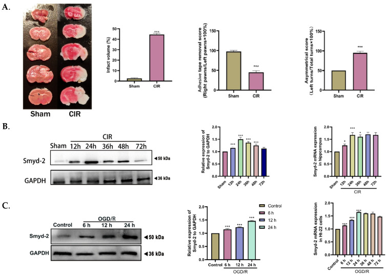
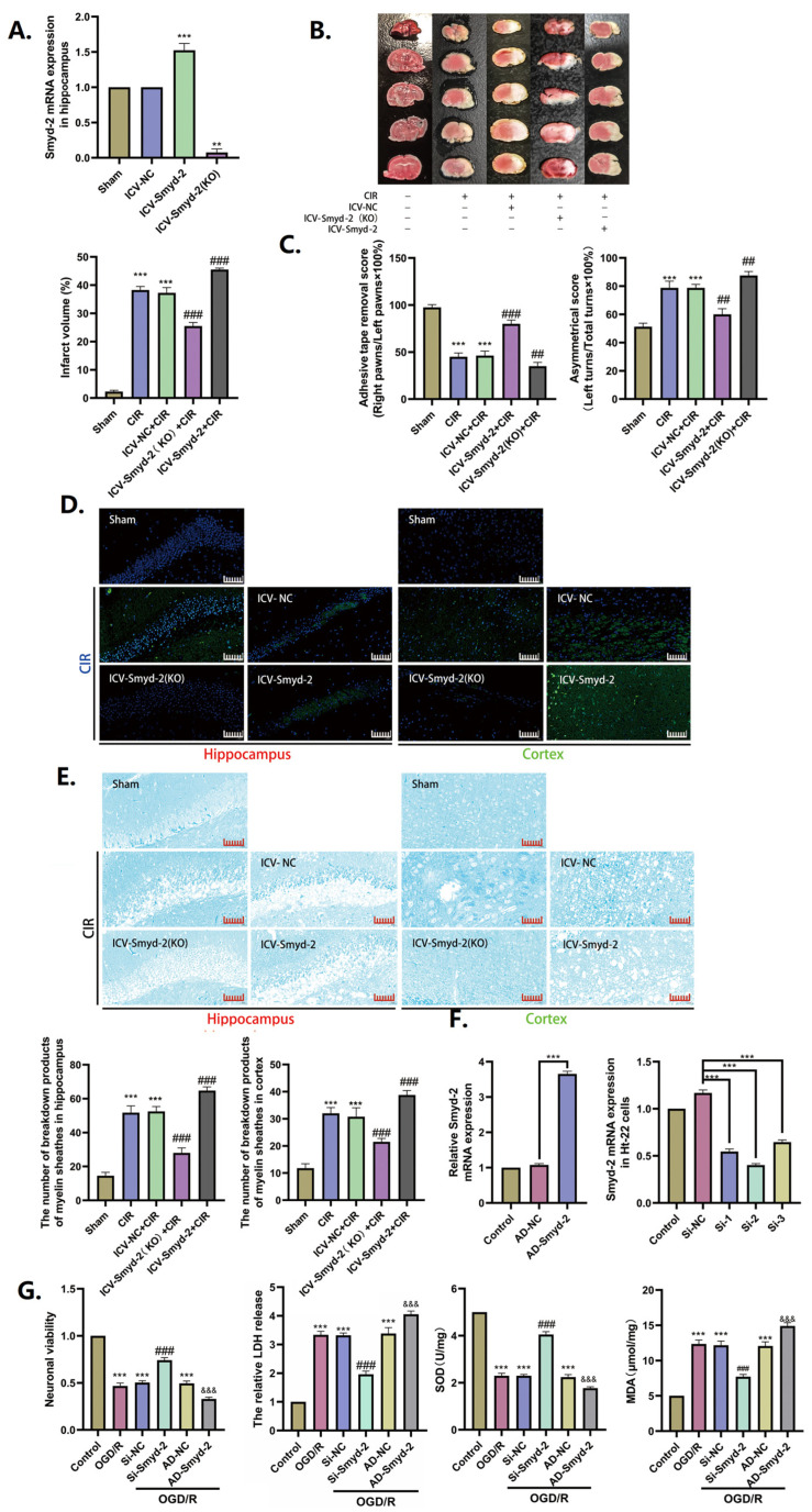
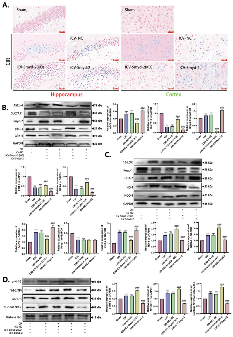
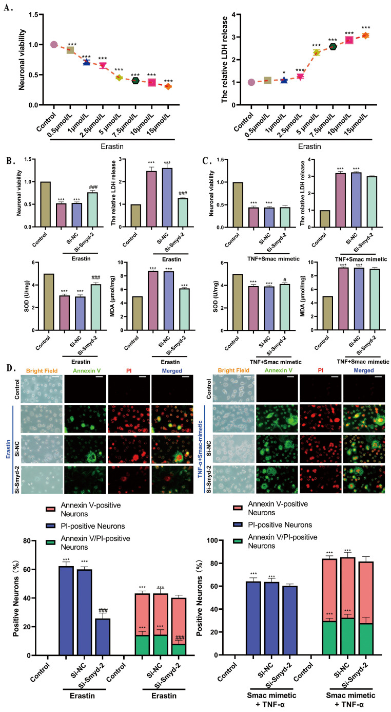
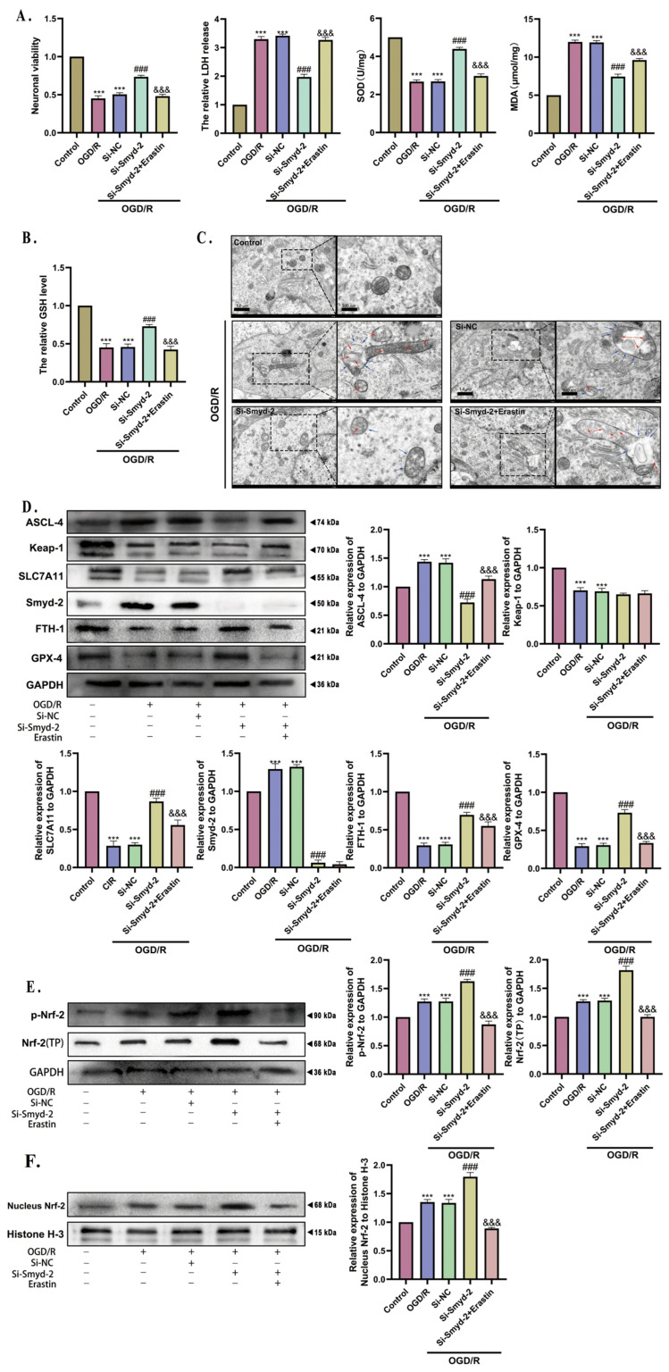
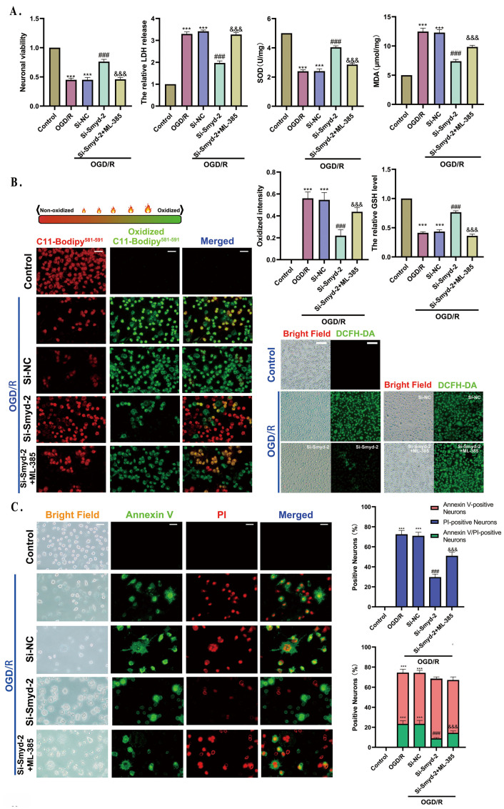
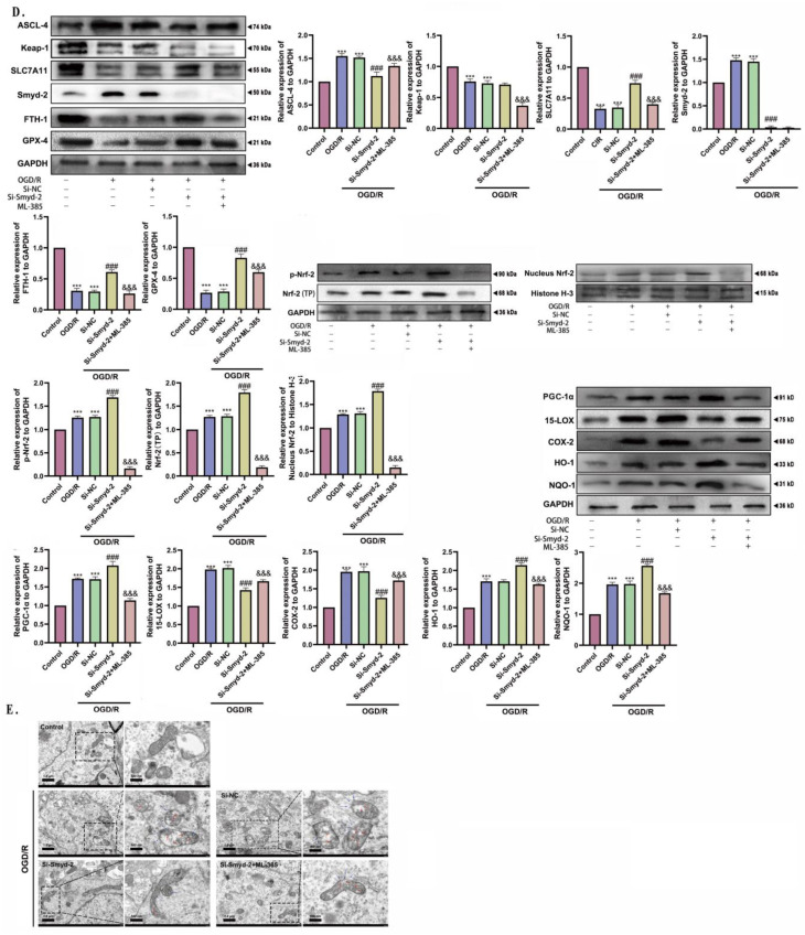
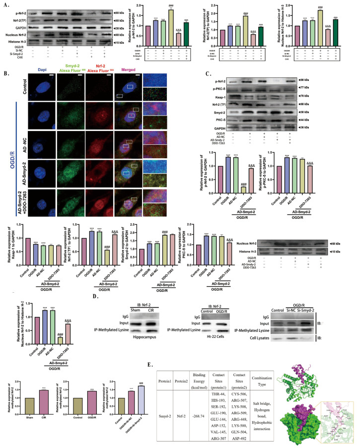
SET and MYND Domain-Containing 2 (Smyd-2), a specific protein lysine methyltransferase (PKMT), influences both histones and non-histones. Its role in cerebral ischemia/reperfusion (CIR), particularly in ferroptosis-a regulated form of cell death driven by lipid peroxidation-remains poorly understood. This study identifies the expression of Smyd-2 in the brain and investigates its relationship with neuronal programmed cell death (PCD). We specifically investigated how Smyd-2 regulates ferroptosis in CIR through its interaction with the Nuclear Factor Erythroid-2-related Factor-2 (Nrf-2)/Kelch-like ECH-associated protein (Keap-1) pathway. Smyd-2 knockout protects HT-22 cells from Erastin-induced ferroptosis but not TNF-α + Smac-mimetic-induced apoptosis/necroptosis. This neuroprotective effect of Smyd-2 knockout in HT-22 cells after Oxygen-Glucose Deprivation/Reperfusion (OGD/R) was reversed by Erastin. Smyd-2 knockout in HT-22 cells shows neuroprotection primarily via the Nuclear Factor Erythroid-2-related Factor-2 (Nrf-2)/Kelch-like ECH-associated protein (Keap-1) pathway, despite the concurrent upregulation of Smyd-2 and Nrf-2 observed in both the middle cerebral artery occlusion (MCAO) and OGD/R models. Interestingly, vivo experiments demonstrated that Smyd-2 knockout significantly reduced ferroptosis and lipid peroxidation in hippocampal neurons following CIR. Moreover, the Nrf-2 inhibitor ML-385 abolished the neuroprotective effects of Smyd-2 knockout, confirming the pivotal role of Nrf-2 in ferroptosis regulation. Cycloheximide (CHX) fails to reduce Nrf-2 expression in Smyd-2 knockout HT-22 cells. Smyd-2 knockout suppresses Nrf-2 lysine methylation, thereby promoting the Nrf-2/Keap-1 pathway without affecting the PKC-δ/Nrf-2 pathway. Conversely, Smyd-2 overexpression disrupts Nrf-2 nuclear translocation, exacerbating ferroptosis and oxidative stress, highlighting its dual regulatory role. This study underscores Smyd-2's potential for ischemic stroke treatment by disrupting the Smyd-2/Nrf-2-driven antioxidant capacity, leading to hippocampal neuronal ferroptosis. By clarifying the intricate interplay between ferroptosis and oxidative stress via the Nrf-2/Keap-1 pathway, our findings provide new insights into the molecular mechanisms of CIR and identify Smyd-2 as a promising therapeutic target.

Keywords: Nrf-2; Smyd-2; ferroptosis; hinge and latch; ischemic stroke; lipid peroxidation.


Conflict of interest statement

The authors declare no conflicts of interest.

Figures

Figure 1
Figure 1
MCAO and OGD/R conduce to Smyd-2 activation in the hippocampus and HT-22 cells. (A). Representative images of TTC staining, quantitative analysis of infarct volume, asymmetrical test scores, and the adhesive removal scores in MCAO mice (n = 5). (B). Smyd-2 expression in mouse brain after CIR (n = 5). (C). Smyd-2 expression in Ht-22 cells challenged with OGD/R (n = 5). * p < 0.05 vs. sham group, *** p < 0.001 vs. sham group; *** p < 0.0001 vs. control group.
Figure 2
Figure 2
The inhibition of Smyd-2 expression delays the progression of CIR impairment. (AC) Effects of Smyd-2 on cerebral infarction volume and neurobehavioral function in MCAO mice (n = 5). (D) Representative images of FJB staining slices of the hippocampus and cortex; scale bars = 40 μm (n = 5). (E) Representative images of LFB staining slices of the hippocampus and cortex and quantitative analysis of the breakdown products of myelin sheathes in the hippocampus and cortex; scale bars = 40 μm (n = 5). (F) The transfection effects of Smyd-2-overexpressing adenovirus and siRNA in Ht-22 cells (n = 5). (G) The neuronal viability, LDH release, SOD level, and MDA level of Ht-22 cells challenged with Smyd-2 siRNA and Smyd-2-overexpressing adenovirus after OGD/R (n = 5). ** p < 0.01 vs. sham group, *** p < 0.001 vs. sham group; ## p < 0.01, ### p < 0.001 vs. CIR group and CIR + IVC-NC group; *** p < 0.0001 vs. control group; ### p < 0.0001 vs. OGD/R group and OGD/R + Si-NC group; &&& p < 0.0001 vs. OGD/R group and OGD/R + AD-NC group.
Figure 3
Figure 3
Smyd-2 knockout counteracts the effect of OGD/R on lipid peroxidation in Ht-22 cells. (A) BODIPY-581/591-C11 staining was applied to analyze and quantify the effect of Smyd-2 siRNA and adenovirus-mediated Smyd-2 on HT-22 cell ferroptosis challenged with OGD/R. The representative images were obtained with an optical microscope at 400× magnification; scale bars = 50 μm. The GSH level of HT-22 cells challenged with AD-Smyd-2 and Si-Smyd-2 after OGD/R. (B) DCFH-DA staining was applied to analyze and quantify the effect of Smyd-2 siRNA and adenovirus-mediated Smyd-2 on HT-22 cell ferroptosis challenged with OGD/R. The representative images were obtained with a confocal microscope at 200× magnification; scale bars = 100 μm (n = 6). (C) Immunofluorescence method was applied to investigate Smyd-2 and GPX-4 protein expression and localization in HT-22 cells and their relation to neuronal ferroptosis after OGD/R. The representative images were obtained with a confocal microscope at 400× magnification; scale bars = 50 μm. *** p < 0.0001 vs. control group; ### p < 0.0001 vs. OGD/R group and OGD/R + Si-NC group; &&& p < 0.0001 vs. OGD/R group and OGD/R + AD-NC group.
Figure 4
Figure 4
Smyd-2 regulates the abnormality in neuronal ferroptosis caused by CIR. (A) Representative images of Perls staining slices of the hippocampus and cortex. Scale bars = 40 μm (n = 5). (BD) Representative images of Western blot and quantitative analysis of the expression of Smyd-2, GPX-4, FTH-1, SLC7A11, ACSL-4, 15-LOX, COX-2, NQO-1, Keap-1, HO-1, nucleus Nrf-2, p-Nrf-2, and Nrf-2 (TP) in the hippocampus. *** p < 0.001 vs. sham group; ### p < 0.001 vs. CIR group and CIR + ICV-NC group.
Figure 5
Figure 5
The effect of Smyd-2 overexpression on programmed cell death of HT-22 cells induced by Erastin and Smac mimetic + TNF-α. (A) The neuronal viability and LDH release of HT-22 cells challenged with Erastin. (B,C) The neuronal viability and LDH release of HT-22 cells challenged with Erastin. (c,d,e,f,g,h,i,j) The neuronal viability, LDH release, SOD level, and MDA level of Si-Smyd-2 HT-22 cells challenged with Erastin and Smac mimetic (n = 6). (D) Annexin V/PI staining was applied to observe and analyze the effect of Erastin and Smac mimetic on different types of programmed cell death in HT-22 cells induced by OGD/R. The representative images were obtained with an optical microscope at 400× magnification; scale bars = 50 μm (n = 6). *** p < 0.0001 vs. control group, * p < 0.05 vs. control group; ### p < 0.0001 vs. OGD/R group and OGD/R + Si-NC group, # p < 0.05 vs. OGD/R + Si-NC group.
Figure 6
Figure 6
The effect of Si-Smyd-2 combined with Erastin on the ferroptosis of HT-22 cells induced by OGD/R. (A) The neuronal viability, LDH release, SOD, and MDA of HT-22 cells challenged with Si-Smyd-2 and Erastin after OGD/R. (B) GSH kit was applied to analyze and quantify the effect of the Si-RNA-mediated Smyd-2 and Erastin on HT-22 cell ferroptosis challenged with OGD/R (n = 5). (C) Pathophysiological and physiological morphologies of mitochondria in each HT-22 cell group were observed by transmission electron microscopy. The red arrows mark the increased electron density of the matrix and fractured and vague cristae. The blue arrows mark vacuoles in mitochondria. The enlarged region bounded by a rectangular dotted box conduces to obtaining a more detailed view of the mitochondria for each experimental condition. The representative images were obtained with an optical microscope at 8k× magnification; scale bars = 1 μm. The representative enlarged images were obtained with an optical microscope at 20k× magnification; scale bars = 500 nm (n = 5). (DF) Representative Western blots and quantitative evaluation of Smyd-2, SLC7A11, ACSL-4, FTH-1, GPX-4, Nrf-2, Keap-1, p-Nrf-2, and nucleus Nrf-2 expression levels in each HT-22 cell group. Data normalized to the loading control GAPDH are expressed as % of control (n = 5). *** p < 0.0001 vs. control group; ### p < 0.0001 vs. OGD/R group and OGD/R + Si-NC group; &&& p < 0.0001 vs. OGD/R + Si-Smyd-2 group.
Figure 7
Figure 7
Effects of Smyd-2 (KO) combined with Nrf-2 inhibitor ML-385 on ferroptosis and lipid peroxidation in MCAO mice and OGD/R-induced HT-22 cells. (A) The neuronal viability, LDH release, SOD, and MDA of HT-22 cells were challenged with Si-Smyd-2 and ML-385 after OGD/R (n = 5). (B) BODIPY-581/591-C11 staining, GSH kit, and DCFH-DA staining were used to analyze and quantify the effect of Si-RNA-mediated Smyd-2 and ML-385 on lipid peroxidation of HT-22 cells after OGD/R. The representative images of BODIPY-581/591-C11 staining were obtained with an optical microscope at 400× magnification; scale bars = 50 μm (n = 5). The representative images of DCFH-DA staining were obtained with an optical microscope at 200× magnification; scale bars = 100 μm (n = 5). (C) Annexin V/PI double fluorescence staining was used to study the effect of Si-RNA-mediated Smyd-2 and ML-385 on a different form of programmed cell death in HT-22 cells induced by OGD/R. The representative images were obtained with a confocal microscope at 400× magnification; scale bars = 50 μm (n = 5). (D) The effect of Si-RNA-mediated Smyd-2 combined with ML-385 on ferroptosis-related proteins in HT-22 cells induced by OGD/R. Representative Western blots and quantitative evaluation of ACSL-4, Keap-1, SLC7A11, Smyd-2, FTH-1, GPX-4, p-Nrf-2, Nrf-2 (TP), nucleus Nrf-2, PGC-1α, COX-2, 15-LOX, NQO-1, and HO-1 expression levels in each HT-22 cell group. Data normalized to the loading control GAPDH are expressed as % of control (n = 5). (E) Pathophysiological and physiological morphologies of mitochondria in each HT-22 cell group were observed by TEM. The red arrows mark the increased electron density of the matrix and fractured and vague cristae. The blue arrows mark vacuoles in mitochondria. The zoom region bounded by a rectangular dotted box allows a more detailed view of mitochondria for each experimental condition. The representative images were obtained with an optical microscope at 8k× magnification; scale bars = 1 μm (n = 5). The representative enlarged images were obtained with an optical microscope at 20k× magnification; scale bars = 500 nm. *** p < 0.001 vs. control group; ### p < 0.001 vs. OGD/R group and Si-NC + OGD/R group; &&& p < 0.001 vs. Si-Smyd-2 + OGD/R group.
Figure 7
Figure 7
Effects of Smyd-2 (KO) combined with Nrf-2 inhibitor ML-385 on ferroptosis and lipid peroxidation in MCAO mice and OGD/R-induced HT-22 cells. (A) The neuronal viability, LDH release, SOD, and MDA of HT-22 cells were challenged with Si-Smyd-2 and ML-385 after OGD/R (n = 5). (B) BODIPY-581/591-C11 staining, GSH kit, and DCFH-DA staining were used to analyze and quantify the effect of Si-RNA-mediated Smyd-2 and ML-385 on lipid peroxidation of HT-22 cells after OGD/R. The representative images of BODIPY-581/591-C11 staining were obtained with an optical microscope at 400× magnification; scale bars = 50 μm (n = 5). The representative images of DCFH-DA staining were obtained with an optical microscope at 200× magnification; scale bars = 100 μm (n = 5). (C) Annexin V/PI double fluorescence staining was used to study the effect of Si-RNA-mediated Smyd-2 and ML-385 on a different form of programmed cell death in HT-22 cells induced by OGD/R. The representative images were obtained with a confocal microscope at 400× magnification; scale bars = 50 μm (n = 5). (D) The effect of Si-RNA-mediated Smyd-2 combined with ML-385 on ferroptosis-related proteins in HT-22 cells induced by OGD/R. Representative Western blots and quantitative evaluation of ACSL-4, Keap-1, SLC7A11, Smyd-2, FTH-1, GPX-4, p-Nrf-2, Nrf-2 (TP), nucleus Nrf-2, PGC-1α, COX-2, 15-LOX, NQO-1, and HO-1 expression levels in each HT-22 cell group. Data normalized to the loading control GAPDH are expressed as % of control (n = 5). (E) Pathophysiological and physiological morphologies of mitochondria in each HT-22 cell group were observed by TEM. The red arrows mark the increased electron density of the matrix and fractured and vague cristae. The blue arrows mark vacuoles in mitochondria. The zoom region bounded by a rectangular dotted box allows a more detailed view of mitochondria for each experimental condition. The representative images were obtained with an optical microscope at 8k× magnification; scale bars = 1 μm (n = 5). The representative enlarged images were obtained with an optical microscope at 20k× magnification; scale bars = 500 nm. *** p < 0.001 vs. control group; ### p < 0.001 vs. OGD/R group and Si-NC + OGD/R group; &&& p < 0.001 vs. Si-Smyd-2 + OGD/R group.
Figure 8
Figure 8
Smyd-2 methylates Nrf-2 (Lys-508) to inhibit OGD/R-induced Nrf-2 (Ser-40) phosphorylation and nuclear translocation. (A) Representative Western blot and quantitative evaluation of p-Nrf-2, Nrf-2 (TP), and nucleus Nrf-2 expression levels in each HT-22 cell group. Data normalized to the loading control GAPDH are expressed as % of control. Data normalized to the loading control GAPDH and histone H-3 are expressed as % of control (n = 5). (B) Confocal images of the localization with immunofluorescence-stained Smyd-2 (green) and immunofluorescence-stained Nrf-2 (red) in various HT-22 cell groups after OGD/R. The zoom region bounded by rectangular boxes represents Smyd-2-Nrf-2 binding in the cytoplasm of HT-22 cells, and the Nrf-2 was transported to the cell nucleus. Scale bars = 10 μm (n = 5). (C) Representative Western blots and quantitative evaluation of Nrf-2 (TP), p-Nrf-2, nucleus Nrf-2, PKC-δ, p-PKC-δ, Smyd-2, and Keap-1 expression levels in each HT-22 cell group. Data normalized to the loading control histone H-3 are expressed as % of GAPDH (n = 5). (D) Quantitative analysis of the expression of the methylation level of Nrf-2 (n = 5). (E) The possible docking sites of two target proteins, Smyd-2/Nrf-2. The binding mode of the complex Nrf-2 with Smyd-2. *** p < 0.0001 vs. control group; ### p < 0.0001 vs. OGD/R + Si-NC group; &&& p < 0.0001 vs. OGD/R group; ^^^ p < 0.0001 vs. OGD/R + CHX group. *** p < 0.0001 vs. control group; ### p < 0.0001 vs. OGD/R group and OGD/R + AD-Smyd-2 group; &&& p < 0.0001 vs. OGD/R + AD-Smyd-2 group.

Similar articles

Cited by

References

    1. Cipolla M.J., Liebeskind D.S., Chan S.L. The importance of comorbidities in ischemic stroke: Impact of hypertension on the cerebral circulation. J. Cereb. Blood Flow. Metab. 2018;38:2129–2149. doi: 10.1177/0271678X18800589. - DOI - PMC - PubMed
    1. van Sloten T.T., Sedaghat S., Carnethon M.R., Launer L.J., Stehouwer C.D.A. Cerebral microvascular complications of type 2 diabetes: Stroke, cognitive dysfunction, and depression. Lancet Diabetes Endocrinol. 2020;8:325–336. doi: 10.1016/S2213-8587(19)30405-X. - DOI - PMC - PubMed
    1. Krinock M.J., Singhal N.S. Diabetes, stroke, and neuroresilience: Looking beyond hyperglycemia. Ann. N. Y. Acad. Sci. 2021;1495:78–98. doi: 10.1111/nyas.14583. - DOI - PubMed
    1. Castro P., Azevedo E., Sorond F. Cerebral Autoregulation in Stroke. Curr. Atheroscler. Rep. 2018;20:37. doi: 10.1007/s11883-018-0739-5. - DOI - PubMed
    1. Maida C.D., Norrito R.L., Daidone M., Tuttolomondo A., Pinto A. Neuroinflammatory Mechanisms in Ischemic Stroke: Focus on Cardioembolic Stroke, Background, and Therapeutic Approaches. Int. J. Mol. Sci. 2020;21:6454. doi: 10.3390/ijms21186454. - DOI - PMC - PubMed

Publication types

MeSH terms

Substances

LinkOut - more resources