. 2024 Nov 28;13(23):1971.
doi: 10.3390/cells13231971.

CD38 Inhibitor 78c Attenuates Pro-Inflammatory Cytokine Expression and Osteoclastogenesis in Macrophages

Affiliations

CD38 Inhibitor 78c Attenuates Pro-Inflammatory Cytokine Expression and Osteoclastogenesis in Macrophages

William Lory et al. Cells. .

Abstract

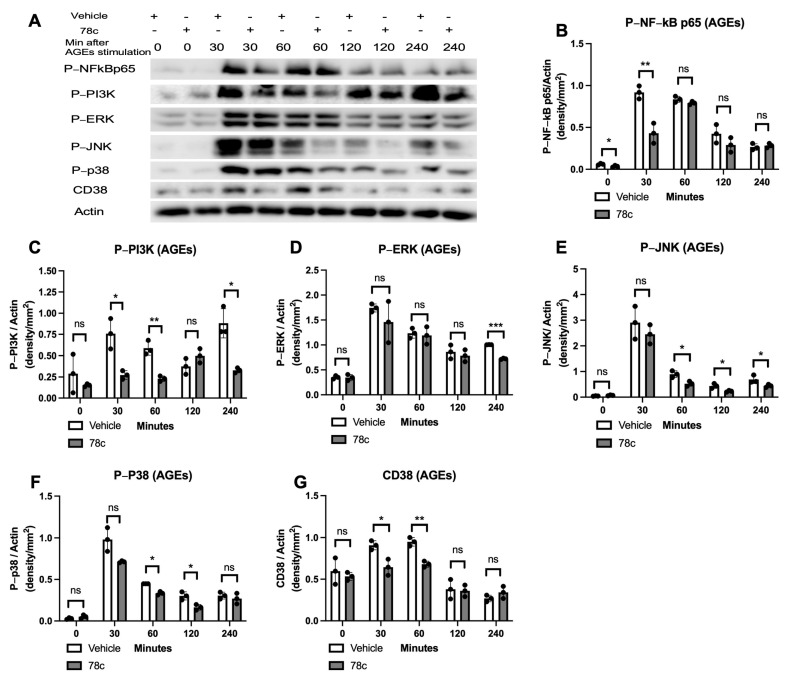
CD38, a nicotinamide adenine dinucleotide (NAD+) glycohydrolase, increases during infection or inflammation. Therefore, we aimed to evaluate the effects of a CD38 inhibitor (78c) on NAD+ levels, IL-1β, IL-6, TNF-α cytokine expressions, and osteoclastogenesis. The results show that treatment with 78c on murine BMMs dose-dependently reduced CD38, reversed the decline of NAD+, and inhibited IL-1β, IL-6, and TNF-α pro-inflammatory cytokine levels induced by oral pathogen Porphyromonas gingivalis (Pg) or Aggregatibacter actinomycetemcomitans (Aa) or by advanced glycation end products (AGEs). Additionally, treatment with 78c dose-dependently suppressed osteoclastogenesis and bone resorption induced by RANKL. Treatment with 78c suppressed CD38, nuclear factor kappa-B (NF-κB), phosphoinositide 3-kinase (PI3K), and mitogen-activated protein kinases (MAPKs) induced by Pg, Aa, or AGEs, and suppressed podosome components (PI3K, Pyk2, Src, F-actin, integrins, paxillin, and talin) induced by RANKL. These results from our studies support the finding that the inhibition of CD38 by 78c is a promising therapeutic strategy to treat inflammatory bone loss diseases. However, treatment with a CD38 shRNA only significantly reduced IL-1β, IL-6, and TNF-α pro-inflammatory cytokine levels induced by AGEs. Compared with controls, it had limited effects on cytokine levels induced by Pg or Aa. Treatment with the CD38 shRNA enhanced RANKL-induced osteoclastogenesis, suggesting that 78c has some off-target effects.

Keywords: CD38; NAD+; bone loss; cytokine; osteoclast; podosome.


Conflict of interest statement

The authors declare no conflicts of interest.

Figures

Figure 1
Figure 1
Inhibition of CD38 by 78c reduced CD38 gene expression, reversed the decline of NAD+, and suppressed IL-1β, IL-6, and TNF-α pro-inflammatory cytokines in murine BMMs infected by oral pathogens or stimulated by AGEs. Murine BMMs were treated with vehicles (diluted DMSO) or 78c (1.25 to 10 μM) with or without infection with Porphyromonas gingivalis (Pg), Aggregatibacter actinomycetemcomitans (Aa), or stimulation by AGEs for 24 h. (A) The mRNA levels of CD38 were evaluated using RT-q-PCR and normalized by β-actin expression. (B) NAD+ levels were measured and calibrated by cell growth and viability. (C) IL-1β, (D) IL-6, and (E) TNF-α levels were measured using ELISA and calibrated by protein concentration in cell lysate. Statistics were analyzed using a two-way ANOVA with Dunnett’s multiple comparisons test (n = 3, * p < 0.05, ** p < 0.01, *** p < 0.001).
Figure 2
Figure 2
Inhibition of CD38 by 78c reduced NF-kB, PI3K, ERK, p38 MAPK, and CD38 protein expressions induced by the oral pathogen Porphyromonas gingivalis (Pg). Murine BMMs were treated with vehicle (diluted DMSO) or 78c (10 μM) with or without infection with Pg from 30 min to 240 min. (A) Protein levels of p-NFκBp65, p-PI3K, p-ERK, p-JNK, p-p38, CD38, and pan-actin in cell lysate were determined using Western blot. Protein densitometry of p-NFκBp65 (B), p-PI3K (C), p-ERK (D), p-JNK (E), p-p38 (F), and CD38 (G) were evaluated. Statistics were assessed using an unpaired t-test to compare vehicle vs. 78c treatment using Welch’s correction (n = 3, ns: no significance, * p < 0.05, ** p < 0.01).
Figure 3
Figure 3
Inhibition of CD38 by 78c reduced NF-kB, PI3K, ERK, JNK, p38 MAPK, and CD38 protein expressions induced by oral pathogen Aggregatibacter actinomycetemcomitans (Aa). Murine BMMs were treated with vehicle (diluted DMSO) or 78c (10 μM) with or without infection with Aa from 30 min to 240 min. (A) Protein levels of p-NFκBp65, p-PI3K, p-ERK, p-JNK, p-p38, CD38, and pan-actin in cell lysate were determined using Western blot. Protein densitometry of p-NFκBp65 (B), p-PI3K (C), p-ERK (D), p-JNK (E), p-p38 (F), and CD38 (G) were evaluated. Statistics were assessed using an unpaired t-test with Welch’s correction (n = 3, ns: no significance, * p < 0.05, ** p < 0.01, *** p < 0.001).
Figure 4
Figure 4
Inhibition of CD38 by 78c reduced NF-kB, PI3K, ERK, JNK, p38 MAPK, and CD38 protein expressions induced by advanced glycation end products (AGEs). Murine BMMs were treated with vehicle (diluted DMSO) or 78c (10 μM) with or without stimulation by AGEs (10 μg/mL) from 30 min to 240 min. (A) Protein levels of p-NFκBp65, p-PI3K, p-ERK, p-JNK, p-p38, CD38, and pan-actin in cell lysate were determined using Western blot. Protein densitometry of p-NFκBp65 (B), p-PI3K (C), p-ERK (D), p-JNK (E), p-p38 (F), and CD38 (G) were evaluated. Statistics were assessed using an unpaired t-test with Welch’s correction (n = 3, ns: no significance, * p < 0.05, ** p < 0.01, *** p < 0.001).
Figure 5
Figure 5
Treatment with 78c suppressed osteoclastogenesis and bone resorption induced by RANKL. Murine bone marrow cells were treated with vehicle (diluted DMSO) or 78c (1.25 to 10 μM) with or without stimulation by RANKL for five days (in a 96-well cell culture plate for osteoclastogenesis assay) or for eight days (in a calcium phosphate-coated 48-well plate for bone resorption assay). (A) Representative images show TRAP-stained cells at the 100× magnification view. Scale bars represent 50 μM. (B) Number of TRAP+ multinucleated (more than three nuclei) osteoclasts/well (96-well). (C) Total areas of osteoclasts/image were quantified. (D) Representative images show osteoclast resorption pits at 200× magnification view. Scale bars represent 100 μM. (E) Total areas of osteoclast resorption pits/images were quantified. Statistics were analyzed using a non-parametric ANOVA with the Kruskal–Wallis correction (n = 4, *** p < 0.001).
Figure 6
Figure 6
Inhibition of CD38 by 78c attenuated CD38, Nfatc1, Ctsk, Acp5, Oscar, Ocstamp, and Dcstamp mRNA expressions induced by RANKL. Murine bone marrow cells were treated with vehicle (diluted DMSO) or 78c (1.25 to 10 μM) with or without stimulation by RANKL for four days. (A) CD38 mRNA, (B) Nfatc1 mRNA, (C) Ctsk mRNA, (D) Acp5 mRNA, (E) Oscar mRNA, (F) Ocstamp mRNA, and (G) Dcstamp mRNA levels were quantified using RT-PCR and normalized by β-actin expression. Statistics were analyzed using an ordinary two-way ANOVA with multiple comparisons (n = 3, * p < 0.05, ** p < 0.01, *** p < 0.001).
Figure 7
Figure 7
Inhibition of CD38 by 78c inhibited podosome components (PI3K, Pyk2, Src, integrin β3, F-actin, paxillin, and talin) induced by RANKL. Murine bone marrow cells were treated with vehicle (diluted DMSO) or 78c (5 μM) with or without RANKL stimulation for four days. (A) Protein levels of p-PI3K, p-Pyk2, p-Src, CD38, and pan-actin protein levels were evaluated using Western blot. Protein densitometry of (B) p-PI3K, (C) p-Pyk2, (D) p-Src, and (E) CD38 were evaluated. (F) Protein levels of integrin β3, F-actin, paxillin, talin, and pan-actin protein levels were evaluated using Western blot. Protein densitometry of integrin β3 (G), F-actin (H), paxillin (I), and talin (J) were evaluated. Statistics were evaluated using an unpaired t-test with Welch’s correction (n = 3, ns: no significance, * p < 0.05, ** p < 0.01).
Figure 8
Figure 8
Effect of a CD38 shRNA on CD38 mRNA, NAD+, IL-1β, IL-6, and TNF-α cytokine levels, NF-κB, PI3K, MAPK, and CD38 Protein levels induced by oral pathogen Porphyromonas gingivalis (Pg), Aggregatibacter actinomycetemcomitans (Aa), or by advanced glycation end products (AGEs) compared with controls. Murine BMMs were treated with a control shRNA or a CD38 shRNA lentiviral vector (MOI 10). At 72 h after lentiviral infection, cells were untreated, infected with Pg or Aa, or stimulated by AGEs for 24 h (for CD38 mRNA assay, NAD+ assay, and IL-1β, IL-6, TNF-α cytokine assays) or 2 h (for Western blot assay). (A) CD38 mRNA levels were evaluated using RT-q-PCR and normalized by β-actin expression. (B) NAD+ levels were measured and calibrated by cell growth and viability. (C) IL-1β, (D) IL-6, and (E) TNF-α levels were measured using ELISA and calibrated by protein concentration in cell lysate. Statistics were analyzed using an unpaired t-test with Welch’s correction (n = 3, ns: no significance, ** p < 0.01, *** p < 0.001). (F) Protein levels of p-NFκBp65, p-PI3K, p-ERK, p-JNK, p-p38, CD38, and pan-actin in cell lysate were determined using Western blot.
Figure 9
Figure 9
Effect of a CD38 shRNA on osteoclastogenesis and bone resorption induced by RANKL. Murine bone marrow cells were treated with a control shRNA or a CD38 shRNA lentiviral vector (MOI 10). At 24 h after lentiviral infection, the cells were unstimulated or stimulated with RANLK for five days (in a 96-well cell culture plate for osteoclastogenesis assay), eight days (in a calcium phosphate-coated 48-well plate for bone resorption assay), or four days (for RT-PCR assay and Western blot assay). (A) Representative images show TRAP-stained cells at the 100× magnification view. Scale bars represent 50 μM. (B) Number of TRAP+ multinucleated (more than three nuclei) osteoclasts/well (96-well) (n = 4). (C) Total areas of osteoclast/image were quantified (n = 4). (D) Representative images show osteoclast resorption pits at 200× magnification view. Scale bars represent 100 μM. (E) CD38 mRNA level, (F) Nfatc1 mRNA level, (G) Ctsk mRNA level, (H) Acp5 mRNA level, (I) Oscar mRNA level, (J) Ocstamp mRNA level, and (K) Dcstamp mRNA levels were quantified using RT-PCR and normalized by β-actin expression. Statistics were analyzed using an ordinary one-way ANOVA with Tukey’s multiple comparisons test (n = 3, * p < 0.05, ** p < 0.01, *** p < 0.001). (L) Protein levels of p-PI3K, p-Pyk2, p-Src, F-actin, integrin β3, paxillin, talin, CD38, and pan-actin protein levels were evaluated using Western blot.

Similar articles

Cited by

References

    1. Piedra-Quintero Z.L., Wilson Z., Nava P., Guerau-de-Arellano M. CD38: An Immunomodulatory Molecule in Inflammation and Autoimmunity. Front. Immunol. 2020;11:597959. doi: 10.3389/fimmu.2020.597959. - DOI - PMC - PubMed
    1. Jablonski K.A., Amici S.A., Webb L.M., Ruiz-Rosado Jde D., Popovich P.G., Partida-Sanchez S., Guerau-de-Arellano M. Novel Markers to Delineate Murine M1 and M2 Macrophages. PLoS ONE. 2015;10:e0145342. doi: 10.1371/journal.pone.0145342. - DOI - PMC - PubMed
    1. Hogan K.A., Chini C.C.S., Chini E.N. The Multi-faceted Ecto-enzyme CD38: Roles in Immunomodulation, Cancer, Aging, and Metabolic Diseases. Front. Immunol. 2019;10:1187. doi: 10.3389/fimmu.2019.01187. - DOI - PMC - PubMed
    1. Kar A., Mehrotra S., Chatterjee S. CD38: T Cell Immuno-Metabolic Modulator. Cells. 2020;9:1716. doi: 10.3390/cells9071716. - DOI - PMC - PubMed
    1. Benzi A., Grozio A., Spinelli S., Sturla L., Guse A.H., De Flora A., Zocchi E., Heeren J., Bruzzone S. Role of CD38 in Adipose Tissue: Tuning Coenzyme Availability? Nutrients. 2021;13:3734. doi: 10.3390/nu13113734. - DOI - PMC - PubMed

Publication types

LinkOut - more resources