. 2024 Nov 29;13(23):1978.
doi: 10.3390/cells13231978.

Herpes Simplex Virus 1 Infection of Human Brain Organoids and Pancreatic Stem Cell-Islets Drives Organoid-Specific Transcripts Associated with Alzheimer's Disease and Autoimmune Diseases

Affiliations

Herpes Simplex Virus 1 Infection of Human Brain Organoids and Pancreatic Stem Cell-Islets Drives Organoid-Specific Transcripts Associated with Alzheimer's Disease and Autoimmune Diseases

Jonathan Sundstrom et al. Cells. .

Abstract

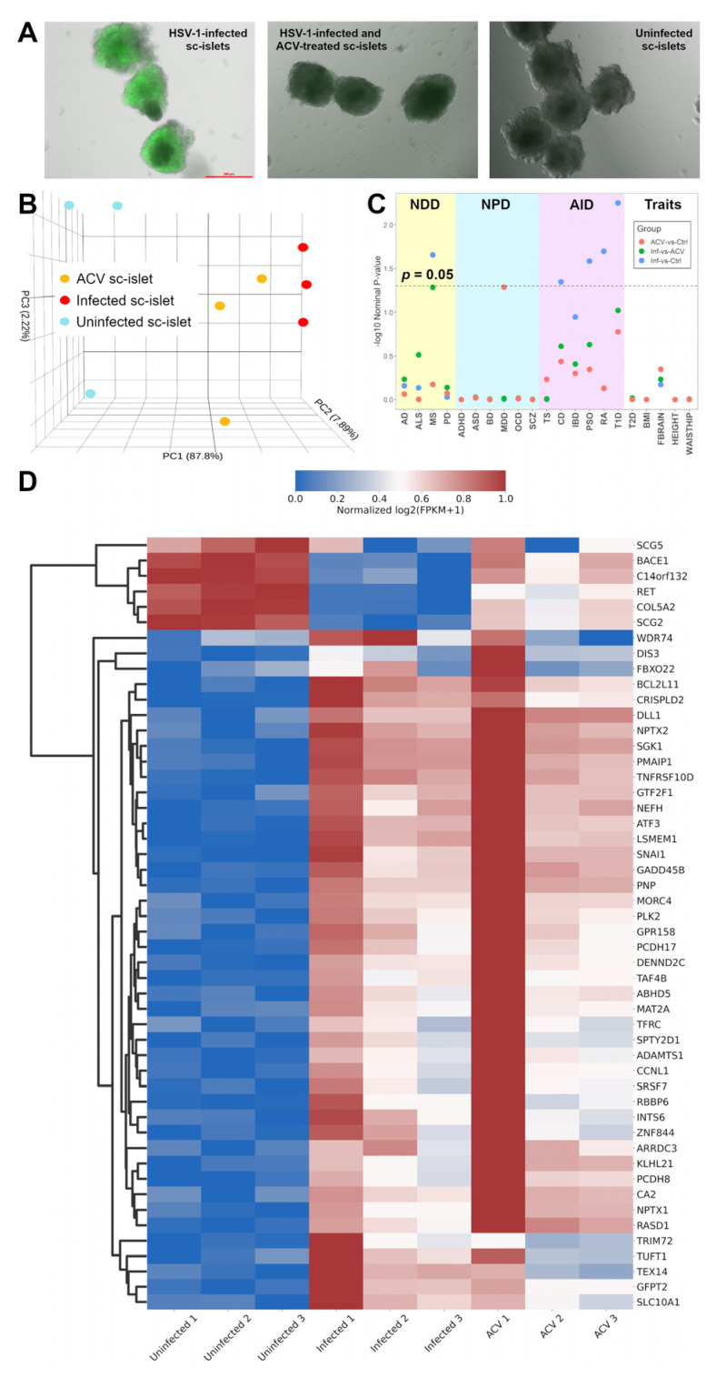
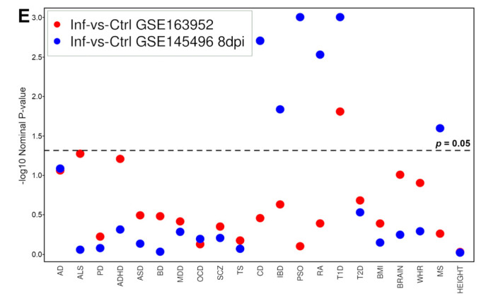
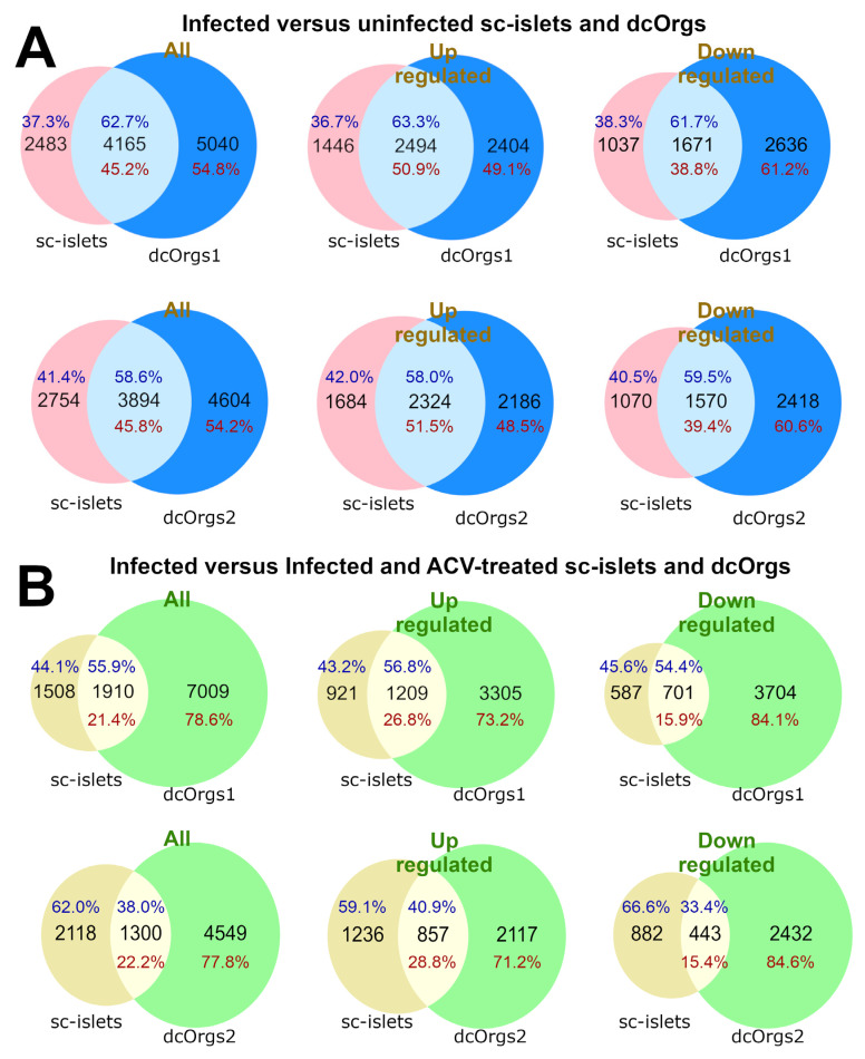
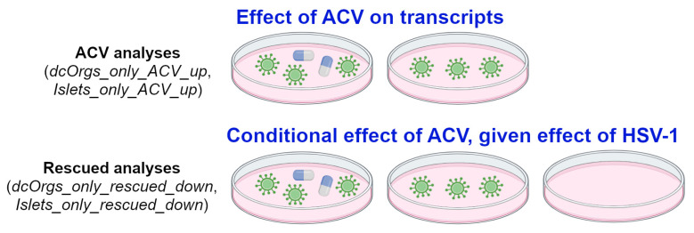
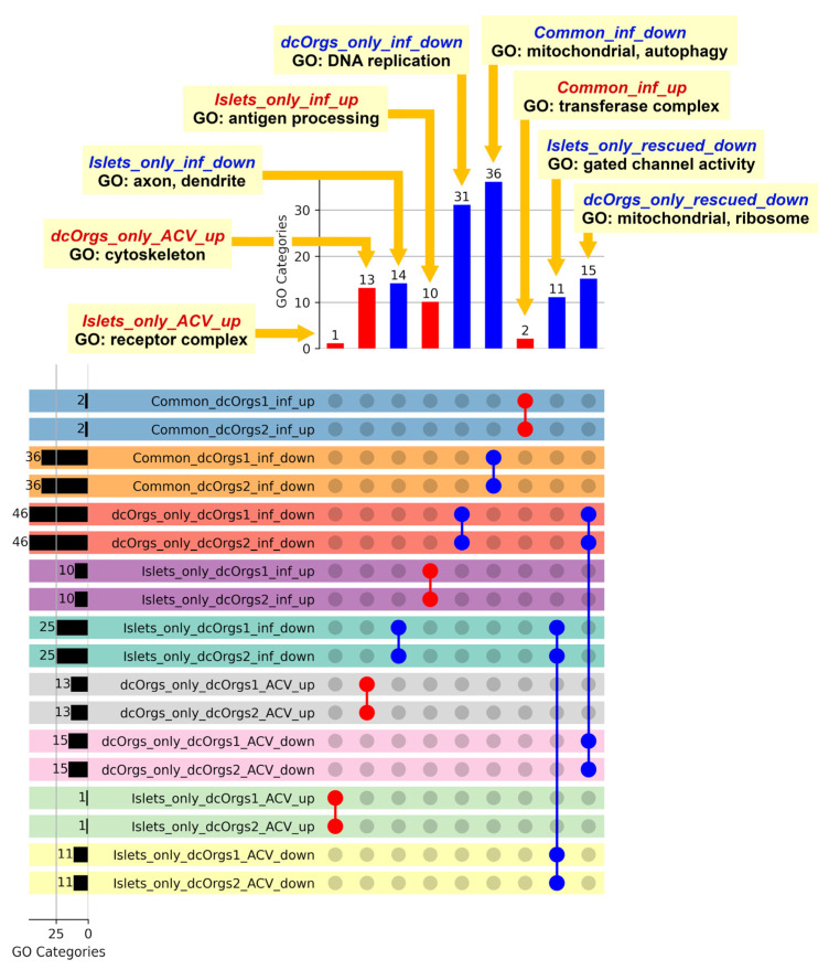
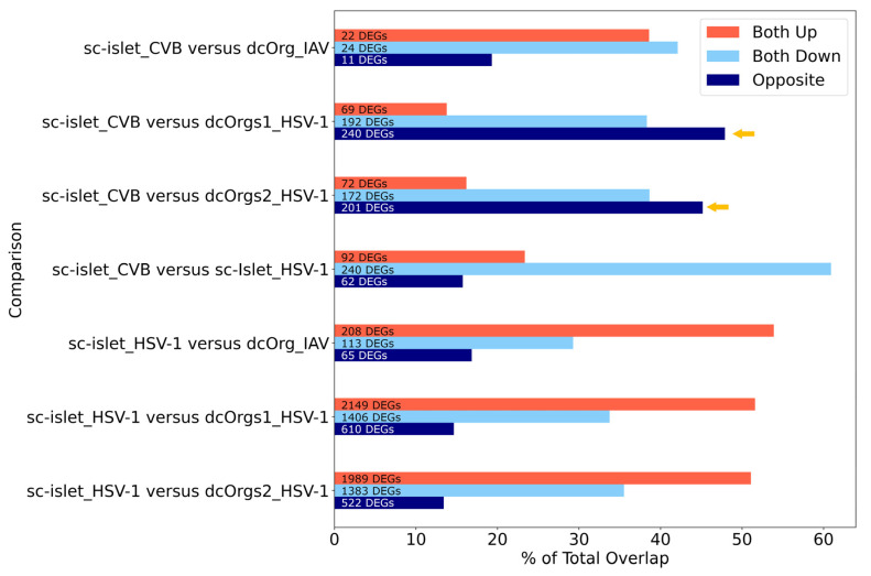
Viral infections leading to inflammation have been implicated in several common diseases, such as Alzheimer's disease (AD) and type 1 diabetes (T1D). Of note, herpes simplex virus 1 (HSV-1) has been reported to be associated with AD. We sought to identify the transcriptomic changes due to HSV-1 infection and anti-viral drug (acyclovir, ACV) treatment of HSV-1 infection in dissociated cells from human cerebral organoids (dcOrgs) versus stem cell-derived pancreatic islets (sc-islets) to gain potential biological insights into the relevance of HSV-1-induced inflammation in AD and T1D. We observed that differentially expressed genes (DEGs) in HSV-1-infected sc-islets were enriched for genes associated with several autoimmune diseases, most significantly, T1D, but also rheumatoid arthritis, psoriasis, Crohn's disease, and multiple sclerosis, whereas DEGs in HSV-1-infected dcOrgs were exclusively enriched for genes associated with AD. The ACV treatment of sc-islets was not as effective in rescuing transcript perturbations of autoimmune disease-associated genes. Finally, we identified gene ontology categories that were enriched for DEGs that were in common across, or unique to, viral treatment of dcOrgs and sc-islets, such as categories involved in the transferase complex, mitochondrial, and autophagy function. In addition, we compared transcriptomic signatures from HSV-1-infected sc-islets with sc-islets that were infected with the coxsackie B virus (CVB) that had been associated with T1D pathogenesis. Collectively, this study provides tissue-specific insights into the molecular effects of inflammation in AD and T1D.

Keywords: Alzheimer’s disease; acyclovir; autoimmune diseases; cerebral organoids; herpes simplex virus 1; innate immune; neurodegenerative diseases; stem cell islets; type 1 diabetes.


Conflict of interest statement

The authors declare no conflicts of interest. The funders had no role in the design of the study; in the collection, analyses, or interpretation of data; in the writing of the manuscript; or in the decision to publish the results.

Figures

Figure 1
Figure 1
Analyses of RNA-seq data from sc-islets showed enrichment of autoimmune disease genes. (A) Images of unfixed HSV-1-infected sc-islets, HSV-1-infected and ACV-treated sc-islets, and uninfected sc-islets after 48 h. (B) PCA plot of the sc-islet RNA-seq data showing clustering of uninfected sc-islets (in blue), infected sc-islets (in red), and HSV-1-infected and ACV-treated sc-islets (in orange) along PC1 (x-axis). (C) Plot showing GSEA results from comparisons of the sc-islet RNA-seq datasets across 21 common diseases. The labels are “NDD” for neurodegenerative diseases, “NPD” for neuropsychiatric disorders, “AID” for autoimmune diseases, and “Traits” for traits and related diseases. The y-axis shows the -log10(nominal p-value from the GSEA result) and the x-axis shows each disease: AD for Alzheimer’s disease, ALS for amyotrophic lateral sclerosis, MS for multiple sclerosis, PD for Parkinson’s disease, ADHD for attention deficit hyperactivity disorder, ASD for autism spectrum disorder, BD for bipolar disorder, MDD for major depressive disorder, OCD for obsessive-compulsive disorder, SCZ for schizophrenia, TS for Tourette’s syndrome, CD for Crohn’s disease, IBD for inflammatory bowel disease, PSO for psoriasis, RA for rheumatoid arthritis, T1D for type 1 diabetes, T2D for type 2 diabetes, BMI for body mass index, FBRAIN for FMRI brain measurements, HEIGHT for height, WAISTHIP for waist-to-hip ratio. The comparison groups are: HSV-1-infected and ACV-treated versus uninfected sc-islets (ACV versus Ctrl, in red), HSV-1-infected versus HSV-1-infected and ACV-treated sc-islets (Inf versus ACV), and HSV-1-infected versus uninfected sc-islets (Inf versus Ctrl, in blue). (D) Heatmap showing the normalized log2 (FPKM+1) expression for the top 50 DEGs sorted by the adjusted p-values for uninfected sc-islets, infected sc-islets, and ACV-treated sc-islets. (E) Plot to show GSEA results from previously published RNA-seq datasets from HSV-1-infected 3D cerebral organoids. For the GSE145496 dataset, we used the 8dpi samples (mock versus HSV-1-infected) for the analyses.
Figure 1
Figure 1
Analyses of RNA-seq data from sc-islets showed enrichment of autoimmune disease genes. (A) Images of unfixed HSV-1-infected sc-islets, HSV-1-infected and ACV-treated sc-islets, and uninfected sc-islets after 48 h. (B) PCA plot of the sc-islet RNA-seq data showing clustering of uninfected sc-islets (in blue), infected sc-islets (in red), and HSV-1-infected and ACV-treated sc-islets (in orange) along PC1 (x-axis). (C) Plot showing GSEA results from comparisons of the sc-islet RNA-seq datasets across 21 common diseases. The labels are “NDD” for neurodegenerative diseases, “NPD” for neuropsychiatric disorders, “AID” for autoimmune diseases, and “Traits” for traits and related diseases. The y-axis shows the -log10(nominal p-value from the GSEA result) and the x-axis shows each disease: AD for Alzheimer’s disease, ALS for amyotrophic lateral sclerosis, MS for multiple sclerosis, PD for Parkinson’s disease, ADHD for attention deficit hyperactivity disorder, ASD for autism spectrum disorder, BD for bipolar disorder, MDD for major depressive disorder, OCD for obsessive-compulsive disorder, SCZ for schizophrenia, TS for Tourette’s syndrome, CD for Crohn’s disease, IBD for inflammatory bowel disease, PSO for psoriasis, RA for rheumatoid arthritis, T1D for type 1 diabetes, T2D for type 2 diabetes, BMI for body mass index, FBRAIN for FMRI brain measurements, HEIGHT for height, WAISTHIP for waist-to-hip ratio. The comparison groups are: HSV-1-infected and ACV-treated versus uninfected sc-islets (ACV versus Ctrl, in red), HSV-1-infected versus HSV-1-infected and ACV-treated sc-islets (Inf versus ACV), and HSV-1-infected versus uninfected sc-islets (Inf versus Ctrl, in blue). (D) Heatmap showing the normalized log2 (FPKM+1) expression for the top 50 DEGs sorted by the adjusted p-values for uninfected sc-islets, infected sc-islets, and ACV-treated sc-islets. (E) Plot to show GSEA results from previously published RNA-seq datasets from HSV-1-infected 3D cerebral organoids. For the GSE145496 dataset, we used the 8dpi samples (mock versus HSV-1-infected) for the analyses.
Figure 2
Figure 2
Comparisons of DEGs from HSV-1 infection in sc-islets versus two dcOrg replicates (dcOrgs1 and dcOrgs2). (A) Venn diagrams showing the numbers of HSV-1-infected versus uninfected DEGs that are common between sc-islets and dcOrgs1, as well as sc-islets and dcOrgs2. The percentages in blue text are calculated using the sc-islet DEGs, and the percentages in red text are calculated using the dcOrg DEGs. (B) Venn diagrams showing the numbers of HSV-1-infected versus HSV-1-infected and ACV-treated DEGs that are in common between sc-islets and dcOrgs1, as well as sc-islets and dcOrgs2. The percentages in blue text are calculated using the sc-islet DEGs, and the percentages in red text are calculated using the dcOrg DEGs.
Figure 3
Figure 3
Schematic of our ACV and rescued analyses.
Figure 4
Figure 4
Shared GO categories across HSV-1-infected, or HSV-1-infected and ACV-treated sc-islets and dcOrgs. The bar graph on the left shows the total number of GO categories for each comparison. The bar graph at the top shows the numbers of shared GO categories across the DEG results, and the UpSet plot shows the DEG results that share these distinct GO categories. Shaded circles indicate the DEG result that shares the GO category; the red shaded circles highlight the up-regulated DEG results with the GO categories that were mentioned in the main text; the blue shaded circles highlight the down-regulated DEG results with the GO categories that were mentioned in the main text. The first four rows in the UpSet plot show the shared GO categories for the up-regulated genes in common due to HSV-1 infection in sc-islets and both sets of dcOrg replicates (Common_dcOrgs1_inf_up and Common_dcOrgs2_inf_up), down-regulated genes in common in HSV-1-infected sc-islets and both dcOrg replicates (Common_dcOrgs1_inf_down and Common_dcOrgs2_inf_down). The next eight rows in the UpSet plot show the shared GO categories for up-regulated or down-regulated genes exclusively in dcOrgs or exclusively in islets. The results for the HSV-1-infected and ACV-treated datasets are shown in the next 12 rows.
Figure 5
Figure 5
Comparisons of DEGs from CVB, IAV, and HSV-1 infection in sc-islets and dcOrgs revealed highest discordance between CVB-infected sc-islets and HSV-1-infected dcOrgs (highlighted using the yellow arrows).

References

    1. Itzhaki R.F., Lin W.R., Shang D., Wilcock G.K., Faragher B., Jamieson G.A. Herpes simplex virus type 1 in brain and risk of Alzheimer’s disease. Lancet. 1997;349:241–244. doi: 10.1016/S0140-6736(96)10149-5. - DOI - PubMed
    1. Cairns D.M., Rouleau N., Parker R.N., Walsh K.G., Gehrke L., Kaplan D.L. A 3D human brain-like tissue model of herpes-induced Alzheimer’s disease. Sci. Adv. 2020;6:eaay8828. doi: 10.1126/sciadv.aay8828. - DOI - PMC - PubMed
    1. Eimer W.A., Vijaya Kumar D.K., Navalpur Shanmugam N.K., Rodriguez A.S., Mitchell T., Washicosky K.J., Gyorgy B., Breakefield X.O., Tanzi R.E., Moir R.D. Alzheimer’s Disease-Associated beta-Amyloid Is Rapidly Seeded by Herpesviridae to Protect against Brain Infection. Neuron. 2018;99:56–63 e3. doi: 10.1016/j.neuron.2018.06.030. - DOI - PMC - PubMed
    1. Olson M.N., Dawes P., Murray L.F., Barton N.J., Sundstrom J., Orszulak A.R., Chigas S.M., Tran K., Aylward A.J., Caliandro M.F., et al. Development of a high-throughput, quantitative platform using human cerebral organoids to study virus-induced neuroinflammation in Alzheimer’s disease. bioRxiv. 2024 doi: 10.1101/2024.03.21.585957. - DOI
    1. Oikarinen S., Martiskainen M., Tauriainen S., Huhtala H., Ilonen J., Veijola R., Simell O., Knip M., Hyoty H. Enterovirus RNA in blood is linked to the development of type 1 diabetes. Diabetes. 2011;60:276–279. doi: 10.2337/db10-0186. - DOI - PMC - PubMed

Publication types

MeSH terms

Associated data