. 2024 Dec 3;13(23):1992.
doi: 10.3390/cells13231992.

Class B Scavenger Receptor CD36 as a Potential Therapeutic Target in Inflammation Induced by Danger-Associated Molecular Patterns

Affiliations

Class B Scavenger Receptor CD36 as a Potential Therapeutic Target in Inflammation Induced by Danger-Associated Molecular Patterns

Irina N Baranova et al. Cells. .

Abstract

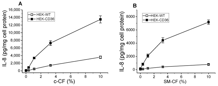
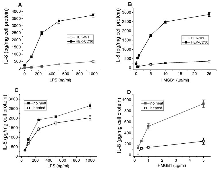
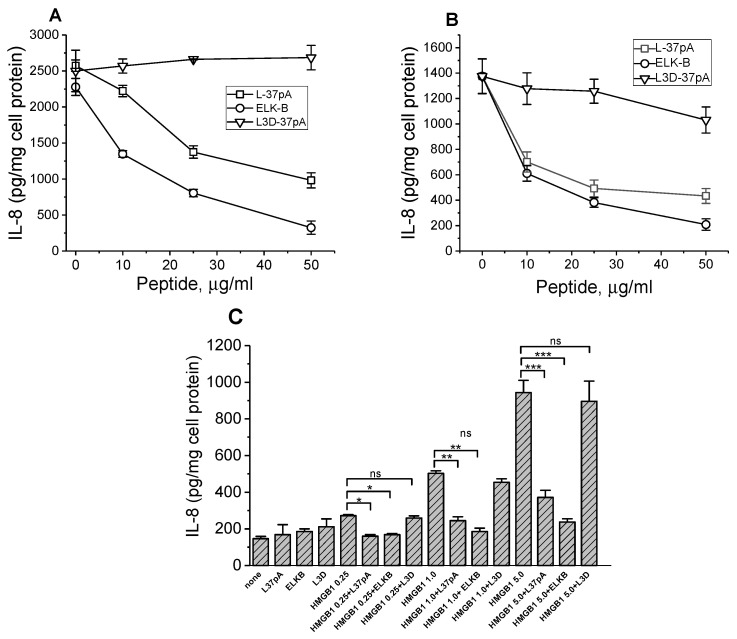
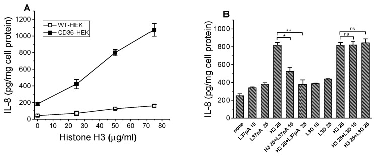
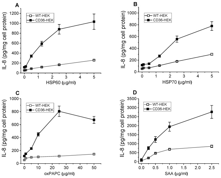
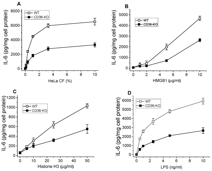
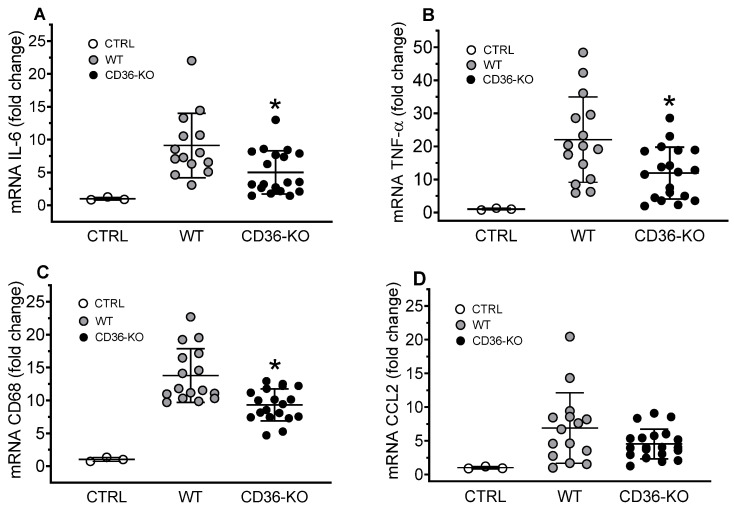
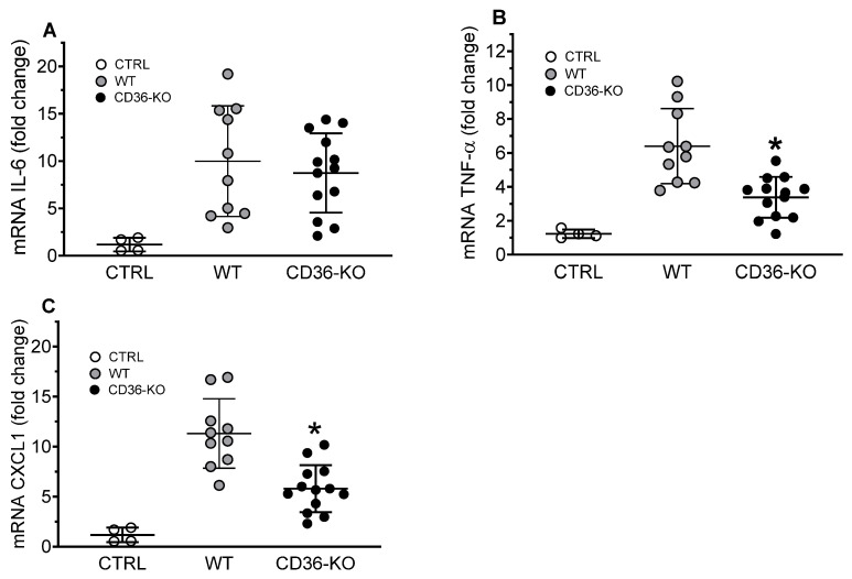
The class B scavenger receptor CD36 is known to bind and mediate the transport of lipid-related ligands and it functions as a pattern recognition receptor (PRR) for a variety of pathogens, including bacteria and viruses. In this study, we assessed CD36's role as a PRR mediating pro-inflammatory effects of several known Danger-Associated Molecular Patterns (DAMPs) used either as a single preparation or as a combination of DAMPs in the form of total cell/skeletal muscle tissue lysates. Our data demonstrated that multiple DAMPs, including HMGB1, HSPs, histone H3, SAA, and oxPAPC, as well as cell/tissue lysate preparations, induced substantially higher (~7-10-fold) IL-8 cytokine responses in HEK293 cells overexpressing CD36 compared to control WT cells. At the same time, DAMP-induced secretion of IL-6 in bone marrow-derived macrophages (BMDM) from CD36-/- mice was markedly (~2-3 times) reduced, as compared to macrophages from normal mice. Synthetic amphipathic helical peptides (SAHPs), known CD36 ligands, efficiently blocked CD36-dependent inflammatory responses induced by both cell and tissue lysates, HMGB1 and histone H3 in CD36+ cells. IP injection of total cellular lysate preparation induced inflammatory responses that were assessed by the expression of liver and lung pro-inflammatory markers, including IL-6, TNF-α, CD68, and CXCL1, and was reduced by ~50% in CD36-deficient mice compared to normal mice. Our findings demonstrate that CD36 is a PRR contributing to the innate immune response via mediating DAMP-induced inflammatory signaling and highlight the importance of this receptor as a potential therapeutic target in DAMP-associated inflammatory conditions.

Keywords: CD36; DAMPs; HMGB1; SAA; heat shock proteins; histones; inflammatory markers; oxPAPC.


Conflict of interest statement

The authors declare no conflicts of interest.

Figures

Figure 1
Figure 1
Dose-dependent IL-8 secretion induced by HeLa cell and skeletal muscle CF in WT and CD36-overexpressing HEK293 cells. WT and CD36-overexpressing HEK293 cells were incubated with increasing concentrations of the CF preparations (see Section 2) from HeLa cells, c-CF (A) or murine skeletal muscle, SM-CF (B) for 20 h. IL-8 levels were quantified in duplicate samples of cell culture supernatants by ELISA. Data represent one of three separate experiments that yielded similar results.
Figure 2
Figure 2
Dose-dependent IL-8 secretion induced by the HMGB1 and LPS in WT and CD36-overexpressing HEK293 cells. Effect of heat treatment on the cytokine-inducing activity of HMGB1 and LPS. WT and CD36-overexpressing HEK293 cells were incubated with increasing concentrations of recombinant HMGB1 (A) or LPS (B) for 20 h. IL-8 levels were quantified in duplicate samples of cell culture supernatants by ELISA. Data represent one of three separate experiments that yielded similar results. The IL-8 levels were determined using duplicate samples of cell culture supernatants collected after a 20 h incubation of cells with either intact or heat-treated (100 °C for 45 min) HMGB1 (C) and LPS (D). The data presented are from one of two separate representative experiments.
Figure 3
Figure 3
Effects of SAHPs on IL-8 secretion induced by the CF preparations and HMGB1 in CD36-overexpressing HEK293 cells. Cells were preincubated with or without increasing doses of L-37pA, ELK-B, or L3D-37pA for 1 h before a 20 h treatment with 1% c-CF (A) or 0.3% SM-CF (B). Cells were preincubated for 1 h with or without 10 µg/mL of L-37pA, ELK-B, or L3D-37pA before a 20 h treatment with increasing doses of HMGB1 (0.25, 1, and 5 µg/mL). IL-8 levels were determined in cell culture supernatants in duplicate (C). Data are from one of at least two representative experiments. * p < 0.05, ** p < 0.01, *** p < 0.001, and ns—nonsignificant, versus no peptide.
Figure 4
Figure 4
Dose-dependent IL-8 secretion induced by the histone H3 in WT and CD36-overexpressing HEK293 cells. Effect of L-37pA on hH3-induced IL-8 secretion in CD36-HEK293 cells. WT and CD36-overexpressing HEK293 cells were incubated with increasing concentrations of histone H3 for 20 h. IL-8 levels were quantified in duplicate samples of cell culture supernatants by ELISA (A). Data represent one of three separate experiments that yielded similar results. CD36-HEK293 cells were preincubated for 1 h with 0, 10 µg/mL, and 25 µg/mL of L-37pA or L3D peptides before a 20 h treatment with 25 µg/mL of histone H3. IL-8 levels were determined in cell culture supernatants in duplicate (B). * p < 0.05, ** p < 0.01, and ns—nonsignificant, versus histone H3 alone. Data are from one of at least two representative experiments.
Figure 5
Figure 5
DAMPs-induced dose-dependent pro-inflammatory responses in WT and CD36-overexpressing HEK293 cells. WT and CD36-overexpressing HEK293 cells were incubated with increasing concentrations of HSP60 (A), HSP70 (B), oxPAPC (C), and SAA (D) for 20 h. IL-8 levels were quantified in duplicate samples of cell culture supernatants by ELISA. Data represent one of two separate experiments that yielded similar results.
Figure 6
Figure 6
Pro-inflammatory responses induced by the various DAMPs in BMDM from WT and CD36-knockout mice. BMDM isolated from WT and CD36−/− mice were incubated with increasing doses of cCF (A), HMGB1 (B), histone H3 (C), and LPS (D) for 20 h. IL-6 levels were quantified in duplicate samples of cell culture supernatants by ELISA. Data represent one of two separate experiments that yielded similar results.
Figure 7
Figure 7
Effects of signaling pathways Inhibitors on CF-induced IL-6 secretion in BMDM from WT and CD36-knockout mice. BMDM isolated from WT and CD36−/− were pre-incubated for 1 h with increasing doses of PD98059 (A), SP600125 (B), SB202190 (C), or PP2 (D). Following pre-incubation, cells were incubated with CF preparation for the next 20 h. IL-6 levels were quantified in duplicate samples of cell culture supernatants by ELISA. Data represent one of two separate experiments that yielded similar results.
Figure 8
Figure 8
Hepatic gene expression of inflammatory markers in WT and CD36-knockout mice IP injected with a TCL preparation. Livers were collected for mRNA extraction and qRT-PCR assay as described in Materials and Methods. Expression levels of IL-6 (A), TNF-α (B), CD68 (C), and CCL2 (D) were normalized by GAPDH expression and are presented as the fold change relative to the PBS-treated WT control mice. Values shown are the means ± STD (n = 12–15 for the WT group, n = 20 for the CD36-KO group). * p < 0.05, versus WT TCL-treated mice.
Figure 9
Figure 9
Pulmonary gene expression of inflammatory markers in WT and CD36-knockout mice IP injected with a TCL preparation. Lungs were collected for mRNA extraction and qRT-PCR assay as described in Materials and Methods. Expression levels of IL-6 (A), TNF-α (B), and CXCL1 (C) were normalized by GAPDH expression and are presented as the fold change relative to PBS-treated WT control mice. Values shown are the means ± STD (n = 10 for the WT group, n = 14 for the CD36-KO group). * p < 0.05, versus WT TCL-treated mice.

References

    1. Silverstein R.L., Febbraio M. CD36, a scavenger receptor involved in immunity, metabolism, angiogenesis, and behavior. Sci. Signal. 2009;2:re3. doi: 10.1126/scisignal.272re3. - DOI - PMC - PubMed
    1. Savill J., Hogg N., Ren Y., Haslett C. Thrombospondin cooperates with CD36 and the vitronectin receptor in macrophage recognition of neutrophils undergoing apoptosis. J. Clin. Investig. 1992;90:1513–1522. doi: 10.1172/JCI116019. - DOI - PMC - PubMed
    1. Baranova I.N., Bocharov A.V., Vishnyakova T.G., Kurlander R., Chen Z., Fu D., Arias I.M., Csako G., Patterson A.P., Eggerman T.L. CD36 is a novel serum amyloid A (SAA) receptor mediating SAA binding and SAA-induced signaling in human and rodent cells. J. Biol. Chem. 2010;285:8492–8506. doi: 10.1074/jbc.M109.007526. - DOI - PMC - PubMed
    1. Mitchell R.W., On N.H., Del Bigio M.R., Miller D.W., Hatch G.M. Fatty acid transport protein expression in human brain and potential role in fatty acid transport across human brain microvessel endothelial cells. J. Neurochem. 2011;117:735–746. doi: 10.1111/j.1471-4159.2011.07245.x. - DOI - PubMed
    1. Hsieh F.L., Turner L., Bolla J.R., Robinson C.V., Lavstsen T., Higgins M.K. The structural basis for CD36 binding by the malaria parasite. Nat. Commun. 2016;7:12837. doi: 10.1038/ncomms12837. - DOI - PMC - PubMed

Publication types

LinkOut - more resources