

Towards Clinical Application: Calcium Waves for In Vitro Qualitative Assessment of Propagated Primary Human Corneal Endothelial Cells
- PMID: 39682760
- PMCID: PMC11640329
- DOI: 10.3390/cells13232012
Towards Clinical Application: Calcium Waves for In Vitro Qualitative Assessment of Propagated Primary Human Corneal Endothelial Cells
Abstract
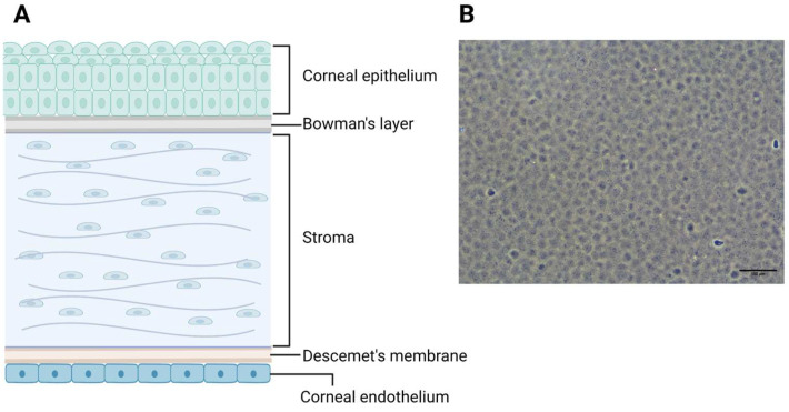
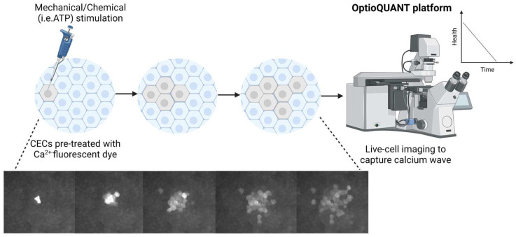
Corneal endothelium cells (CECs) regulate corneal hydration between the leaky barrier of the corneal endothelium and the ionic pumps on the surface of CECs. As CECs do not regenerate, loss of CECs leads to poor vision and corneal blindness. Corneal transplant is the only treatment option; however, there is a severe shortage of donor corneas globally. Cell therapy using propagated primary human CECs is an alternative approach to corneal transplantations, and proof of functionality is crucial for validating such CECs. Expression markers like Na-K-ATPase and ZO-1 are typical but not specific to CECs. Assessing the barrier function of the expanded CECs via electrical resistance (i.e., TEER and Ussing's chamber) involves difficult techniques and is thus impractical for clinical application. Calcium has been demonstrated to affect the paracellular permeability of the corneal endothelium. Its absence alters morphology and disrupts apical junctions in bovine CECs, underscoring its importance. Calcium signaling patterns such as calcium waves affect the rate of wound healing in bovine CECs. Therefore, observing calcium waves in expanded CECs could provide valuable insights into their health and functional integrity. Mechanical or chemical stimulations, combined with Ca2+-sensitive fluorescent dyes and time-lapse imaging, can be used to visualize these waves, which could potentially be used to qualify expanded CECs.
Keywords: calcium wave; cell therapy; cornea; corneal endothelial cells; corneal endothelium.
Conflict of interest statement
All authors have no proprietary or commercial interest in any materials involved in this article. Kun-Han Lin and Poh Loong Soong are co-founders and shareholders of Ternion Biosciences and do not have any conflicts of interest in this manuscript. The funders had no role in the design of the study; in the collection, analyses, or interpretation of data; in the writing of the manuscript; or in the decision to publish the results.
Figures
References
-
- Muller L.J., Pels L., Vrensen G.F. Novel aspects of the ultrastructural organization of human corneal keratocytes. Investig. Ophthalmol. Vis. Sci. 1995;36:2557–2567. - PubMed
Publication types
MeSH terms
Substances
Grants and funding
LinkOut - more resources
Full Text Sources
Research Materials
Miscellaneous
