. 2024 Dec 6;13(23):2016.
doi: 10.3390/cells13232016.

Riluzole Reverses Blood-Testis Barrier Loss to Rescue Chemotherapy-Induced Male Infertility by Binding to TRPC

Affiliations

Riluzole Reverses Blood-Testis Barrier Loss to Rescue Chemotherapy-Induced Male Infertility by Binding to TRPC

Rufei Huang et al. Cells. .

Abstract

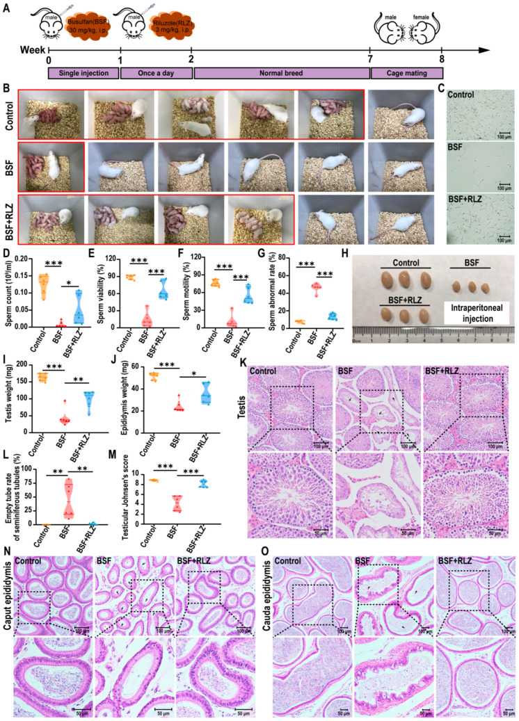
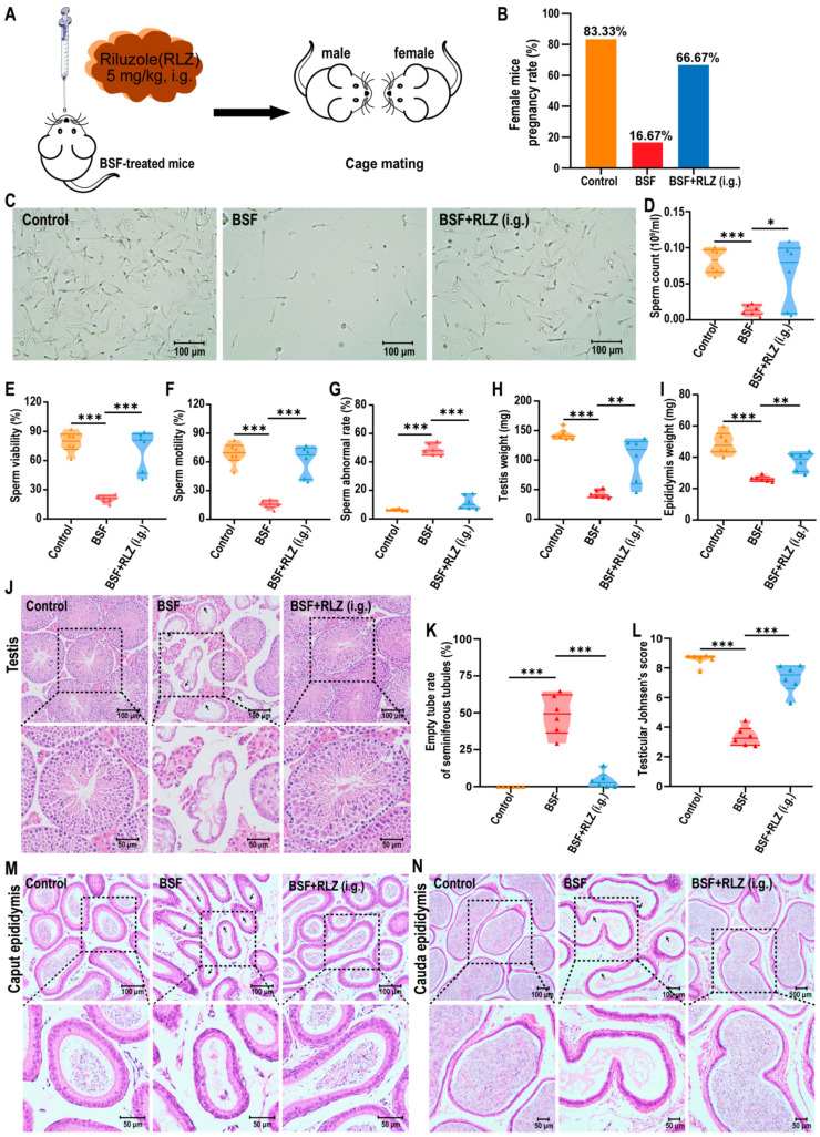
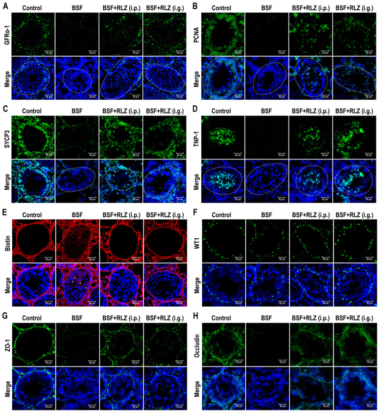
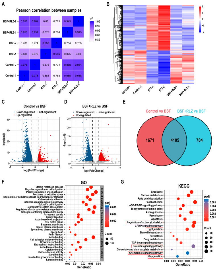
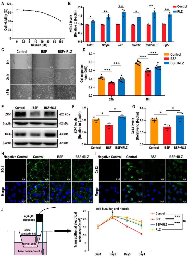
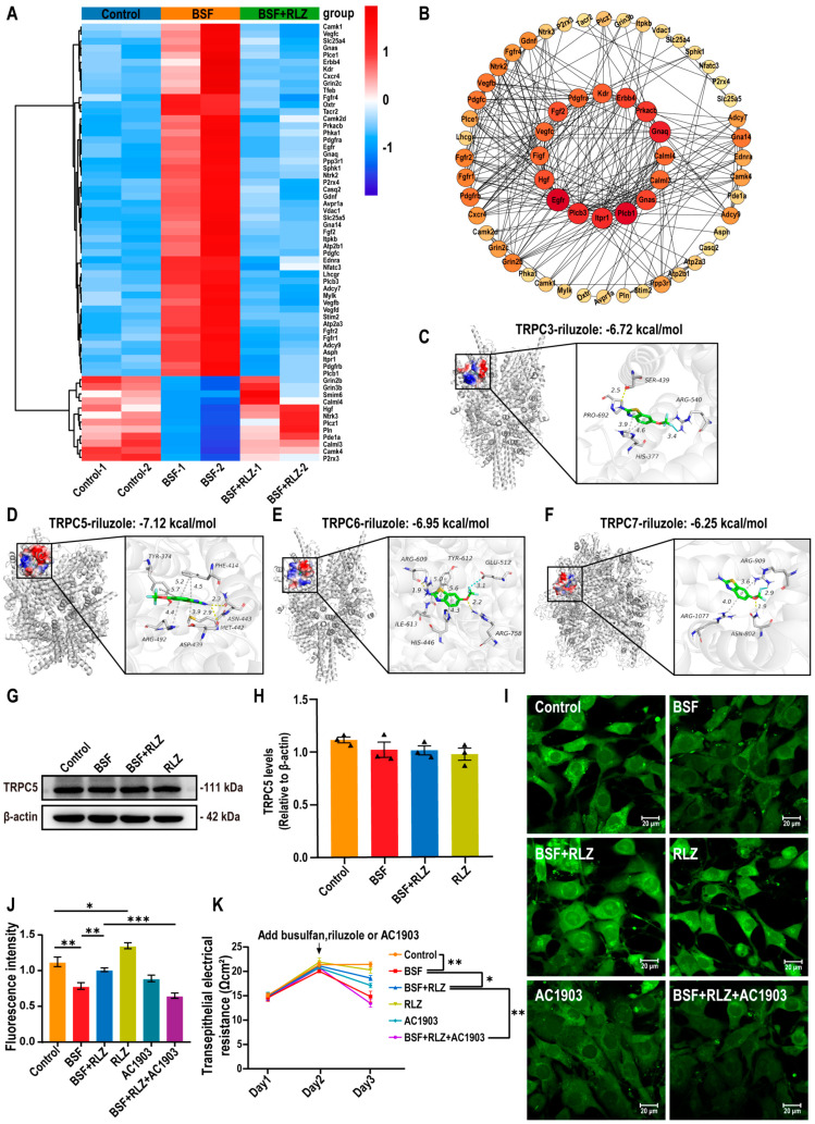
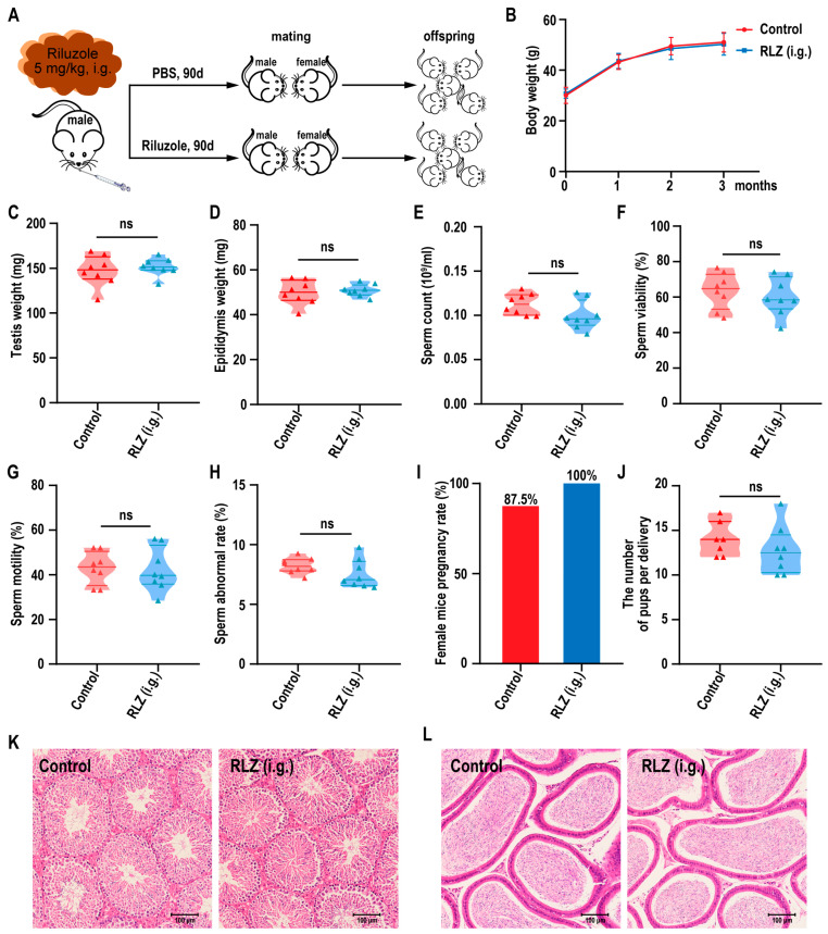
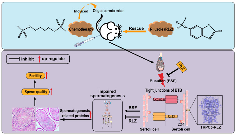
Cancer treatments, including cytotoxic therapy, often result in male infertility, necessitating the development of safe and effective strategies to preserve male reproductive potential during chemotherapy. Notably, our study uncovers the potential of repurposing riluzole, an FDA-approved drug for amyotrophic lateral sclerosis (ALS), in enhancing spermatogenesis. Hence, this research aims to explore the feasibility of utilizing riluzole to alleviate male infertility induced by busulfan (BSF), a commonly used chemotherapy drug. We established a BSF-induced oligospermia model in 4-week-old male mice and found that riluzole could effectively counter the detrimental effects of BSF on sperm production in mice with oligospermia. By restoring blood-testis barrier (BTB) functionality, riluzole improves sperm quality and reduces testicular atrophy. Through transcriptomic and molecular docking analyses, we identify transient receptor potential canonical subfamily member 5 (TRPC5) as a potential target for riluzole-mediated regulation of blood-testis barrier function. These findings propose riluzole as a promising therapeutic option for chemotherapy-induced male infertility, thereby addressing the fertility challenges associated with cancer treatments. Moreover, repurposing riluzole could streamline the drug development process, providing a cost-effective approach with reduced risk compared to developing entirely new drugs.

Keywords: Sertoli cells; TRPC5; blood–testis barrier; male infertility; riluzole; spermatogenesis.


Conflict of interest statement

The authors declare no conflicts of interest.

Figures

Figure 1
Figure 1
Effects of intraperitoneal administration of riluzole on sperm quality and recovery of damaged reproductive organs in oligospermic mice. (A) Illustration of experimental scheme. (B) Fertility of oligospermic mice induced by BSF after riluzole treatment (n = 6). (C) Representative micrographs of sperms released from the cauda epididymis of BSF-induced oligospermic mice after riluzole therapy. Scale bar, 100 μm. (DG) Count, viability, motility, and abnormal rate of sperms in the cauda epididymis (n = 6). Sperm counts over 1000 were analyzed using CASA. (H) Morphology of the testes at week 8 from oligospermic mice after riluzole treatment. (I,J) Testis and epididymis weight of oligospermic mice after riluzole treatment (n = 6). (K) Representative histopathology of the testes of mice with oligospermia after riluzole therapy. The black arrow represents low numbers of germ cells in the seminiferous tubule lumen. Scale bar, 100 μm. (L) Rate of empty tubes in seminiferous tubules after riluzole therapy (n = 6). (M) Testicular Johnsen’s score of seminiferous epithelium (n = 6). The seminiferous epithelium was evaluated according to the description of Johnsen’s score standard. (N,O) Representative histopathology of the caput epididymis and cauda epididymis of mice with oligospermia after riluzole therapy. The black arrow represents a lower density of sperm in the epididymis. Scale bar, 100 μm. The bottom images show an enlargement of the black dashed square in each figure. Values are expressed here as mean ± SEM. One-way analysis of variance (ANOVA) was used to analyze statistical differences; *** p < 0.001, ** p < 0.01, * p <0.05 compared to oligospermic mice treated with the vehicle (BSF).
Figure 2
Figure 2
Intragastric gavage administration of riluzole rescued fertility and sperm quality in oligospermic mice. (A) Schematic diagram of intragastric gavage administration to BSF-treated mice. (B) The fertility rate of mice with oligospermia induced by BSF after 8 weeks of riluzole treatment via intragastric gavage (5 mg/kg·bw) (n = 6). (C) Representative micrographs of sperms. (DG) Count, viability, motility, and abnormal rate of sperms in the cauda epididymis of oligospermic mice (n = 6). The data were analyzed for more sperm counts over 1000 using CASA. (H,I) Testis and epididymis weight in mice (n = 6). (J) Representative histopathology of the testes. The black arrow represents low numbers of germ cells in the seminiferous tubule lumen. Scale bar, 100 μm. (K,L) Empty tube rate and testicular Johnsen’s score of seminiferous tubules in oligospermic mice after riluzole therapy (n = 6). (M,N) Representative histopathology of the caput epididymis and cauda epididymis. The black arrow represents a lower density of sperm in the epididymis. Scale bar, 100 μm. The bottom images show an enlargement of the black dashed square in each figure. Values are expressed as mean ± SEM. One-way analysis of variance (ANOVA) was used to analyze statistical differences; *** p < 0.001, ** p < 0.01, * p <0.05 compared to BSF.
Figure 3
Figure 3
Effects of riluzole on proliferation and differentiation of spermatogonia and BTB integrity. (AD) Immunofluorescence staining showed the expressions of spermatogenesis-related proteins (GFRα-1, PCNA, SYCP3, and TNP-1) in the testis of BSF-induced oligospermic mice after riluzole treatment. Nuclei were stained with DAPI (blue). Scale bar, 50 μm. (E) Assessment of BTB integrity using the sulfo-NHS-LC-biotin assay. The nuclei were stained with DAPI (blue). White arrows indicate the presence of biotin in the lumen of seminiferous tubules, indicating BTB disruption. Scale bars, 50 μm. (FH) Immunofluorescence staining showed the expressions of WT1, a marker for Sertoli cells, and proteins related to the blood–testis barrier (ZO-1 and Occludin) in the testes. Nuclei were stained with DAPI (blue). Scale bar, 50 μm.
Figure 4
Figure 4
Changes in the transcriptome profile of testis after riluzole treatment. (A) The Pearson correlation coefficient was exhibited as coefficient values to determine the correlation of the transcriptional profiles between each sample. (B) Cluster analysis of differentially expressed gene (DEG) expression changes in testis between each group. The color bar indicates gene expression on a log2 scale. Red represents up-regulated and blue represents down-regulated gene expression. (C,D) Volcano plot of the DEGs of testes between each group. The criterion for screening DEGs had adjusted padj < 0.05 and |log2FoldChange| ≥ 1. Up-regulated genes are represented by red dots, and down-regulated genes are represented by blue dots. Gray dots indicate genes with no difference in expression. (E) Venn diagram analysis of the differential gene. (F,G) GO and KEGG pathway enrichment analysis of the DEGs. The abscissa in the figure is the ratio of the number of differential genes annotated to the GO Term or KEGG pathway to the total number of differential genes, and the ordinate is the GO Term or KEGG pathway. The size of the point represents the number of genes annotated to the GO Term or KEGG pathway. The color of the dot indicates the enrichment degree (padj) of the pathway. The pathways highlighted in the red box are the major enriched pathways that are closely associated with Sertoli cell function and the blood–testis barrier. Control group: normal mice; BSF group: BSF-induced oligospermic mice; BSF+RLZ group: BSF-induced oligospermic mice treated with riluzole.
Figure 5
Figure 5
Effects of riluzole on Sertoli cell (SC) functionality in vitro. (A) The cell viability of SCs treated with different concentrations of riluzole. (B) Expression levels of SC-related secretory factors (Gdnf, Bmp4, Scf, Cxcl12, Inhibin B, and Fgf2) in SCs treated with 10 μM riluzole for 24 h, measured by qPCR with β-actin as the internal reference. (C) Scratch assay was used to detect the effect of 10 μM riluzole and/or 200 μM BSF on SC migration at 24 and 48 h. Representative images were captured under a microscope. Scale bar, 200 μm. (D) Cell migration rate was calculated based on average scratch distance using Image J analysis after 24 and 48 h of treatment (n = 12). (EG) Western blot analysis of ZO-1 and Cx43 expression in SCs treated with 10 μM riluzole and/or 200 μM BSF, with β-actin as the loading control. (H,I) Immunofluorescent staining for ZO-1 and Cx43 in SCs treated with 10 μM riluzole and/or 200 μM BSF. As a negative control, immunofluorescence staining was performed without the primary antibody. Nuclei were stained with DAPI (blue). Scale bar, 20 μm. (J) Transepithelial electrical resistance (TER) measurement to assess Sertoli cell TJ-permeability barrier function. All data were obtained from three independent experiments and presented as mean ± SEM. Statistical significance was determined by two-tailed Student’s t-test (B) or one-way ANOVA (D,F,G,J); *** p < 0.001, ** p < 0.01, * p < 0.05, ns: no significance.
Figure 6
Figure 6
Riluzole activated TRPC5 and regulated intracellular calcium levels. (A) The heat map illustrates the alterations in gene expression within the calcium signaling pathway of the testis following riluzole treatment. The red represents up-regulated and the blue represents down-regulated gene expressions. The data were obtained from the same RNA sequencing dataset as presented in Figure 4. (B) Protein–protein interaction (PPI) analysis of the DEGs involved in the calcium signaling pathway from testes. Network visualized with Cytoscape. (CF) The binding mode of TRPC3/5/6/7 with riluzole. (C) TRPC3-riluzole; (D) TRPC5-riluzole; (E) TRPC6-riluzole; (F) TRPC7-riluzole. On the left is the electrostatic surface of the complex, and on the right is the detail binding mode of the complex. Yellow dash, gray dash, and blue dash show the hydrogen bond, π-stacking interaction, and halogen bond, respectively. (G,H) Western blot analysis of Sertoli cells (SCs) treated with 200 μM BSF and/or 10 μM riluzole to detect TRPC5 expression. β-actin was used as the internal reference. (I,J) Changes in intracellular calcium concentration. (K) TER to assess changes in the function of the SC tight junction–permeability barrier. All data were obtained from three independent experiments and presented as mean ± SEM. One-way analysis of variance (ANOVA) was used to analyze statistical differences; *** p < 0.001, ** p < 0.01, * p < 0.05.
Figure 7
Figure 7
Assessment of the reproductive ability of male mice exposed to riluzole for 90 days via intragastric gavage. (A) Illustration of experimental scheme. (B) Body weight of male mice after riluzole exposed through intragastric gavage at a daily dose of 5 mg/kg for a duration of 90 days (n = 8). (C,D) Testis and epididymis weight in male mice exposed to riluzole (n = 8). (EH) Sperm count, viability, motility, and abnormal rate in male mice exposed to riluzole (n = 8). The data were analyzed for more sperm counts over 1000 using CASA. (I) Fertility rate in male mice exposed to riluzole (n = 8). (J) Number of offspring of male mice exposed to riluzole. (K,L) Representative histopathology of the testis and epididymis of male mice exposed to riluzole. Scale bar, 100 μm. Values are presented as mean ± SEM. Two-tailed Student’s t-test was used to analyze statistical differences; ns: no significance.
Figure 8
Figure 8
Schematic diagram of the potential mechanism of RLZ against BSF-induced reproductive injury. Riluzole modulates the blood–testis barrier (BTB), enhancing spermatogenesis and restoring fertility in mice with busulfan (BSF)-induced oligospermia through its interaction with TRPC5.

Similar articles

Cited by

References

    1. Ávila C., Vinay J.I., Arese M., Saso L., Rodrigo R. Antioxidant Intervention against Male Infertility: Time to Design Novel Strategies. Biomedicines. 2022;10:3058. doi: 10.3390/biomedicines10123058. - DOI - PMC - PubMed
    1. Martínez M., Marquès M., Salas-Huetos A., Babio N., Domingo J.L., Salas-Salvadó J. Lack of association between endocrine disrupting chemicals and male fertility: A systematic review and meta-analysis. Environ. Res. 2023;217:114942. doi: 10.1016/j.envres.2022.114942. - DOI - PubMed
    1. Choy J.T., Amory J.K. Nonsurgical Management of Oligozoospermia. J. Clin. Endocrinol. Metab. 2020;105:e4194–e4207. doi: 10.1210/clinem/dgaa390. - DOI - PMC - PubMed
    1. Choy J.T., Eisenberg M.L. Male infertility as a window to health. Fertil. Steril. 2018;110:810–814. doi: 10.1016/j.fertnstert.2018.08.015. - DOI - PubMed
    1. Perrone P., Lettieri G., Marinaro C., Longo V., Capone S., Forleo A., Pappalardo S., Montano L., Piscopo M. Molecular Alterations and Severe Abnormalities in Spermatozoa of Young Men Living in the “Valley of Sacco River” (Latium, Italy): A Preliminary Study. Int. J. Environ. Res. Public Health. 2022;19:11023. doi: 10.3390/ijerph191711023. - DOI - PMC - PubMed

Publication types

MeSH terms

LinkOut - more resources