. 2024 Dec 6;13(23):3431.
doi: 10.3390/plants13233431.

A Bifunctional Nuclease Promotes the Infection of Zucchini Yellow Mosaic Virus in Watermelon by Targeting P3

Affiliations

A Bifunctional Nuclease Promotes the Infection of Zucchini Yellow Mosaic Virus in Watermelon by Targeting P3

Baoshan Kang et al. Plants (Basel). .

Abstract

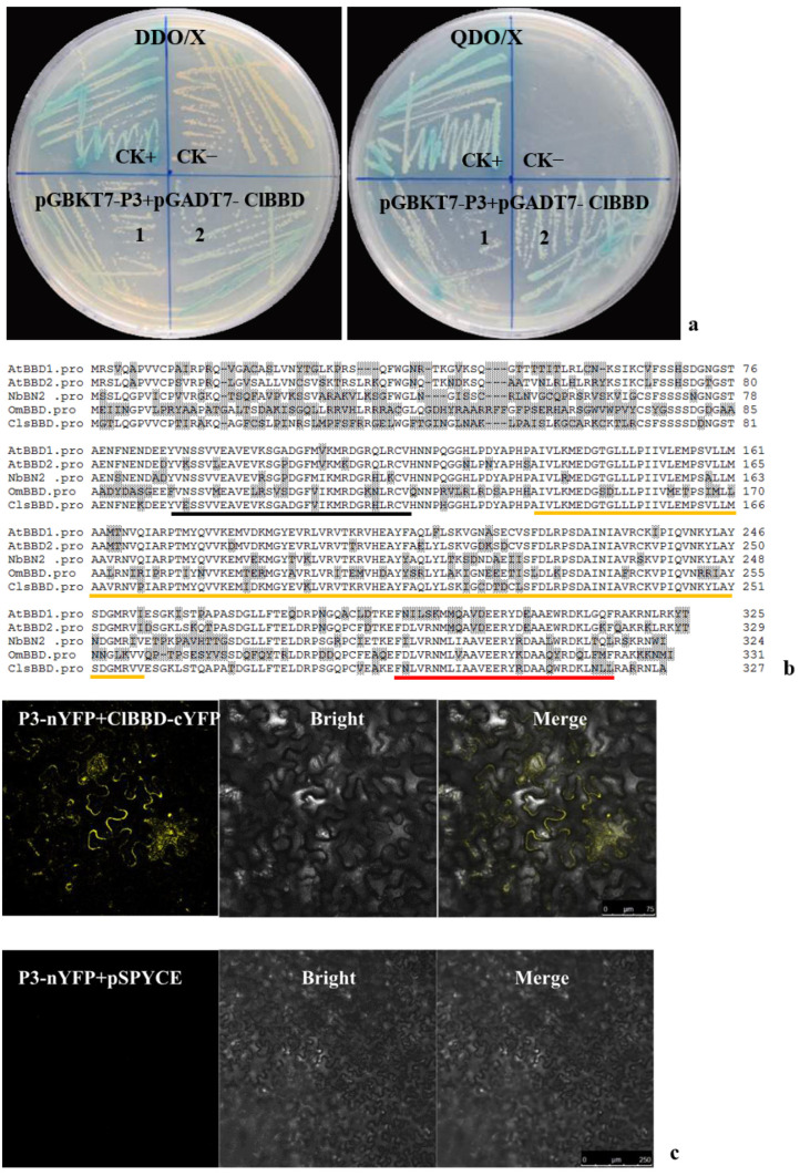
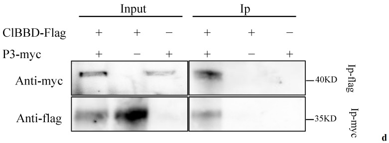
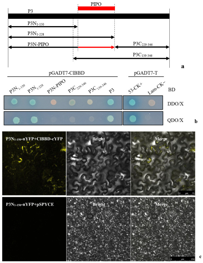
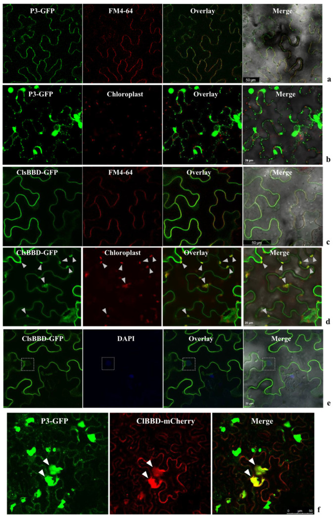
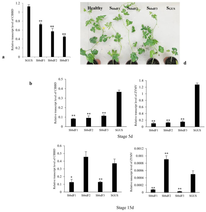
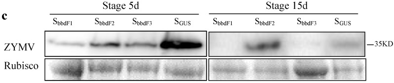
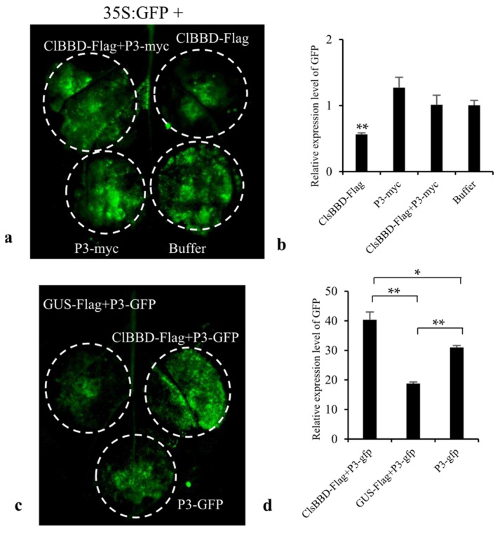
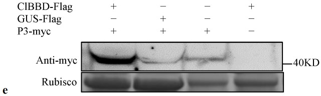
Potyviral P3 is involved in viral replication, movement, and pathogenicity; however, its biochemical function is unknown. In this study, the P3 of the zucchini yellow mosaic virus (ZYMV) interacted with ClBBD, a protein with high ortholog bifunctional nuclease activity, in watermelon. The binding site was shown via yeast two-hybrid screening and BiFC assay to be located at the N-terminus of P3 rather than P3N-PIPO. ClBBD localized predominantly to the chloroplast and plasma membrane. ZYMV P3 was also present in the nucleus and cytoplasm as aggregates. When co-expressed with P3 in tobacco, ClBBD formed aggregates with P3 in the cytoplasm. The knockdown of ClBBD using the VIGS vector pV190 and challenge with ZYMV revealed a positive correlation between viral accumulation and ClBBD expression, indicating that ClBBD reduces the resistance of watermelon to ZYMV. Furtherly, we found that when P3 and ClBBD were transiently co-expressed in tobacco, the level of P3 was significantly higher than that when it was expressed alone or co-expressed with GUS. It inferred that ClBBD may be able to stabilize the expression of P3. Overall, the results suggest that the interaction of P3 with ClBBD promotes virus infection, and ClBBD may be involved in stabilizing the expression level of P3.

Keywords: P3; bifunctional nuclease; interaction; watermelon; zucchini yellow mosaic virus.


Conflict of interest statement

The authors declare no conflicts of interest.

Figures

Figure 1
Figure 1
The interaction of ZYMV P3 with ClBBD of watermelon. (a) Yeast cells containing pGBKT7-P3 were co-transformed with pGADT7-ClBBD on DDO/X (SD/-Leu/-Trp/X-α-Gal) and QDO/X (SD/-Leu/-Trp/-His/-Ade /x-α-gal) mediums. CK+, positive control for pGADT7-T + pGBKT7-53; CK−, negative control for pGADT7-T + pGBKT7-Lam. (b) Alignment of the deduced amino acid sequences of ClBBD with other plant homologs using the Clustal W algorithm. Different amino acids are highlighted in gray. Three domains, a highly conserved region (HCR) domain, an unknown function 151 (DUF151) domain, and a UV-responsive (UVR) domain, are underlined with black, orange, and red colors, respectively. (c,d) Verification of the interaction between P3 and ClBBD via bimolecular fluorescence complementation assays in N. benthamiana and co-immunoprecipitation (Co-IP) assays. YFP fluorescence was detected in the N. benthamiana leaves agroinfiltrated with pSPYNE-P3/pSPYCE-ClBBD using pSPYNE-P3/pSPYCE as negative control (c). Myc-tagged ZYMV P3 was co-expressed with Flag-tagged ClBBD in N. benthamiana. Proteins were immunoprecipitated (Ip) from total extracts using anti-myc or anti-flag antibodies and followed by Western blot using tag-specific antibodies (d).
Figure 1
Figure 1
The interaction of ZYMV P3 with ClBBD of watermelon. (a) Yeast cells containing pGBKT7-P3 were co-transformed with pGADT7-ClBBD on DDO/X (SD/-Leu/-Trp/X-α-Gal) and QDO/X (SD/-Leu/-Trp/-His/-Ade /x-α-gal) mediums. CK+, positive control for pGADT7-T + pGBKT7-53; CK−, negative control for pGADT7-T + pGBKT7-Lam. (b) Alignment of the deduced amino acid sequences of ClBBD with other plant homologs using the Clustal W algorithm. Different amino acids are highlighted in gray. Three domains, a highly conserved region (HCR) domain, an unknown function 151 (DUF151) domain, and a UV-responsive (UVR) domain, are underlined with black, orange, and red colors, respectively. (c,d) Verification of the interaction between P3 and ClBBD via bimolecular fluorescence complementation assays in N. benthamiana and co-immunoprecipitation (Co-IP) assays. YFP fluorescence was detected in the N. benthamiana leaves agroinfiltrated with pSPYNE-P3/pSPYCE-ClBBD using pSPYNE-P3/pSPYCE as negative control (c). Myc-tagged ZYMV P3 was co-expressed with Flag-tagged ClBBD in N. benthamiana. Proteins were immunoprecipitated (Ip) from total extracts using anti-myc or anti-flag antibodies and followed by Western blot using tag-specific antibodies (d).
Figure 2
Figure 2
Identification of the key interacting region of P3 with ClBBD via Y2H and BiFC. (a) Schematic representations of different parts of P3 and P3N-PIPO. The numbers represent the total number of amino acids contained in the part; (b) yeast cells containing different parts of P3 were co-transformed with pGADT7-ClBBD on DDO/X and QDO/X cultures for determining the interacting region with ClBBD; pGADT7-T co-transformed with pGBKT7-53 and pGBKT7-Lam to be as positive control (CK+) and negative control (CK−), respectively; (c) confirmation of the interacting region of P3N1–150 via BiFC using pSPYNE-P31–150/pSPYCE as negative control.
Figure 3
Figure 3
Subcellular localization of ClBBD or ZYMV P3 in N. benthamiana leaf epidermal cells. GFP fluorescence is shown as green. The red fluorescence indicates autofluorescence from chloroplasts, mCherry, or FM4-64. The amphiphilic dye FM4-64 and DAPI were used to stain the plasma membrane (PM) and nucleus, respectively. (a) P3-GFP showed punctate pattern along the cell wall and overlaid with FM4-64; (b) P3-GFP formed irregular aggregates in the cytoplasm and could not overlay with chloroplasts; (c,d) ClBBD-GFP overlaid with the signal of FM4-64-labeled PM (c) and chloroplasts are indicated with white arrows (d); (e) the weak signal of ClBBD-GFP in nucleus is indicated with white boxes; (f) C-terminal GFP/mCherry-fused proteins (P3-GFP, ClBBD-mCherry) were transiently co-expressed in N. benthamiana via agroinfiltration. In these cells, ClBBD formed aggregates and was overlaid with P3-GFP (shown by white arrows).
Figure 4
Figure 4
The viral accumulation in watermelon with knockdown of the expression of ClBBD via VIGS. (a) The mRNA levels of ClsBBD in three knockdown lines were significantly reduced at 15 days post-VIGS; (b) the mRNA levels of ZYMV and ClBBD in three knockdown lines at 10 days post-inoculation with ZYMV at Stages 5d and 15d, respectively; (c) detection of the viral accumulation at 10 dpi with ZYMV via Western blot; (d) the knockdown plants showed better growth than SGUS plant at 25 dpi with ZYMV at Stage 5d. Bars represent mean ± SD calculated from three independent biological samples. Asterisks indicate the significant difference to SGUS control; * and ** represent p < 0.05 and 0.01, respectively.
Figure 4
Figure 4
The viral accumulation in watermelon with knockdown of the expression of ClBBD via VIGS. (a) The mRNA levels of ClsBBD in three knockdown lines were significantly reduced at 15 days post-VIGS; (b) the mRNA levels of ZYMV and ClBBD in three knockdown lines at 10 days post-inoculation with ZYMV at Stages 5d and 15d, respectively; (c) detection of the viral accumulation at 10 dpi with ZYMV via Western blot; (d) the knockdown plants showed better growth than SGUS plant at 25 dpi with ZYMV at Stage 5d. Bars represent mean ± SD calculated from three independent biological samples. Asterisks indicate the significant difference to SGUS control; * and ** represent p < 0.05 and 0.01, respectively.
Figure 5
Figure 5
The test of ClBBD regulating the expression of P3. (a) Transient co-expression of ClBBD-Flag, ClBBD-Flag plus P3-myc, P3-myc, or Buffer with 35S:GFP in N. benthamiana. (b) RT-qPCR analysis of relative GFP mRNA levels. (c) Transient co-expression of ClBBD-Flag or GUS-Flag with P3-GFP, and only P3-GFP in N. benthamiana. (d) qRT-PCR analysis of relative P3-GFP mRNA levels. (e) Western blot assay of the P3 protein level through expressing transiently of P3-myc alone, or co-expression of P3-myc with ClBBD-Flag, or co-expression of P3-myc with GUS-Flag in N. benthamiana. The constructs GUS-Flag, ClBBD-Flag, P3-GFP and P3-myc were generated using the pCAMBIA1300 vector, and 35S:GFP was constructed using PCB301 vector. Bars represent mean ± SD calculated from three independent biological samples. Asterisks indicate the significant difference; * and ** represent p < 0.05 and 0.01, respectively.
Figure 5
Figure 5
The test of ClBBD regulating the expression of P3. (a) Transient co-expression of ClBBD-Flag, ClBBD-Flag plus P3-myc, P3-myc, or Buffer with 35S:GFP in N. benthamiana. (b) RT-qPCR analysis of relative GFP mRNA levels. (c) Transient co-expression of ClBBD-Flag or GUS-Flag with P3-GFP, and only P3-GFP in N. benthamiana. (d) qRT-PCR analysis of relative P3-GFP mRNA levels. (e) Western blot assay of the P3 protein level through expressing transiently of P3-myc alone, or co-expression of P3-myc with ClBBD-Flag, or co-expression of P3-myc with GUS-Flag in N. benthamiana. The constructs GUS-Flag, ClBBD-Flag, P3-GFP and P3-myc were generated using the pCAMBIA1300 vector, and 35S:GFP was constructed using PCB301 vector. Bars represent mean ± SD calculated from three independent biological samples. Asterisks indicate the significant difference; * and ** represent p < 0.05 and 0.01, respectively.

References

    1. Desbiez C., Lecoq H. Zucchini yellow mosaic virus. Plant Pathol. 1997;46:809–829. doi: 10.1046/j.1365-3059.1997.d01-87.x. - DOI
    1. Urcuqui-Inchima S., Haenni A.L., Bernardi F. Potyvirus proteins: A wealth of functions. Virus Res. 2001;74:157–175. doi: 10.1016/S0168-1702(01)00220-9. - DOI - PubMed
    1. Chung B.Y., Miller W.A., Atkins J.F., Firth A.E. An overlapping essential gene in the Potyviridae. Proc. Natl. Acad. Sci. USA. 2008;105:5897–5902. doi: 10.1073/pnas.0800468105. - DOI - PMC - PubMed
    1. Cui X., Wei T., Chowda-Reddy R.V., Sun G., Wang A. The Tobacco etch virus P3 protein forms mobile inclusions via the early secretory pathway and traffics along actin microfilaments. Virology. 2010;397:56–63. doi: 10.1016/j.virol.2009.11.015. - DOI - PubMed
    1. Johansen I.E., Lund O.S., Hjulsager C.K., Laursen J. Recessive resistance in Pisum sativum and potyvirus pathotype resolved in a gene-for-cistron correspondence between host and virus. J. Virol. 2001;75:6609–6614. doi: 10.1128/JVI.75.14.6609-6614.2001. - DOI - PMC - PubMed

LinkOut - more resources