. 2024 Nov 21;29(23):5491.
doi: 10.3390/molecules29235491.

Peroxiredoxin 4 Ameliorates T-2 Toxin-Induced Growth Retardation in GH3 Cells by Inhibiting Oxidative Stress and Apoptosis

Affiliations

Peroxiredoxin 4 Ameliorates T-2 Toxin-Induced Growth Retardation in GH3 Cells by Inhibiting Oxidative Stress and Apoptosis

Qirong Lu et al. Molecules. .

Abstract

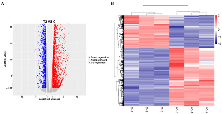
T-2 toxin, a highly toxic type A trichothecene, is a secondary fungal metabolite produced by various Fusarium species. The consumption of food and feed contaminated with T-2 toxin is a major factor contributing to growth retardation, posing significant risks to both human and animal health. However, the specific targets and mechanisms that mitigate T-2 toxin-induced growth retardation remain unclear. In this study, transcriptomic analysis was employed to identify key differentially expressed genes associated with the alleviation of T-2 toxin-induced growth retardation. Peroxiredoxin 4 (PRDX4), a gene linked to oxidative stress and apoptosis, was found to be one of the most downregulated in T-2 toxin-treated GH3 cells, an in vitro model of growth retardation. The experiments demonstrated that T-2 toxin significantly increased reactive oxygen species' production, apoptosis, and cell cycle arrest while reducing the activity of antioxidant enzymes (superoxide dismutase and glutathione peroxidase) and PRDX4 expression in GH3 cells. Furthermore, PRDX4 silencing exacerbated T-2 toxin-induced oxidative stress and apoptosis, whereas PRDX4 overexpression effectively mitigated these effects. These findings highlight the protective role of PRDX4 in counteracting T-2 toxin-induced oxidative stress and apoptosis, suggesting that PRDX4 can serve as a therapeutic target for the treatment of T-2 toxin-induced growth retardation.

Keywords: T-2 toxin; apoptosis; growth retardation; oxidative stress; peroxiredoxin 4.


Conflict of interest statement

The authors declare no conflicts of interest.

Figures

Figure 1
Figure 1
Cytotoxicity of T-2 toxin toward GH3 cells as measured using CCK-8 and LDH assays. GH3 cells were treated with 0–160 nM T-2 toxin for 24 h. (A) Cell viability was measured using the CCK-8 assay. (B) LDH activity in the cell supernatant was measured using the LDH assay. All data are presented as the average of three replicates. p < 0.05 (*) was defined as significant difference, p < 0.01 (**) was defined as extremely significant difference.
Figure 2
Figure 2
Transcriptomic analysis of differentially expressed genes in T-2 toxin-treated GH3 cells. (A) Volcano plot of differentially expressed genes. (B) Cluster analysis of differentially expressed genes.
Figure 3
Figure 3
GO annotation and KEGG analysis of differentially expressed genes in T-2 toxin-treated GH3 cells. (A) GO annotation of differentially expressed genes. (B) KEGG analysis of differentially expressed genes.
Figure 4
Figure 4
T-2 toxin induced growth retardation in GH3 cells by promoting apoptosis. (A) Immunoblotting of apoptosis-related proteins in T-2 toxin-treated GH3 cells. Protein density was quantified using ImageJ software (version 1.8.0). (B) Flow cytometry analysis of cell cycle and apoptosis in GH3 cells treated with T-2 toxin. All data are presented as the average of three replicates. p < 0.05 (*) was defined as significant difference, p < 0.01 (**) was defined as extremely significant difference.
Figure 5
Figure 5
T-2 toxin induced growth retardation in GH3 cells by promoting oxidative stress. (A) Flow cytometry analysis of ROS content in GH3 cells treated with T-2 toxin. (B) The GSH-Px activities in GH3 cells treated with T-2 toxin was measured using GSH-Px assay kit. (C) The total SOD activities in GH3 cells treated with T-2 toxin was measured using the total SOD assay kit. All data are presented as the average of three replicates. p < 0.05 (*) was defined as significant difference, p < 0.01 (**) was defined as extremely significant difference.
Figure 6
Figure 6
PRDX4 overexpression alleviates T-2 toxin-induced apoptosis. (A) Immunoblotting of apoptosis-related proteins in PRDX4-overexpressing and T-2 toxin-treated GH3 cells. Protein density was quantified using ImageJ software (version 1.8.0). (B) Flow cytometry analysis of cell cycle and apoptosis in GH3 cells treated with T-2 toxin and PRDX4 overexpression. All data are presented as the average from three replicates. p < 0.05 (*) was defined as significant difference, p < 0.01 (**) was defined as extremely significant difference.
Figure 7
Figure 7
PRDX4 overexpression alleviates T-2 toxin-induced oxidative stress. (A) Flow cytometry analysis of ROS content in GH3 cells treated with T-2 toxin and PRDX4 overexpression. (B) The GSH-Px activities in GH3 cells treated with T-2 toxin and PRDX4 overexpression were measured using a GSH-Px assay kit. (C) The total SOD activities in GH3 cells treated with T-2 toxin and PRDX4 overexpression were measured using a total SOD assay kit. All data are presented as the average of three replicates. p < 0.01 (**) was defined as extremely significant difference.
Figure 8
Figure 8
PRDX4 siRNA accelerates T-2 toxin-induced apoptosis. (A) Immunoblotting of apoptosis-related proteins in PRDX4 siRNA-transfected and T-2 toxin-treated GH3 cells. Protein density was quantified using ImageJ software (version 1.8.0). (B) Flow cytometry analysis of cell cycle and apoptosis in GH3 cells treated with T-2 toxin and PRDX4 siRNA. All data are presented as the average of three replicates. p < 0.05 (*) was defined as significant difference, p < 0.01 (**) was defined as extremely significant difference.
Figure 9
Figure 9
PRDX4 siRNA accelerates T-2 toxin-induced oxidative stress. (A) Flow cytometry analysis of ROS content in GH3 cells treated with T-2 toxin and PRDX4 siRNA. (B) The GSH-Px activities in GH3 cells treated with T-2 toxin and PRDX4 siRNA were measured using a GSH-Px assay kit. (C) The total SOD activities in GH3 cells treated with T-2 toxin and PRDX4 siRNA were measured using a total SOD assay kit. All data are presented as the average of three replicates. p < 0.01 (**) was defined as extremely significant difference.
Figure 10
Figure 10
Schematic representation of the proposed mechanism by which T-2 toxin induces growth retardation in GH3 cells. T-2 toxin significantly reduces PRDX4 expression, induces oxidative stress by increasing the ROS content and decreasing the total SOD and GSH-Px activities, and induces apoptosis by increasing p53, cleaved caspase 3/caspase 3, and the Bax/Bcl-2 ratio. These findings highlight PRDX4’s protective role in mitigating the oxidative stress and apoptosis caused by T-2 toxin in GH3 cells.

Similar articles

Cited by

References

    1. Guo J., Ye X., Zhao Y., Huang D., Wu Q., Ihsan A., Wang X. Nrf-2α and mitophagy underlie enhanced mitochondrial functions and biogenesis induced by t-2 toxin in gh3 cells. Food Chem. Toxicol. 2023;174:113687. doi: 10.1016/j.fct.2023.113687. - DOI - PubMed
    1. Wu Q., Qin Z., Kuca K., You L., Zhao Y., Liu A., Musilek K., Chrienova Z., Nepovimova E., Oleksak P., et al. An update on t-2 toxin and its modified forms: Metabolism, immunotoxicity mechanism, and human exposure assessment. Arch. Toxicol. 2020;94:3645–3669. doi: 10.1007/s00204-020-02899-9. - DOI - PubMed
    1. Gruber-Dorninger C., Jenkins T., Schatzmayr G. Global mycotoxin occurrence in feed: A ten-year survey. Toxins. 2019;11:375. doi: 10.3390/toxins11070375. - DOI - PMC - PubMed
    1. Janik E., Niemcewicz M., Podogrocki M., Ceremuga M., Stela M., Bijak M. T-2 toxin-the most toxic trichothecene mycotoxin: Metabolism, toxicity, and decontamination strategies. Molecules. 2021;26:6868. doi: 10.3390/molecules26226868. - DOI - PMC - PubMed
    1. Wang P., Sun L.H., Wang X., Wu Q., Liu A. Effective protective agents against the organ toxicity of t-2 toxin and corresponding detoxification mechanisms: A narrative review. Anim. Nutr. 2024;16:251–266. doi: 10.1016/j.aninu.2023.12.001. - DOI - PMC - PubMed

LinkOut - more resources