Review
. 2024 Nov 26;29(23):5594.
doi: 10.3390/molecules29235594.

Emerging Fluorescent Nanoparticles for Non-Invasive Bioimaging

Affiliations
Review

Emerging Fluorescent Nanoparticles for Non-Invasive Bioimaging

Asma Khalid et al. Molecules. .

Abstract

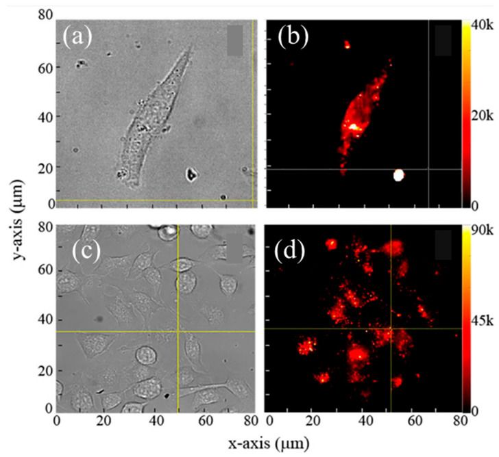
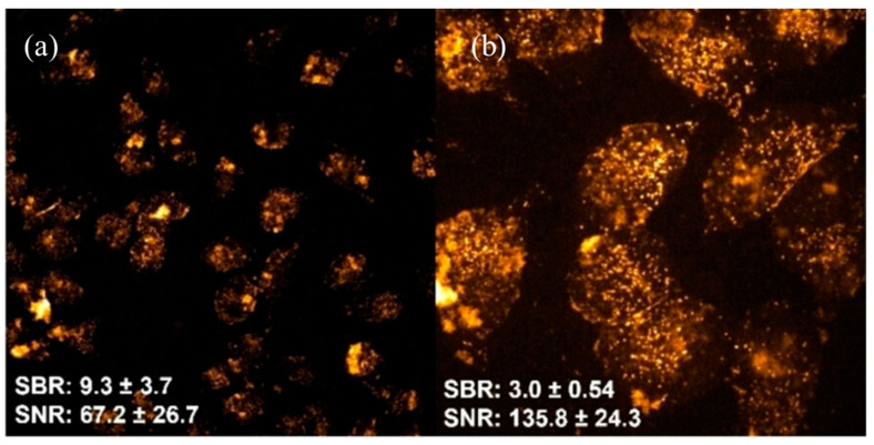
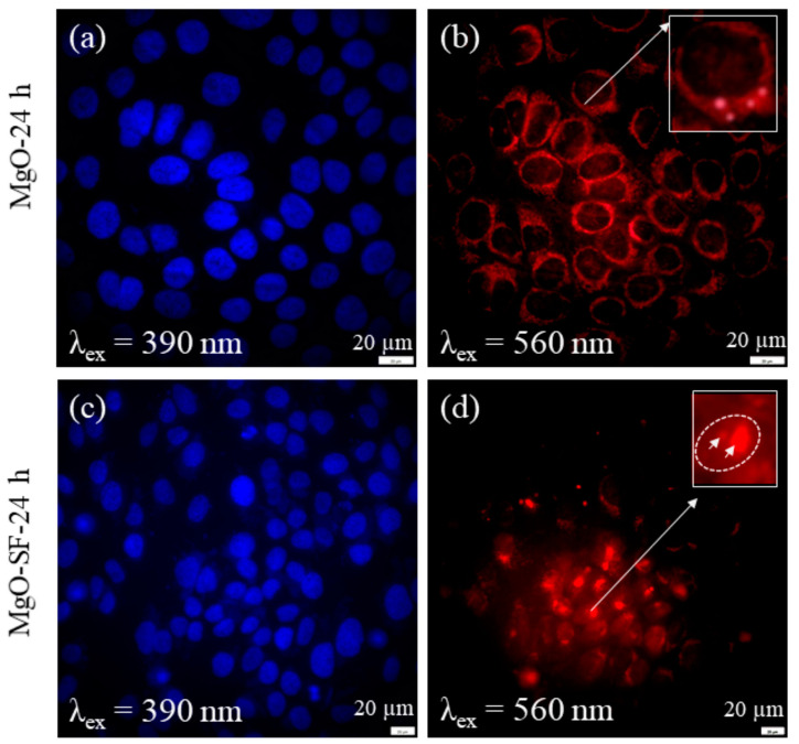
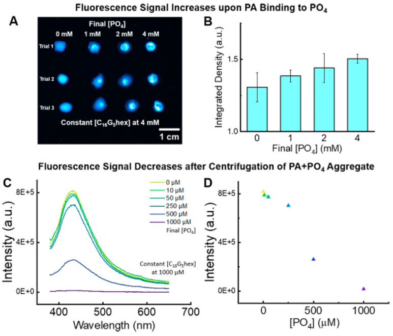
Fluorescence-based techniques have great potential in the field of bioimaging and could bring tremendous progress in microbiology and biomedicine. The most essential element in these techniques is fluorescent nanomaterials. The use of fluorescent nanoparticles as contrast agents for bioimaging is a large topic to cover. The purpose of this mini-review is to give the reader an overview of biocompatible and biodegradable fluorescent nanoparticles that are emerging nanomaterials for use in fluorescent bioimaging. In addition to the biocompatibility of these nanomaterials, biodegradability is considered a necessity for short-term sustainable bioimaging. Firstly, the main requirements for bioimaging are raised, and a few existing fluorescent nanoprobes are discussed. Secondly, a few inert biocompatible fluorescent nanomaterials for long-term bioimaging that have been, to some extent, demonstrated as fluorescent probes are reviewed. Finally, a few biocompatible and biodegradable nanomaterials for short-term bioimaging that are evolving for bioimaging applications are discussed. Together, these advancements signal a transformative leap toward sustainability and functionality in biomedical imaging.

Keywords: biocompatibility; biodegradability; bioimaging; fluorescence; nanoparticles.


Conflict of interest statement

The authors declare no conflicts of interest.

Figures

Figure 1
Figure 1
Some nanoparticles used in bioimaging. QD: quantum dots, UCNPs: up-conversion NPs, Pd: palladium, Ag: silver, ND: nanodiamond, SiC: silicon carbide, C-dots: carbon dots, SiO2: silica, GNPs: gold NPs, P-dots: polymer dots, NC: nanocellulose, ZnO: zinc oxide, MgO: magnesium oxide. In this review, fluorescent NPs are broadly classified into two main categories of importance for bioimaging: biocompatible and (ii) biocompatible and biodegradable NPs.
Figure 2
Figure 2
Representative images showing the uptake of epirubicin-loaded nanodiamonds (NDs) in chemoresistant hepatic cancer stem cells (HCSCs). Panel (a) displays the bright-field image of HCSCs, highlighting cell morphology. Panel (b) illustrates the fluorescence distribution of epirubicin-loaded NDs within the cells, confirming intracellular uptake. Panel (c) presents the merged image of panels (a,b), providing a comprehensive view of ND localization within HCSCs. Scale bar: 10 μm. Adapted from Wang et al. (2014) [23].
Figure 3
Figure 3
Surface plasmon resonance (SPR) scattering images and absorption spectra of HaCaT noncancerous cells (left column), HOC cancerous cells (middle column), and HSC cancerous cells (right column) after incubation with anti-EGFR antibody-conjugated gold NPs. SPR scattering highlights distinct binding patterns, with GNPs accumulating specifically on the surface of cancer cells due to overexpressed EGFR, creating sharp spectral peaks near 545 nm. Noncancerous cells exhibit less specific binding, resulting in broader spectra. These results demonstrate the potential of GNPs for distinguishing cancerous and noncancerous cells. Image reprinted from [37].
Figure 4
Figure 4
Bright-field and fluorescence overlay image of mesenchymal stem cells labeled with SiC NPs after 11 days of incubation at (a) 20× and (b) 63× magnifications. The persistent fluorescence signal highlights the stability and retention of SiC NPs within the cellular environment, demonstrating their suitability for long-term imaging applications in biological systems. Image reprinted from [50].
Figure 5
Figure 5
(a) Bright-field microscopic and (b) fluorescence microscopy images showing cellulose nanoparticles (CNPs) localized within human skin keratinocyte cells. (c) Bright-field and (d) fluorescence microscopy images illustrating the internalization of cellulose nanofibers (CNFs) within skin cells. These images demonstrate the photostable intrinsic fluorescence of cellulose-based materials, enabling their potential use in non-invasive cellular imaging. Image reprinted from [61].
Figure 6
Figure 6
Confocal fluorescence images of fixed HeLa cells incubated with polymer dots (P-dots) functionalized with a guanidine-mimic monomer (Pdot-BGN-B) at a concentration of 12.5 μg/mL for 6 h at (a) 20× and (b) 63×. Values of signal-to-noise ratio (SNR) and signal-to-background ratio (SBR) were calculated based on the average fluorescence intensity from 45 cells imaged with a (a) 20× and (b) 63× objective lens. Fluorescence was excited at 488 nm, and emission was recorded within the 570–620 nm range, showcasing the efficient cellular uptake and bright fluorescence of the P-dots. Image reprinted from [64].
Figure 7
Figure 7
Confocal fluorescence images of living HeLa cells incubated with carbon dots (5 μg/mL) after (a) 90 s and (b) 5 min of incubation. The excitation wavelength was set at 488 nm, and the emission range was recorded between 500 and 600 nm. These images highlight the carbon dots’ suitability for cellular imaging, demonstrating their stable fluorescence and effective localization within the cells. Scale bar = 30 μm. Image reprinted from [75].
Figure 8
Figure 8
In vivo images of human skin (green) treated with ZnO commercial formulation (red). En face optical sections of the skin are displayed from top left to bottom right at depths of 0 (S), 3, 14, 22, 30, and 48 µm from the skin surface. ZnO-nano predominantly remained on the topmost layer of stratum corneum within a several-micrometer layer. No penetration of ZnO-nano into the cells or extracellular space was observed. Image reprinted from [90].
Figure 9
Figure 9
Enhanced cellular uptake and improved intracellular mobilities of MgO-SF spheres compared to MgO NPs. Wide-field fluorescence images of HaCaT cells incubated for 24 h with (a,b) MgO NPs and (c,d) MgO-SF NPs under 390 and 560 nm excitation. White box highlights the very weak fluorescence from three MgO NPs present in the cell membrane. The arrows in inset (d) indicate two fluorescent, bright red MgO-SF spheres. Image reprinted from [97].
Figure 10
Figure 10
Application of peptide amphiphile micelles (PAMs) intrinsic fluorescence to protein-inspired phosphate sensing. (A) Fluorescence gel microscopy images of PAM droplets with increasing final concentration of phosphate. (B) Integrated fluorescence density of droplet images averaged across three trials quantifies the increase in fluorescence intensity with increasing amounts of phosphate. (C) Emission spectra of the supernatant after centrifugation of 1000 μM PAMs with increasing amounts of phosphate. Concentration ranges from 0 µM (yellow) to 1000 µM (purple)As phosphate complexes increase with droplets, the emission decreases. (D) Fluorescence intensity of the peak maximums at 430 nm for each phosphate concentration, which linearly decreases as the added phosphate increases. Image reprinted from [115].

Similar articles

Cited by

References

    1. Weijer C.J. Visualizing signals moving in cells. Science. 2003;300:96–100. doi: 10.1126/science.1082830. - DOI - PubMed
    1. Vahrmeijer A.L., Hutteman M., van der Vorst J.R., van de Velde C.J.H., Frangioni J.V. Image-guided cancer surgery using near-infrared fluorescence. Nat. Rev. Clin. Oncol. 2013;10:507–518. doi: 10.1038/nrclinonc.2013.123. - DOI - PMC - PubMed
    1. Gao Z., Hao Y., Zheng M., Chen Y. A fluorescent dye with large Stokes shift and high stability: Synthesis and application to live cell imaging. RSC Adv. 2017;7:7604–7609. doi: 10.1039/C6RA27547H. - DOI
    1. Egawa T., Koide Y., Hanaoka K., Komatsu T., Terai T., Nagano T. Development of a fluorescein analogue, TokyoMagenta, as a novel scaffold for fluorescence probes in red region. Chem. Commun. 2011;47:4162–4164. doi: 10.1039/c1cc00078k. - DOI - PubMed
    1. Wagner A.M., Knipe J.M., Orive G., Peppas N.A. Quantum dots in biomedical applications. Acta Biomater. 2019;94:44–63. doi: 10.1016/j.actbio.2019.05.022. - DOI - PMC - PubMed

MeSH terms

LinkOut - more resources