. 2024 Nov 21;25(23):12498.
doi: 10.3390/ijms252312498.

Endothelial Dysfunction and Impaired Wound Healing Following Radiation Combined Skin Wound Injury

Affiliations

Endothelial Dysfunction and Impaired Wound Healing Following Radiation Combined Skin Wound Injury

Li Wang et al. Int J Mol Sci. .

Abstract

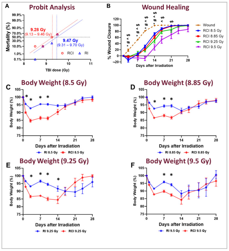
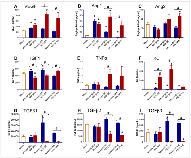
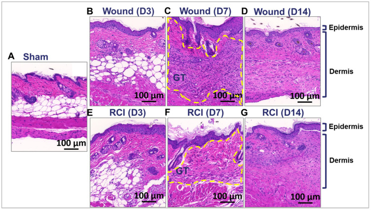
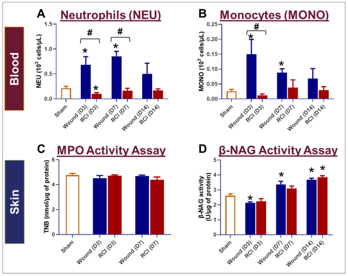
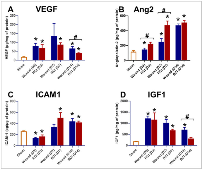
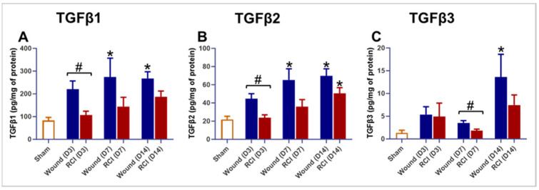
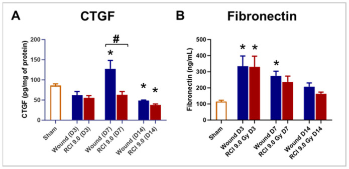
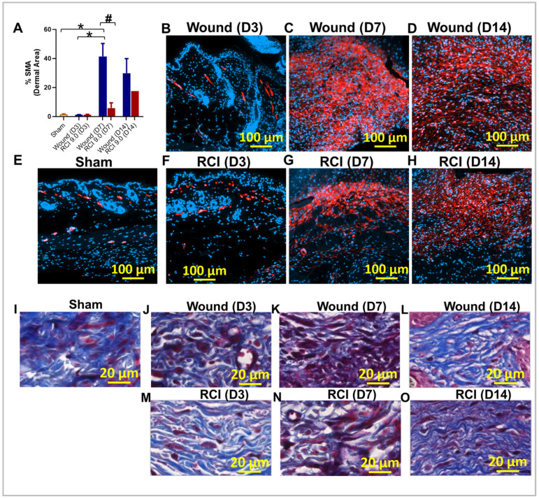
Currently, there are no U.S. Food and Drug Administration (FDA)-approved medical countermeasures (MCMs) for radiation combined injury (RCI), partially due to limited understanding of its mechanisms. Our previous research suggests that endothelial dysfunction may contribute to a poor prognosis of RCI. In this study, we demonstrated an increased risk of mortality, body weight loss, and delayed skin wound healing in RCI mice compared to mice with skin wounds alone or radiation injury (RI) 30 days post-insult. Furthermore, we evaluated biomarkers of endothelial dysfunction, inflammation, and impaired wound healing in mice at early time points after RCI. Mice were exposed to 9.0 Gy total-body irradiation (TBI) followed by skin wound. Samples were collected on days 3, 7, and 14 post-TBI. Endothelial dysfunction markers were measured by ELISA, and skin wound healing was assessed histologically. Our results show that endothelial damage and inflammation are more severe and persistent in the RCI compared to the wound-alone group. Additionally, RCI impairs granulation tissue formation, reduces myofibroblast presence, and delays collagen deposition, correlating with more severe endothelial damage. TGF signaling may play a key role in this impaired healing. These findings suggest that targeting the endothelial dysfunction and TGF-β pathways may provide potential therapeutic strategies for improving delayed wound healing in RCI, which could subsequently influence outcomes such as survival after RCI.

Keywords: TGFβ expression; collagen deposition; endothelial dysfunction; granulation tissue formation; impaired wound healing; myofibroblast; radiation combined skin wound injury; systemic and local proinflammatory response.


Conflict of interest statement

The authors declare no conflicts of interest. The funders had no role in the design of the study; in the collection, analyses, or interpretation of data; in the writing of the manuscript; or in the decision to publish the results. The opinions and assertions expressed herein are those of the author(s) and do not reflect the official policy or position of the Uniformed Services University of the Health Sciences or the Department of Defense.

Figures

Figure 1
Figure 1
Effect of different radiation doses on mouse survival, wound healing, and body weight loss following skin wound alone, RI, and RCI. Mice in both the RI and RCI groups were exposed to varying radiation doses: 8.5 Gy, 8.85 Gy, 9.25 Gy, and 9.5 Gy. (Panel A) Radiation dose–response curves for RI mice (blue) and RCI mice (red) are shown. The LD50/30 (the radiation dose expected to cause lethality to 50% of an exposed population within 30 days) was determined using a probit analysis. For RI mice, the LD50/30 is 9.47 Gy (95% confidence interval: 9.31–9.70 Gy), while for RCI mice, the LD50/30 is 9.28 Gy (95% confidence interval: 9.13–9.46 Gy). (Panel B) The delay in skin wound healing was observed in a dose-dependent manner: wounds alone (dashed line, bronze); 8.5 Gy RCI (solid line, blue); 8.85 Gy RCI (solid line, red); 9.25 Gy RCI (solid line, green); and 9.5 Gy RCI (solid line, purple). Significant differences in wound healing were observed: * p < 0.05 for the wound-alone group vs. 8.5 Gy RCI; # p < 0.05 for the wound-alone group vs. 8.85 Gy RCI; ^ p < 0.05 for the wound-alone group vs. 9.25 Gy RCI; $ p < 0.05 for the wound-alone group vs. 9.5 Gy RCI, determined by two-way ANOVA followed by Dunnett’s multiple comparison test. The sample size ranged from N = 7 to 20 per group per time point. (Panel CF) RCI mice showed more body weight loss compared to RI mice across all tested doses: 8.5 Gy, 8.85 Gy, 9.25 Gy, and 9.5 Gy (RI: blue; RCI: red). Significant differences in weight loss were observed (* p < 0.05 for RI vs. RCI), determined by two-way ANOVA followed by Sidak’s multiple comparison test. The sample size ranged from N = 7 to 20 per group per time point. Data in panel B to panel F are expressed as mean ± standard error of the mean (SEM).
Figure 2
Figure 2
Effect of RCI on serum markers associated with endothelial dysfunction and inflammation. Mice were subjected to sham injury, wounds alone, and 9.0 Gy RCI, and sera were collected on days 3, 7, and 14 post-injury. Various markers associated with endothelial dysfunction and inflammation were assessed using ELISA. The markers analyzed include (Panel A) vascular endothelial growth factor (VEGF); (Panel B) Angiopoietin-1 (Ang1); (Panel C) Angiopoietin-2 (Ang2); (Panel D) insulin-like growth factor 1 (IGF1); (Panel E) tumor necrosis factor alpha (TNFα); (Panel F) keratinocyte-derived cytokine (KC, also known as CXCL-1); (Panel G) transforming growth factor-beta 1 (TGFβ1); (Panel H) TGFβ2; and (Panel I) TGFβ3. Data are expressed as mean ± SEM. Significant differences in the mentioned markers were observed: * p < 0.05 for Sham vs. Wound/RCI on days 3, 7, and 14 post-injury, determined by one-way ANOVA followed by Dunnett’s multiple comparison test; # p < 0.05 for Wound vs. RCI at the same time point, determined by two-way ANOVA followed by Sidak’s multiple comparison test. The sample size ranged from N = 3 to 9 per group per time point.
Figure 3
Figure 3
Effect of RCI on granulation tissue formation during skin wound healing. Mice were subjected to sham injury (Panel A), wounds alone (Panel BD), and 9.0 Gy RCI (Panel EG). Skin tissues surrounding the wounds were collected on days 3, 7, and 14 post-injury. Hematoxylin and eosin (H&E) staining was performed on formalin-fixed and paraffin-embedded skin cross sections, showing nuclei in blue-purple and cytoplasm in pink-red. Representative images of the skin peri-wound area display two main layers: the epidermis and the dermis. Granulation tissue formed on day 7 post-injury is outlined with a dotted yellow line. The scale bar represents 100 μm.
Figure 4
Figure 4
Impact of RCI on peripheral blood neutrophil and monocyte counts, and neutrophil and macrophage infiltration in skin wounds. Mice were subjected to sham injury, wounds alone, and 9.0 Gy RCI. Blood and skin tissues surrounding the wounds were collected on days 3, 7, and 14 post-injury. Complete blood count (CBC) with a differential analysis was performed on EDTA-treated blood to evaluate the types of blood cells, specifically (Panel A) neutrophils (NEU) and (Panel B) monocytes (MONO). To assess NEU and macrophage (Mϕ) infiltration in the wounds, the activity of (Panel C) Myeloperoxidase (MPO) and (Panel D) β-N-Acetylglucosaminidase (β-NAG) were measured in skin tissue lysates. Data are expressed as mean ± SEM. Significant differences were observed, indicated as * p < 0.05 for Sham vs. Wound/RCI on days 3, 7, and 14 post-injury, determined by one-way ANOVA followed by Dunnett’s multiple comparison test, and # p < 0.05 for Wound vs. RCI at the same time point, determined by two-way ANOVA followed by Sidak’s multiple comparison test. The sample size ranged from N = 4 to 9 per group per time point.
Figure 5
Figure 5
Effect of RCI on skin tissue endothelial dysfunction markers. Mice were subjected to sham injury, wounds alone, and 9.0 Gy RCI, and skin tissues surrounding the wounds were collected on days 3, 7, and 14 post-injury. Various markers associated with endothelial dysfunction were assessed in skin tissue lysates using ELISA. The markers analyzed include (Panel A) vascular endothelial growth factor (VEGF); (Panel B) Angiopoietin-2 (Ang2); (Panel C) intercellular adhesion molecule 1 (ICAM1); and (Panel D) insulin-like growth factor 1 (IGF1). Data are expressed as mean ± SEM. Significant differences in these markers were observed: * p < 0.05 for Sham vs. Wound/RCI on days 3, 7, and 14 post-injury, determined by one-way ANOVA followed by Dunnett’s multiple comparison test, and # p < 0.05 for Wound vs. RCI at the same time point, determined by two-way ANOVA followed by Sidak’s multiple comparison test. The sample size ranged from N = 3 to 9 per group per time point.
Figure 6
Figure 6
Effect of RCI on skin tissue levels of transforming growth factor-beta (TGFβ) 1-3. Mice were subjected to sham injury, wounds alone, and 9.0 Gy RCI, and skin tissues surrounding the wounds were collected on days 3, 7, and 14 post-injury. Skin tissue levels of (Panel A) TGFβ1, (Panel B) TGFβ2, and (Panel C) TGFβ3 were measured using the Bio-Plex multiplexing system. Data are expressed as mean ± SEM. Significant differences in TGFβ1-3 were observed: * p < 0.05 for Sham vs. Wound/RCI on days 3, 7, and 14 post-injury, determined by one-way ANOVA followed by Dunnett’s multiple comparison test, and # p < 0.05 for Wound vs. RCI at the same time point, determined by two-way ANOVA followed by Sidak’s multiple comparison test. The sample size was N = 4 per group per time point.
Figure 7
Figure 7
Effect of RCI on skin tissue levels of connective tissue growth factor (CTGF) and fibronectin. Mice were subjected to sham injury, wounds alone, and 9.0 Gy RCI, and skin tissues surrounding the wounds were collected on days 3, 7, and 14 post-injury. Levels of (Panel A) CTGF and (Panel B) fibronectin were measured in tissue lysates by EKISA. Data are expressed as mean ± SEM. Significant differences in these factors were observed: * p < 0.05 for Sham vs. Wound/RCI on days 3, 7, and 14 post-injury, determined by one-way ANOVA followed by Dunnett’s multiple comparison test, and # p < 0.05 for Wound vs. RCI at the same time point, determined by two-way ANOVA followed by Sidak’s multiple comparison test. The sample size ranged from N = 4 to 9 per group per time point.
Figure 8
Figure 8
Effect of RCI on fibroblast differentiation and collagen deposition during skin wound healing. Mice were subjected to sham injury (Panel E,I), wounds alone (Panel BD,JL), and 9.0 Gy RCI (Panel FH,MO). Skin tissues surrounding the wounds were collected on days 3, 7, and 14 post-injury. (Panel AH) Immunohistochemical (IHC) staining using anti-alpha-smooth muscle actin (anti-α-SMA) antibody was performed on formalin-fixed and paraffin-embedded skin cross sections, highlighting α-SMA in red and nuclei in blue. (Panel A) Quantitative analysis of the percentage of α-SMA (% SMA) signal in the dermal area is shown. Data are expressed as mean ± SEM. Significant differences in % SMA were observed: * p < 0.05 for difference among Sham, Wound, and RCI on days 3, 7, and 14 post-injury, determined by one-way ANOVA followed by Tukey’s multiple comparison test, and # p < 0.05 for Wound vs. RCI at the same time point, determined by two-way ANOVA followed by Sidak’s multiple comparison test. The sample size ranged from N = 3 to 4 per group per time point (except for N = 1 for RCI at the day 14 post-injury time point). The scale bar represents 100 μm. (Panel IO) Masson’s trichrome staining was performed on formalin-fixed and paraffin-embedded skin cross sections, showing collagen fibers in blue, nuclei in black, and cytoplasm in red. The scale bar represents 20 μm.

Similar articles

Cited by

References

    1. Wang L., Lin B., Zhai M., Cui W., Hull L., Zizzo A., Li X., Kiang J.G., Xiao M. Deteriorative Effects of Radiation Injury Combined with Skin Wounding in a Mouse Model. Toxics. 2022;10:785. doi: 10.3390/toxics10120785. - DOI - PMC - PubMed
    1. Wang L., Zhai M., Lin B., Cui W., Hull L., Li X., Anderson M.N., Smith J.T., Umali M.V., Jiang S., et al. PEG-G-CSF and L-Citrulline Combination Therapy for Mitigating Skin Wound Combined Radiation Injury in a Mouse Model. Radiat. Res. 2021;196:113–127. doi: 10.1667/RADE-20-00151.1. - DOI - PMC - PubMed
    1. DiCarlo A.L., Ramakrishnan N., Hatchett R.J. Radiation combined injury: Overview of NIAID research. Health Phys. 2010;98:863–867. doi: 10.1097/HP.0b013e3181a6ee32. - DOI - PMC - PubMed
    1. Kiang J.G., Blakely W.F. Combined radiation injury and its impacts on radiation countermeasures and biodosimetry. Int. J. Radiat. Biol. 2023;99:1055–1065. doi: 10.1080/09553002.2023.2188933. - DOI - PMC - PubMed
    1. Wang J., Boerma M., Fu Q., Hauer-Jensen M. Radiation responses in skin and connective tissues: Effect on wound healing and surgical outcome. Hernia. 2006;10:502–506. doi: 10.1007/s10029-006-0150-y. - DOI - PubMed

MeSH terms

LinkOut - more resources