. 2024 Nov 22;25(23):12547.
doi: 10.3390/ijms252312547.

Neutrophils in the Spotlight-An Analysis of Neutrophil Function and Phenotype in ARDS

Affiliations

Neutrophils in the Spotlight-An Analysis of Neutrophil Function and Phenotype in ARDS

Richard F Kraus et al. Int J Mol Sci. .

Abstract

Acute respiratory distress syndrome (ARDS) is a complex disease pattern in which pathogenesis polymorphonuclear neutrophil granulocytes (PMN) play a key role. In previous experiments, we could show that interaction with collagen III (an important component of pulmonary tissue) is a possible trigger of neutrophil reactive oxygen species (ROS) production. To investigate possible correlations, further elucidate ARDS pathophysiology, and maybe find pharmacological targets, we evaluated PMNs from blood (circulating PMNs: cPMNs) and tracheal secretion (tPMNs) from patients with and without ARDS with regard to function and phenotype. Blood samples and tracheal secretions were obtained from intensive care patients with and without ARDS. Isolation of cPMN was performed by density-gradient gravity sedimentation without centrifugation. For tPMN isolation, endotracheal aspirate was filtered, and tPMNs were separated from the remaining aspirate using a particle filter. Specific surface epitopes (CD66b, CD62L, fMLP-receptor, LOX-1, CD49d, CD29, CD11b) of the isolated PMN cells were labeled with antibody-coupled dyes and analyzed by flow cytometry. Neutrophil ROS production before and after activation with N-formyl-methyl-leucyl-phenylalanine (fMLP) and tumor necrosis factor α (TNFα) was quantified using rhodamine-123. In addition, a qualitative cytological hematoxylin-eosin (HE) staining was performed with a portion of the secretion. tPMNs were observed in both bloody and mucosal tracheal secretions from ARDS patients. The epitope distribution on cPMNs and tPMNs differed significantly in patients with and without ARDS: tPMNs generally showed increased expression of CD66b, LOX-1 and fMLP-receptor compared to cPMNs, and decreased expression of CD62L. The CD49d levels of all cPMNs were at the same level as tPMNs in ARDS, whereas CD49d expression was increased on tPMNs without ARDS. ROS production was significantly stimulated by fMLP/TNFα in cPMNs regardless of the patient group, while it was similarly increased in tPMNs with and without stimulation. Increased expression of CD66b, LOX-1 and fMLP-receptor on tPMNs indicated a higher activity status compared to cPMNs. Increased CD49d expression on tPMNs without ARDS marks different PMN surface changes in lung disease. PMNs appear to be in a more activated state in lung secretions than in blood, as indicated by higher CD66b and lower CD62L expression, higher constitutive ROS production and lower excitability with fMLP and TNFα. In the context of possible CD49d-triggered ROS production, it is noteworthy that CD49d is downregulated in secretion from patients with ARDS compared to patients without. This phenotypic and functional PMN characterization can provide valuable diagnostic and therapeutic information for the intensive care treatment of ARDS patients.

Keywords: ARDS; ROS; flow cytometry; intensive care; neutrophil; surface epitope.


Conflict of interest statement

The authors declare no conflicts of interest.

Figures

Figure 1
Figure 1
(a) Serous endotracheal aspirate (b) Mucuous endotracheal aspirate (c) Sanguineous endotracheal aspirate.
Figure 2
Figure 2
Cytological staining of endotracheal aspirate. (a) Hematoxylin-eosin staining of mucosal endotracheal aspirate: Black arrows = rod-nucleated and segment-nucleated PMNs, Black circle = alveolar macrophage. The varying intensity of the pink coloration is due to the mucous consistency of the endotracheal aspirate, resulting in intensely pink-colored areas in the lower left edge of the image and lighter areas in the upper right area of the image. Purple threads of mucus can also be seen. (b) HE staining of mucosal endotracheal aspirate. Black arrows = rod-nucleated and segment-nucleated PMNs, Black circles = alveolar macrophages, probably with phagocytosed. The loosened and less cell-dense appearance of this image is due to the staining technique. (c) Papanicolaou stain of mucosal endotracheal aspirate. Respiratory epithelia may also be present among the cells (Red arrows = squamous epithelial cells). There are also dark cell nuclei, e.g., of PMNs (black arrows), as well as numerous dark blue mucus threads interspersing the image. (d) HE staining of a sanguineous endotracheal aspirate. PMNs colored in dark purple (black arrows) dominate the aspirate next to the pink-colored blood. Macrophages and plasma cells are also present between the PMNs. (e) Papanicolaou staining of sanguineous endotracheal aspirate. Mucus threads can be seen in strong dark blue. Red arrows mar brightly colored erythrocytes. PMNs are marked with black arrows.
Figure 3
Figure 3
Results of flow cytometry measurements Data are shown as median ± interquartile ranges or mean ± 95% confidence interval and p-values. (a) Comparison of the percentage of avital PMN, (b) Comparison of MFI(CD66b), (c) Comparison of MFI(CD62L). (d) Comparison of MFI(LOX-1), (e) Comparison of MFI(CD49d), (f) Comparison of MFI(fMLP-R).
Figure 4
Figure 4
Comparison of MFI(DHR) subdivided into activating substances and study groups. Data are shown as median ± interquartile ranges and p-values.
Figure 5
Figure 5
The results of this work, in conjunction with results from previous publications [24,26], support hypotheses that the interaction of PMNs with type III collagen via interaction with CD49d/CD29 may play an important role in the promotion of ARDS.
Figure 6
Figure 6
Workflow of the experiments.
Figure 7
Figure 7
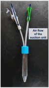
Illustration of the tracheal suction set. The dark blue arrows illustrate the direction of the airflow.
Figure 8
Figure 8
Preparation methods for the aspirate depend on the original quality of the tPMN isolation.
Figure 9
Figure 9
Experimental setup for flow cytometric analysis.
Figure 10
Figure 10
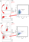
(a) Analysis of surface epitopes of (a) cPMNs or (b) tPMNs using APC-conjugated antibody against LOX-1. Abbreviations: Granulocytes = PMNs; SSC-H = side scatter height; FSC-H = forward scatter height.
Figure 11
Figure 11
(a) Analysis of (a) cPMN and (b) tPMN ROS production (Red arrows illustrate the further procession of selected cells. Abbreviations: Granulocytes = PMNs; SSC-H = side scatter height; FSC-H = forward scatter height; FL3-H = fluorescence 3; Live = living cells.

References

    1. Manzano F., Yuste E., Colmenero M., Aranda A., García-Horcajadas A., Rivera R., Fernández-Mondéjar E. Incidence of acute respiratory distress syndrome and its relation to age. J. Crit. Care. 2005;20:274–280. doi: 10.1016/j.jcrc.2005.05.008. - DOI - PubMed
    1. Bos L.D.J., Ware L.B. Acute respiratory distress syndrome: Causes, pathophysiology, and phenotypes. Lancet. 2022;400:1145–1156. doi: 10.1016/S0140-6736(22)01485-4. - DOI - PubMed
    1. Pham T., Rubenfeld G.D. Fifty Years of Research in ARDS. The Epidemiology of Acute Respiratory Distress Syndrome. A 50th Birthday Review. Am. J. Respir. Crit. Care Med. 2017;195:860–870. doi: 10.1164/rccm.201609-1773CP. - DOI - PubMed
    1. Read by QxMD Acute Respiratory Distress Syndrome: The Berlin Definition. [(accessed on 9 September 2023)]. Available online: https://read.qxmd.com/read/22797452/acute-respiratory-distress-syndrome-....
    1. Tomashefski J.F. Pulmonary pathology of acute respiratory distress syndrome. Clin. Chest Med. 2000;21:435–466. doi: 10.1016/S0272-5231(05)70158-1. - DOI - PubMed

Substances

LinkOut - more resources