. 2024 Nov 22;25(23):12552.
doi: 10.3390/ijms252312552.

Plasmalogens Improve Lymphatic Clearance of Amyloid Beta from Mouse Brain and Cognitive Functions

Affiliations

Plasmalogens Improve Lymphatic Clearance of Amyloid Beta from Mouse Brain and Cognitive Functions

Alexander Shirokov et al. Int J Mol Sci. .

Abstract

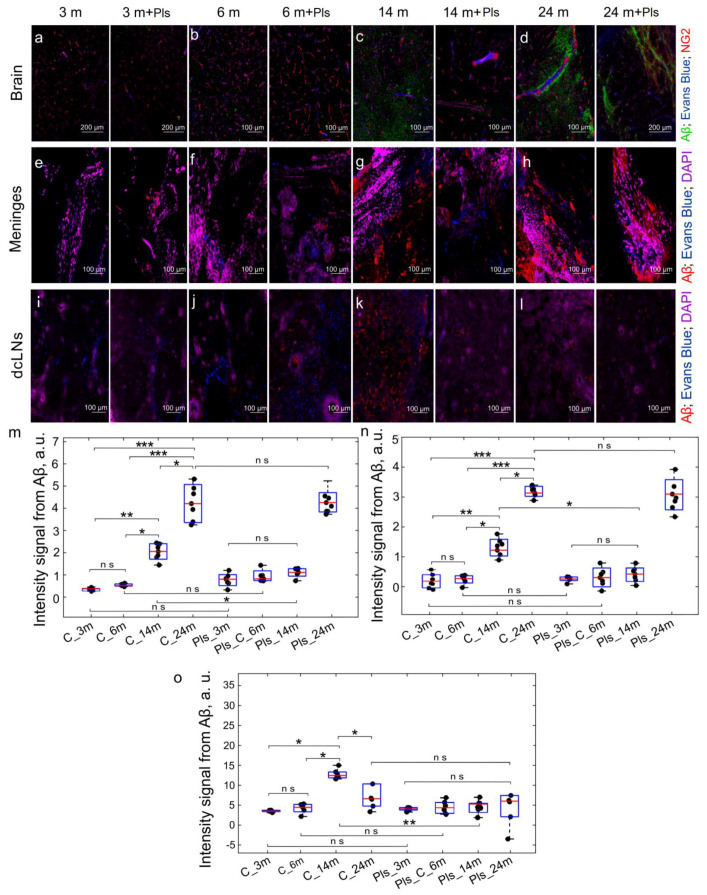
Amyloid beta (Aβ) is a neuronal metabolic product that plays an important role in maintaining brain homeostasis. Normally, intensive brain Aβ formation is accompanied by its effective lymphatic removal. However, the excessive accumulation of brain Aβ is observed with age and during the development of Alzheimer's disease (AD) leading to cognitive impairment and memory deficits. There is emerging evidence that plasmalogens (Pls), as one of the key brain lipids, may be beneficial for AD and cognitive aging. Here, we studied the effects of Pls on cognitive functions and the lymphatic clearance of Aβ from the brain of AD mice and mice of different ages. The results showed that Pls effectively reduce brain Aβ levels and facilitate learning in aged but not old mice. In AD mice, Pls improve the lymphatic clearance of Aβ that is accompanied by an increase in general motor activity and an improvement of the emotional status and learning ability. Thus, these findings suggest that Pls could be a promising candidate for the alternative or concomitant therapy of AD and age-related brain diseases to enhance the lymphatic clearance of Aβ from the brain and cognitive functions.

Keywords: Alzheimer’s disease; age; amyloid beta; cognitive functions; lymphatic clearance; plasmalogens.


Conflict of interest statement

The authors declare no conflicts of interest.

Figures

Figure 1
Figure 1
Schematic illustration of the study design: (a) Pls (phospholipids from bovine brain) were administered for 21 days into the right lateral ventricle through a chronic catheter; (b,c) the effects of Pls on clearance of the soluble forms of Aβ and cognitive functions were studied in two functional models, including (b) an injection model of early AD (unilateral administration of Aβ into the hippocampus) and (c) the age model reflecting natural age-related changes in Aβ deposition in brain tissues. Before and after a 21-day course of Pls, studies of behavior of mice as well as qualitative and quantitative analysis of Aβ in brain tissues were conducted using enzyme immunoassay (EIA) and confocal imaging of Aβ in the brain, the meninges, and in the deep cervical lymph nodes (dcLNs).
Figure 2
Figure 2
The effects of Pls on Aβ clearance from the brain and the meninges to the peripheral lymphatics in AD mice: (ad) Representative images of Aβ (green) in the brain from the tested groups. The blood vessels are filled with Evans Blue (blue) and labeled with NG2 (red); (eh) Representative images of Aβ (red) in the meninges from the tested groups; (il) Representative images of Aβ (red) in dcLNs from the tested groups. In (el), the blood vessels are filled with Evans Blue (blue), the cell nuclei are labeled with DAPI (violet); (mo) Quantitative analysis of the intensity of fluorescent signal from Aβ labeled with primary and secondary antibodies in the brain (m), the meninges (n), and in dcLNs (o), n = 7 in each group, *—p < 0.05, ***—p < 0.001, the ANOVA test with the post hoc Duncan test.
Figure 3
Figure 3
The effects of Pls on cognitive functions in AD mice and mice of different ages: (ae) Assessment of locomotor activity and anxiety using the open-field test (a) in the AD groups (b,c) without Pls and after a 21-day course of Pls as well as in middle-aged, aged, and old mice without Pls (d,e); (fi) Evaluation of spatial memory using the Y-maze test (f,g) in the AD groups (h) without Pls and after a 21-day course of Pls and well as in middle-aged, aged, and old mice without Pls (i); Analysis of recognition memory using the new object recognition test (j) in the AD groups (k) without Pls and after a 21-day course of Pls as well as in middle-aged, aged, and old mice without Pls (l); n = 8 in the AD groups and n = 7 in the age groups, *—p < 0.05, **—p < 0.01, ***—p < 0.001, ns means not significant, the ANOVA with the post hoc Tukey HSD Test.
Figure 4
Figure 4
The effects of Pls on the development of Pavlov’s conditioned reflex in AD mice and mice of different ages: (ac) the training of mice is based on the presentation of an unconditional signal (green light), after which the animal receives food (reward) if it accidentally finds the correct window, in which food (seeds) falls out when the head is howled. The number of training sessions required for the mouse to form a stable conditioned reflex is assessed (when the light is turned on, the mouse must quickly (within 15 s) find the correct window and receive food (reward)). The number of rewards (food) received is also assessed in the final session; (d,e) Assessment of Pavlov’s conditioned reflex in AD mice and mice of different ages, n = 8 in each group, *—p < 0.05, **—p < 0.01, ***—p < 0.001, ns means not significant, the ANOVA test with the post hoc Duncan test.
Figure 5
Figure 5
The effects of Pls on Aβ clearance from the brain and the meninges to the peripheral lymphatics in AD mice: (ad) Representative images of Aβ (green) in the brain from the tested groups. The blood vessels are filled with Evans Blue (blue) and labeled with NG2 (red); (eh) Representative images of Aβ (red) in the meninges from the tested groups; (il) Representative images of Aβ (red) in dcLNs from the tested groups. In (el), the blood vessels are filled with Evans Blue (blue), the nuclei are labeled with DAPI (violet); (mo) Quantitative analysis of Aβ levels in the brain (m), the meninges (n), and in dcLNs (o), n = 7 in each group, *—p < 0.05, **—p < 0.01, ***—p < 0.001, ns means not significant, the Mann–Whitney U Test.

Similar articles

Cited by

References

    1. Zhang Y., Chen H., Li R., Sterling K., Song W. Amyloid β-based therapy for Alzheimer’s disease: Challenges, successes and future. Sig. Transduct. Target. Ther. 2023;8:248. doi: 10.1038/s41392-023-01484-7. - DOI - PMC - PubMed
    1. Ma C., Hong F., Yang S. Amyloidosis in Alzheimer’s Disease: Pathogeny, Etiology, and Related Therapeutic Directions. Molecules. 2022;27:1210. doi: 10.3390/molecules27041210. - DOI - PMC - PubMed
    1. An J., Kim K., Lim H.J., Kim H.Y., Shin J., Park I., Cho I., Kim H.Y., Kim S., McLean C., et al. Early onset diagnosis in Alzheimer’s disease patients via amyloid-β oligomers-sensing probe in cerebrospinal fluid. Nat. Commun. 2024;15:1004. doi: 10.1038/s41467-024-44818-x. - DOI - PMC - PubMed
    1. Gouras G.K., Almeida C.G., Takahashi R.H. Intraneuronal Abeta accumulation and origin of plaques in Alzheimer’s disease. Neurobiol. Aging. 2005;26:1235–1244. doi: 10.1016/j.neurobiolaging.2005.05.022. - DOI - PubMed
    1. Naslund J., Haroutunian V., Mohs R., Davis K.L., Davies P., Greengard P., Buxbaum J.D. Correlation between elevated levels of amyloid beta-peptide in the brain and cognitive decline. JAMA. 2000;283:1571–1577. doi: 10.1001/jama.283.12.1571. - DOI - PubMed

LinkOut - more resources