. 2024 Nov 25;25(23):12629.
doi: 10.3390/ijms252312629.

Unravelling the Signature Follicular Fluid Metabolites in Dairy Cattle Follicles Growing Under Negative Energy Balance: An In Vitro Approach

Affiliations

Unravelling the Signature Follicular Fluid Metabolites in Dairy Cattle Follicles Growing Under Negative Energy Balance: An In Vitro Approach

Muhammad Shahzad et al. Int J Mol Sci. .

Abstract

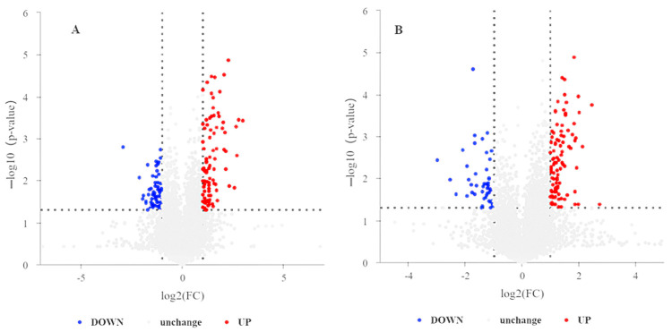
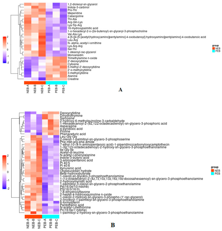
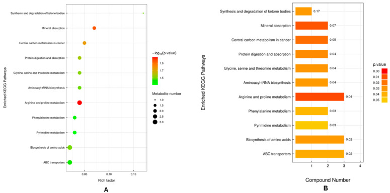
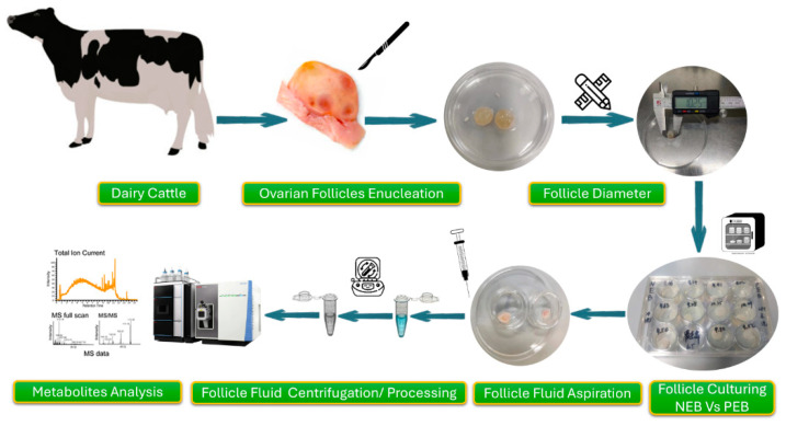
The astringent selection criteria for milk-oriented traits in dairy cattle have rendered these animals prone to various metabolic disorders. Postpartum lactational peak and reduced feed intake lead to negative energy balance in cattle. As a compensatory mechanism, cattle start mobilizing fat reserves to meet the energy demand for vital body functions. Consequently, diminished glucose concentrations and elevated ketone body levels lead to poor ovarian function. The impaired follicular development and subpar oocyte quality diminish the conception rates, which poses significant economic repercussions. Follicular fluid is integral to the processes of follicular growth and oocyte development. Hence, the present study was performed to identify potential alterations in metabolites in the follicular fluid under in vitro culture conditions mimicking negative energy balance. Our results revealed nine distinct metabolites exhibiting differential expression in follicular fluid under negative energy balance. The differentially expressed metabolites were predominantly associated with pathways related to amino acid metabolism, lipid metabolism, signal transduction mechanisms, and membrane transport, alongside other biological processes. The identified signature metabolites may be further validated to determine oocyte fitness subjected to in vitro fertilization or embryo production from slaughterhouse source ovaries.

Keywords: dairy cattle; follicular fluid; metabolites; negative energy balance.


Conflict of interest statement

The authors declare that they have no conflict of interest.

Figures

Figure 1
Figure 1
Chromatogram illustrates the total ion current (TIC) with respect to the retention time of metabolite in QC samples both in positive/+ (A) and negative (B) ions of electrospray ionization (ESI) modes. The signal intensity is displayed on the y-axis in million counts, and the retention time is displayed on the x-axis in min. The QC1, QC2, and QC3 are represented by the colors blue, pink, and red in the spectrum, respectively.
Figure 2
Figure 2
PCA of differential metabolites in follicular fluid from dairy cows under different metabolic states. (A) PCA plot illustrates metabolite distribution in positive ion samples. (B) PCA plot displaying metabolite profiles from negative ion metabolites. Green squares represent NEB metabolites, blue circles represent PEB metabolites, and pink triangles represent QC. The x and y axis show the first two principal components (PC1 and PC2) which explain the greatest variance in the data. The label “t[1]” on the x-axis represents the first principal component (PC1) of the Principal Component Analysis (PCA).
Figure 3
Figure 3
The figure shows correlation profiles of QC samples in metabolomic analysis of FF from dairy cows. (A) Correlation matrix for positive ion mode QC samples. (B) Correlation matrix for negative ion mode QC samples. Each subplot represents pairwise comparisons between three QC samples (QC-1, QC-2, QC-3), with correlation coefficients shown in blue. The high correlation coefficients (≥0.998) indicate strong reproducibility and stability of the metabolomic measurements across both ionization modes.
Figure 4
Figure 4
Distribution of metabolite classes (colors) identified in FF of dairy cows. The pie chart illustrates the relative proportions of various metabolite classes, expressed as percentages (%).
Figure 5
Figure 5
Volcano plots show the differential metabolite expression in FF under NEB and PEB. (A) Positive ion mode and (B) negative ion mode metabolite profiles. The x-axis represents log2 FC. The y-axis shows −log10 of p-value. Different color dots represent metabolites that were significantly upregulated (red), significantly downregulated (blue), and with no significant change (grey). The fold change (FC) threshold is represented by vertical dotted lines, whereas the significance threshold is represented by the horizontal dotted line (p-value).
Figure 6
Figure 6
Heatmap showing hierarchical clustering of differentially expressed metabolites in FF NEB and PEB groups. (A) Metabolites detected in positive ion mode. (B) Metabolites detected in negative ion mode. The color scale denotes the relative abundance of metabolites, with red indicating higher levels and blue indicating lower levels. Each horizontal row represents a distinct metabolite, and each vertical column represents a sample (NEB-A, NEB-B, NEB-C, PEB-A, PEB-B, PEB-C). The dendrograms on the left-hand side represent the hierarchical clustering of metabolites based on their expression patterns.
Figure 7
Figure 7
KEGG pathway enrichment analysis of differentially expressed metabolites in FF of dairy cattle under varying energy balance conditions. (A) The bubble plot represents the enriched pathways. The x-axis gauges the rich factor (ratio of differentially expressed metabolites in a pathway to the total metabolites in that pathway). The y-axis enlists the enriched KEGG pathways. The bubble size indicates the number of metabolites, and the color represents the significance level (−log10 (p-value)). (B) Bar plot of the same enriched pathways, where the x-axis shows the number of compounds involved in each pathway, and bars are colored according to p-value significance.
Figure 8
Figure 8
Schematic diagram of the experiment illustrating follicle enucleation, diameter measurement, culturing, FF collection, and metabolite analysis.

Similar articles

References

    1. Brito L., Bédère N., Douhard F., Oliveira H., Arnal M., Peñagaricano F., Schinckel A., Baes C.F., Miglior F. Genetic selection of high-yielding dairy cattle toward sustainable farming systems in a rapidly changing world. Animal. 2021;15:100292. doi: 10.1016/j.animal.2021.100292. - DOI - PubMed
    1. Gutierrez-Reinoso M.A., Aponte P.M., Garcia-Herreros M. Genomic analysis, progress and future perspectives in dairy cattle selection: A review. Animals. 2021;11:599. doi: 10.3390/ani11030599. - DOI - PMC - PubMed
    1. Hayes E., Wallace D., O’Donnell C., Greene D., Hennessy D., O’Shea N., Tobin J., Fenelon M. Trend analysis and prediction of seasonal changes in milk composition from a pasture-based dairy research herd. J. Dairy Sci. 2023;106:2326–2337. doi: 10.3168/jds.2021-21483. - DOI - PubMed
    1. USDA Development of the Average Annual Milk Production Per Cow in the United States Since 1924. [(accessed on 19 September 2024)]; Available online: https://quickstats.nass.usda.gov/
    1. Gross J.J. Dairy cow physiology and production limits. Anim. Front. Rev. Mag. Anim. Agric. 2023;13:44. doi: 10.1093/af/vfad014. - DOI - PMC - PubMed

LinkOut - more resources