Review
. 2024 Nov 25;25(23):12661.
doi: 10.3390/ijms252312661.

Backstage Heroes-Yeast in COVID-19 Research

Affiliations
Review

Backstage Heroes-Yeast in COVID-19 Research

Wojciech Grabiński et al. Int J Mol Sci. .

Abstract

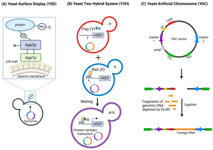
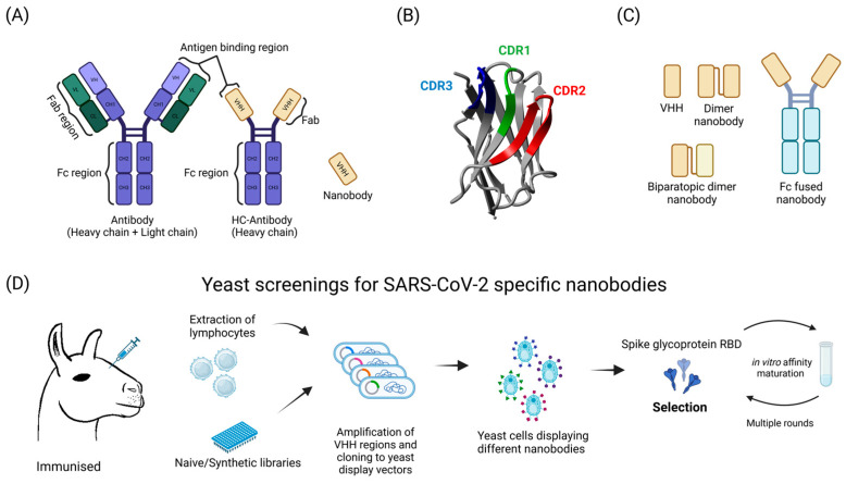
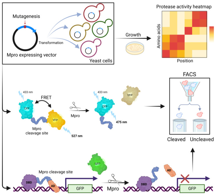
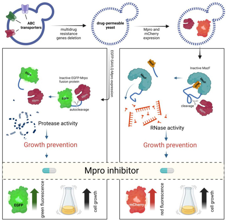
The extremely rapid development of understanding and technology that led to the containment of the COVID-19 pandemic resulted from collaborative efforts in the fields of Betacoronavirus pandemicum (SARS-CoV-2) biology, pharmacology, vaccinology, and medicine. Perhaps surprisingly, much of the research was conducted using simple and efficient yeast models. In this manuscript, we describe how yeast, eukaryotic microorganisms, have been used to research this global challenge, focusing on the therapeutic potential of the studies discussed herein. Thus, we outline the role of yeast in studying viral protein interactions with the host cell proteome, including the binding of the SARS-CoV-2 virus spike protein to the human ACE2 receptor and its modulation. The production and exploration of viral antigens in yeast systems, which led to the development of two approved COVID-19 vaccines, are also detailed. Moreover, yeast platforms facilitating the discovery and production of single-domain antibodies (nanobodies) against SARS-CoV-2 are described. Methods guiding modern and efficient drug discovery are explained at length. In particular, we focus on studies designed to search for inhibitors of the main protease (Mpro), a unique target for anti-coronaviral therapies. We highlight the adaptability of the techniques used, providing opportunities for rapid modification and implementation alongside the evolution of the SARS-CoV-2 virus. Approaches introduced in yeast systems that may have universal potential application in studies of emerging viral diseases are also described.

Keywords: COVID-19; SARS-CoV-2; Saccharomyces cerevisiae; vaccines; yeast surface display; yeast two-hybrid system.


Conflict of interest statement

The authors declare no conflicts of interest. The funders had no role in the design of the study; in the collection, analyses, or interpretation of data; in the writing of the manuscript; or in the decision to publish the results.

Figures

Figure 1
Figure 1
Overview of Yeast-Based Techniques Used in COVID-19 Research. (A) Yeast Surface Display (YSD). YSD allows the display of recombinant proteins on the surface of Saccharomyces cerevisiae. The figure shows the protein of interest linked to Aga2p, which is attached to the cell wall via disulfide bonds with Aga1p. (B) The Yeast Two-Hybrid (Y2H) system. Y2H is used to study protein—protein interactions. The figure depicts two haploid yeast cells, each expressing a different fusion protein: Bait (X) with a DNA-binding domain (DBD) and Prey (Y) with an activation domain (AD). Upon mating, the X and Y interact, bringing the DBD and AD into proximity and leading to the activation of a reporter gene (HIS3). (C) Yeast Artificial Chromosomes (YACs). YACs are vectors used to clone large DNA fragments. The figure shows the construction of a YAC by digesting the vector, followed by ligation with foreign DNA. MYC—a Myc-tag, HA—hemagglutinin-tag, TEL—telomere, CEN—centromere. Figure created with BioRender.com.
Figure 2
Figure 2
Structure and discovery of SARS-CoV-2 specific nanobodies using yeast systems. (A) Antibodies. A conventional antibody consists of two heavy chains and two light chains, with antigen-binding regions located on the Fab (antigen-binding fragment) region. The Fc (crystallizable fragment) region is responsible for immune system activation. Unlike conventional antibodies, heavy-chain-only antibodies (HC-antibodies) lack light chains. Nanobodies consist only of the antigen-binding region (VHH). (B) Nanobody structure. The nanobody is a small, single-domain antibody. It contains three complementarity-determining regions (CDR1, CDR2, CDR3) responsible for antigen recognition and binding. The 3D structure of the nanobody was obtained from the Protein Data Bank (PDB) with the structure ID 6OBC. The visualization was created using the Yasara software, Version 20.8.15 (www.yasara.org). (C) Nanobody types. VHH—a single nanobody unit. Dimer nanobody—two nanobodies linked together, potentially increasing binding affinity. Biparatopic dimer nanobody—a dimer formed by two nanobodies that bind different epitopes on the same antigen. Fc-fused nanobody—a nanobody fused to an Fc region, combining the small size and specificity of nanobodies with the effector functions of conventional antibodies. (D) Discovering SARS-CoV-2 specific nanobodies using yeast display systems. The process begins with the immunization of camelids, such as llamas, which leads to the production of heavy-chain-only antibodies (HC-antibodies). Lymphocytes are then extracted from these animals, and the VHH regions, responsible for antigen binding, are amplified and cloned into yeast display vectors, creating libraries of nanobodies. Alternatively, the synthetic or naive nanobody libraries can be used. Yeast cells are transformed to display various nanobodies on their surface. The displayed nanobodies are then screened for their ability to bind to the receptor-binding domain (RBD) of the SARS-CoV-2 spike glycoprotein. Selected nanobodies undergo in vitro affinity maturation, involving multiple rounds of selection, to enhance their binding affinity to the antigen, resulting in highly specific nanobodies against SARS-CoV-2. Figure created with BioRender.com.
Figure 3
Figure 3
Yeast-based assays for SARS-CoV-2 main protease (Mpro) activity and mutagenesis studies. An Mpro-expressing vector undergoes mutagenesis to introduce various mutations in the protein. The mutated vectors are then transformed into yeast cells, resulting in yeast strains with different Mpro variants. Yeast cells expressing different Mpro variants are grown on selective media (top right). The growth rates are used to generate a heatmap, indicating protease activity across different amino acid positions and mutations. The FRET assay (middle) is used to measure Mpro activity. Mpro cleaves a substrate linking cyan fluorescent protein (CFP) and yellow fluorescent protein (YFP). The cleavage results in a loss of energy transfer from CFP to YFP, leading to a change in fluorescence emission from 527 nm (YFP) to 475 nm (CFP). The cleaved and uncleaved populations can be sorted using fluorescence-activated cell sorting (FACS). In cleavage assays using GFP (bottom), Mpro cleaves within a fusion protein that links a DNA-binding domain (DBD) and an activation domain (AD) of a transcription factor activating the green fluorescent protein (GFP) expression. Upon cleavage by Mpro, GFP expression is lost, indicating Mpro activity. Figure created with BioRender.com.
Figure 4
Figure 4
Yeast-based systems for screening SARS-CoV-2 main protease (Mpro) inhibitors. Yeast strains are genetically modified by the deletion of multidrug resistance genes, creating drug-permeable yeast suitable for high-throughput screening. Yeast cells express a fusion protein consisting of enhanced green fluorescent protein (EGFP) and Mpro, linked by a specific cleavage site (SAVLQ). Active Mpro cleaves the fusion protein, separating EGFP from Mpro, which prevents the growth of yeast cells. In the presence of an effective Mpro inhibitor, cleavage does not occur, resulting in green fluorescence and restored yeast cell growth, indicating the inhibition of Mpro activity (left panel). Yeast cells express the bacterial toxin MazF linked to the Mpro cleavage site and tagged with mCherry, a red fluorescent protein. Active Mpro cleaves the fusion protein, releasing MazF, which is an RNase that degrades RNA, leading to yeast growth inhibition. When an Mpro inhibitor is present, cleavage does not occur, preventing MazF release and allowing yeast cell growth. The yeast cells exhibit red fluorescence if the inhibitor is effective, signifying the prevention of RNase-induced cell death (right panel). Figure created with BioRender.com.

Similar articles

References

    1. Kashani N.R., Azadbakht J., Ehteram H., Kashani H.H., Rajabi-Moghadam H., Ahmad E., Nikzad H., Hosseini E.S. Molecular and Clinical Investigation of COVID-19: From Pathogenesis and Immune Responses to Novel Diagnosis and Treatment. Front. Mol. Biosci. 2022;9:770775. doi: 10.3389/fmolb.2022.770775. - DOI - PMC - PubMed
    1. McDonnell L., Moore A., Micou M., Day C., Grossman E., Meaders C. CRISPR in Your Kitchen: An At-Home CRISPR Kit to Edit Genes in Saccharomyces Cerevisiae Used during a Remote Lab Course. J. Microbiol. Biol. Educ. 2022;23:e00321-21. doi: 10.1128/jmbe.00321-21. - DOI - PMC - PubMed
    1. Gaikani H.K., Stolar M., Kriti D., Nislow C., Giaever G. From Beer to Breadboards: Yeast as a Force for Biological Innovation. Genome Biol. 2024;25:10. doi: 10.1186/s13059-023-03156-9. - DOI - PMC - PubMed
    1. Gastelum S., Michael A.F., Bolger T.A. Saccharomyces Cerevisiae as a Research Tool for RNA-Mediated Human Disease. Wiley Interdiscip. Rev. RNA. 2023;15:e1814. doi: 10.1002/wrna.1814. - DOI - PubMed
    1. Franco L.V.R., Bremner L., Barros M.H. Human Mitochondrial Pathologies of the Respiratory Chain and ATP Synthase: Contributions from Studies of Saccharomyces Cerevisiae. Life. 2020;10:304. doi: 10.3390/life10110304. - DOI - PMC - PubMed

MeSH terms

LinkOut - more resources