. 2024 Nov 26;25(23):12702.
doi: 10.3390/ijms252312702.

Genetic Manipulation of Caveolin-1 in a Transgenic Mouse Model of Aortic Root Aneurysm: Sex-Dependent Effects on Endothelial and Smooth Muscle Function

Affiliations

Genetic Manipulation of Caveolin-1 in a Transgenic Mouse Model of Aortic Root Aneurysm: Sex-Dependent Effects on Endothelial and Smooth Muscle Function

Tala Curry-Koski et al. Int J Mol Sci. .

Abstract

Marfan syndrome (MFS) is a systemic connective tissue disorder stemming from mutations in the gene encoding Fibrillin-1 (Fbn1), a key extracellular matrix glycoprotein. This condition manifests with various clinical features, the most critical of which is the formation of aortic root aneurysms. Reduced nitric oxide (NO) production due to diminished endothelial nitric oxide synthase (eNOS) activity has been linked to MFS aortic aneurysm pathology. Caveolin-1 (Cav1), a structural protein of plasma membrane caveolae, is known to inhibit eNOS activity, suggesting its involvement in MFS aneurysm progression by modulating NO levels. In this study, we examined the role of Cav1 in aortic smooth muscle and endothelial function, aortic wall elasticity, and wall strength in male and female MFS mice (FBN1+/Cys1041Gly) by generating developing Cav1-deficient MFS mice (MFS/Cav1KO). Our findings reveal that Cav1 ablation leads to a pronounced reduction in aortic smooth muscle contraction in response to phenylephrine, attributable to an increase in NO production in the aortic wall. Furthermore, we observed enhanced aortic relaxation responses to acetylcholine in MFS/Cav1KO mice, further underscoring Cav1's inhibitory impact on NO synthesis within the aorta. Notably, van Gieson staining and chamber myography analyses showed improved elastin fiber structure and wall strength in male MFS/Cav1KO mice, whereas these effects were absent in female counterparts. Cav1's regulatory influence on aortic root aneurysm development in MFS through NO-mediated modulation of smooth muscle and endothelial function, with notable sex-dependent variations.

Keywords: Marfan syndrome; aortic aneurysm; caveolin-1; endothelium; smooth muscle.


Conflict of interest statement

The authors declare no conflict of interest.

Figures
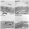
Figure 1
Figure 1
Genetic Cav1 deletion disrupts the formation of caveolae invagination in the aortic wall. Representative transition electron microscopy images of 9-month-old aortic sections in CTRL (A), MFS (B), Cav1KO (C), and MFS/Cav1KO (D) mice visualize the structure of caveolae invaginations in the aortic wall and shows that deletion of Cav1 gene impacts the structural integrity and membrane distribution of caveolae.
Figure 2
Figure 2
Cav1 deletion does not affect the progression of aortic root growth in MFS mice. Presented bar graphs showcase measurements of aortic root diameter using the myograph chambers. Aortic root diameters are increased in both male and female MFS mice compared to age- and sex-matched healthy CTRL mice. Cav1 gene deletion has no impact on artic root growth in both male and female MFS mice aorta. (Means ± SE, N = 6–9 mic/group, Two-Way ANOVA followed by Tukey’s pairwise comparison, p ≤ 0.05).
Figure 3
Figure 3
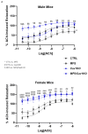
Cav1 deletion increases endothelium-dependent vasorelaxation in MFS mice. Measurements of acetylcholine (Ach)-induced relaxation in aortic rings isolated from different experimental groups using isometric small chamber myography. (A) Dose–response curves (50 pM–1 µM) for acetylcholine (Ach)-induced relaxation in aortic rings isolated from 9-month-old male and female CTRL, MFS, CTRL/Cav1KO, and MFS/Cav1KO mice. Aortic rings were pre-contracted with sub-max of PE prior to cumulative application of Ach. Relaxation responses were compared to values for CTRL aortic rings that were arbitrarily set as 100% of relaxation. All values were normalized to peak values for PE-induced contraction for each aortic ring (* CTRL vs. MFS, # CTRL vs. CTRL/Cav1KO, and $ MFS vs. MFS/Cav1KO, * p < 0.05, ** p < 0.001, $ p < 0.05, $$ p < 0.001, $$$ p < 0.0001, $$$$ p < 0.00001, ## p < 0.001, ### p < 0.0001, #### p < 0.00001). (B) The Emax values for Ach were derived from the dose–response curves. (N = 9–12 mice/group). Aortic relaxation in response to Ach is significantly decreased in aortic rings isolated from male MFS mice compared to age- and sex-matched healthy CTRL aorta. Female MFS aortic rings show a higher peak relaxation compared to age-matched male MFS mice. Cav1 gene deletion leads to an increase in maximum Ach-induced aortic vasorelaxation (Emax) in male and female MFS/Cav1KO mice compared to age- and sex-matched MFS groups, indicating significant increases in aortic wall NO production in the absence of Cav1 protein. (C) EC50 values for Ach are markedly increased in male MFS mice compared to sex- and age-matched CTRL, but no difference is observed in age-matched female CTRL and MFS aorta. Deletion of Cav1 results in a significant drop in Ach EC50 values in age-matched male MFS/Cav1KO aorta compared to MFS mice, indicating an increased sensitivity of aortic endothelial layer to Ach treatment in male mice aortic rings. However, in female mice, Cav1 deletion does not impact aortic endothelial cell sensitivity to Ach. (D) Deletion of Cav1 increased Ach-induced vasorelaxation in female CTRL mice aorta, with no changes observed in male CTRL subjects. In this bar graph, the average values for Ach-induced vasorelaxation in male CTRL aorta are arbitrarily set as 100% of relaxation. (Means ± SE, N = 9–12 mic/group, Two-Way ANOVA followed by Tukey’s pairwise comparison, p ≤ 0.05).
Figure 3
Figure 3
Cav1 deletion increases endothelium-dependent vasorelaxation in MFS mice. Measurements of acetylcholine (Ach)-induced relaxation in aortic rings isolated from different experimental groups using isometric small chamber myography. (A) Dose–response curves (50 pM–1 µM) for acetylcholine (Ach)-induced relaxation in aortic rings isolated from 9-month-old male and female CTRL, MFS, CTRL/Cav1KO, and MFS/Cav1KO mice. Aortic rings were pre-contracted with sub-max of PE prior to cumulative application of Ach. Relaxation responses were compared to values for CTRL aortic rings that were arbitrarily set as 100% of relaxation. All values were normalized to peak values for PE-induced contraction for each aortic ring (* CTRL vs. MFS, # CTRL vs. CTRL/Cav1KO, and $ MFS vs. MFS/Cav1KO, * p < 0.05, ** p < 0.001, $ p < 0.05, $$ p < 0.001, $$$ p < 0.0001, $$$$ p < 0.00001, ## p < 0.001, ### p < 0.0001, #### p < 0.00001). (B) The Emax values for Ach were derived from the dose–response curves. (N = 9–12 mice/group). Aortic relaxation in response to Ach is significantly decreased in aortic rings isolated from male MFS mice compared to age- and sex-matched healthy CTRL aorta. Female MFS aortic rings show a higher peak relaxation compared to age-matched male MFS mice. Cav1 gene deletion leads to an increase in maximum Ach-induced aortic vasorelaxation (Emax) in male and female MFS/Cav1KO mice compared to age- and sex-matched MFS groups, indicating significant increases in aortic wall NO production in the absence of Cav1 protein. (C) EC50 values for Ach are markedly increased in male MFS mice compared to sex- and age-matched CTRL, but no difference is observed in age-matched female CTRL and MFS aorta. Deletion of Cav1 results in a significant drop in Ach EC50 values in age-matched male MFS/Cav1KO aorta compared to MFS mice, indicating an increased sensitivity of aortic endothelial layer to Ach treatment in male mice aortic rings. However, in female mice, Cav1 deletion does not impact aortic endothelial cell sensitivity to Ach. (D) Deletion of Cav1 increased Ach-induced vasorelaxation in female CTRL mice aorta, with no changes observed in male CTRL subjects. In this bar graph, the average values for Ach-induced vasorelaxation in male CTRL aorta are arbitrarily set as 100% of relaxation. (Means ± SE, N = 9–12 mic/group, Two-Way ANOVA followed by Tukey’s pairwise comparison, p ≤ 0.05).
Figure 4
Figure 4
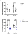
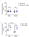
Cav1 deletion reduces KCl-induced aortic contraction in MFS mice. Bar graphs present maximum force generation in isolated aortic rings in response to KCl (60 mM) in the myograph chambers. (A) KCl-induced aortic contraction is reduced in female MFS mice compared to age- and sex-matched controls. No genotype-associated differences are observed in age-matched male CTRL and MFS. The maximum force generated in response to KCl in male CTRL aortic rings was arbitrarily set as 100% of generated force in aortic rings. (B) Cav1 deletion reduces KCl-induced smooth muscle contraction in male and female MFS/Cav1KO mice compared to age- and sex-matched MFS aorta. (Means ± SE, N = 9–12 mic/group, Two-Way ANOVA followed by Tukey’s pairwise comparison, p ≤ 0.05).
Figure 5
Figure 5
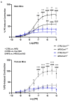
Cav1 deletion decreases phenylephrine-induced aortic contraction in CTRL and MFS mice. Measurements of mouse aortic ring contractions in response to sub-max concentration (1 µM) of phenylephrine (PE) in the myograph chambers. (A) Dose–response curves (1 nM–50 µM) for PE-induced contraction in aortic rings isolated from 9-month-old male and female CTRL, MFS, CTRL/Cav1KO, and MFS/Cav1KO mice. All values were normalized to peak values for KCl-induced contraction for each aortic ring. Recorded contraction responses were compared to values for CTRL aortic rings that were arbitrarily set as 100% of generated contractile force. (* CTRL vs. MFS, # CTRL vs. CTRL/Cav1KO, and $ MFS vs. MFS/Cav1KO, * p < 0.05, ** p < 0.001, *** p < 0.0001, **** p < 0.00001, $$$ p < 0.0001, $$$$ p < 0.00001, ### p < 0.0001, #### p < 0.00001).). (B) In both male and female MFS mice, aortic ring contractions in response to PE are significantly lower compared to healthy CTRL aorta. When we compared age-matched male and female MFS aorta, we observed a significant reduction in female MFS aortic contraction compared to male MFS mice. (C) Cav1 deletion reduces PE-induced aortic contraction in male MFS mice, but not in females, indicating a sex-specific effect of Cav1 deletion on aortic wall contraction. However, it is important to notice that Cav1 deletion reduces PE-induced contraction in male MFS aorta to levels that are similar to the values observed in female MFS/Cav1KO mice. (Means ± SE, N = 7–10 mic/group, Two-Way ANOVA followed by Tukey’s pairwise comparison, p ≤ 0.05).
Figure 5
Figure 5
Cav1 deletion decreases phenylephrine-induced aortic contraction in CTRL and MFS mice. Measurements of mouse aortic ring contractions in response to sub-max concentration (1 µM) of phenylephrine (PE) in the myograph chambers. (A) Dose–response curves (1 nM–50 µM) for PE-induced contraction in aortic rings isolated from 9-month-old male and female CTRL, MFS, CTRL/Cav1KO, and MFS/Cav1KO mice. All values were normalized to peak values for KCl-induced contraction for each aortic ring. Recorded contraction responses were compared to values for CTRL aortic rings that were arbitrarily set as 100% of generated contractile force. (* CTRL vs. MFS, # CTRL vs. CTRL/Cav1KO, and $ MFS vs. MFS/Cav1KO, * p < 0.05, ** p < 0.001, *** p < 0.0001, **** p < 0.00001, $$$ p < 0.0001, $$$$ p < 0.00001, ### p < 0.0001, #### p < 0.00001).). (B) In both male and female MFS mice, aortic ring contractions in response to PE are significantly lower compared to healthy CTRL aorta. When we compared age-matched male and female MFS aorta, we observed a significant reduction in female MFS aortic contraction compared to male MFS mice. (C) Cav1 deletion reduces PE-induced aortic contraction in male MFS mice, but not in females, indicating a sex-specific effect of Cav1 deletion on aortic wall contraction. However, it is important to notice that Cav1 deletion reduces PE-induced contraction in male MFS aorta to levels that are similar to the values observed in female MFS/Cav1KO mice. (Means ± SE, N = 7–10 mic/group, Two-Way ANOVA followed by Tukey’s pairwise comparison, p ≤ 0.05).
Figure 6
Figure 6
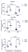
Cav1 deletion decreases mouse aortic contraction by increasing NO production. Bar graphs present maximum (Emax) aortic contraction in response to sub-maximum concentration (1 µM) of phenylephrine (PE) in CTRL, MFS, and MFS/Cav1KO mice in the absence or presence of L-NAME (200 µM), which is a general inhibitor of NO synthesis. (A) Pre-treatment of aortic rings with L-NAME increases aortic root contraction in age-matched male CTRL and MFS aorta. (B) Pre-treatment of aortic rings with L-NAME also increases aortic root contraction in age-matched female CTRL and MFS aorta. (C) L-NAME increases PE-induced aortic contraction in male and female MFS/Cav1KO mice. (Means ± SE, N = 6–9 mic/group, Two-Way ANOVA followed by Tukey’s pairwise comparison, p ≤ 0.05).
Figure 7
Figure 7
Cav1 deletion-mediated increases in aortic NO production are not through iNOS. Presented bar graphs showcase the impact of 1400 W, a potent inhibitor of inducible NOS (iNOS) on maximum (Emax) PE-induced contraction in aortic rings isolated from male and female MFS/Cav1KO mice. (A) Pre-treatment of MFS/Cav1KO mice aortic rings with the iNOS inhibitor 1400 W (1 µM) has no effects on PE-induced aortic contraction in age-matched male and female MFS/Cav1KO mice, indicating that the excessive increase in aortic endogenous NO production in the absence of Cav1 is not mediated by iNOS activation. (B) Pre-treatment of CTRL and MFS mice aortic rings with the iNOS inhibitor 1400 W (1 µM) significantly increased PE-induced vasoconstriction in both male and female MFS mice aorta, indicating that iNOS-mediated NO response elevated in male and female MFS aorta. (Means ± SE, N = 6–8 mic/group, Two-Way ANOVA followed by Tukey’s pairwise comparison, p ≤ 0.05).
Figure 8
Figure 8
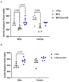
Cav1 deletion markedly improves aortic wall strength in male MFS/Cav1KO mice. Scatter plot graphs show the force generated (mN) at the rupture point. The rupture point of these aortic segments represents the maximum force generated by each segment at the point of maximum stretch, just before the aortic wall ruptures. (A) As expected, aortic wall rupture point is significantly reduced in both male and female MFS groups compared to age- and sex-matched healthy CTRL mice. Deletion of Cav1 gene increases the aortic ring rupture points in male CTRL mice, with no effects observed in female CTRL subjects. (B) Similarly, Cav1 deletion further increases aortic wall strength (rupture point) only in male MFS mice, indicating a sex-dependent effect. (Means ± SE, N = 4 mic/group, Two-Way ANOVA followed by Tukey’s pairwise comparison, p ≤ 0.05).
Figure 9
Figure 9
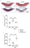
Cav1 deletion improves aortic wall elastin structure in male MFS/Cav1KO mice. Data presents measurements of aortic wall elastin fragment counts and length within the aortic wall sections isolated from CTRL, MFS, and MFS/Cav1KO mice. (A) Representative images showing Van Gieson staining of 10 µm aortic sections isolated from male CTRL, MFS, CTRL/Cav1KO, and MFS/Cav1KO mice, showing elastin in dark purple in the medial layer of mouse aortic wall (scale bar = 50 µm). (B) Quantification of elastin fiber counts shows a significant decrease in fragment counts in male MFS/Cav1KO aorta compared to MFS mice. (C) Similarly, Cav1 deletion reduces elastin fiber fragment length in MFS mice, indicating a marked improvement in elastin structure within the aortic wall of MFS/Cav1KO mice. (Means ± SE, N = 4 mic/group, Two-Way ANOVA followed by Tukey’s pairwise comparison, p ≤ 0.05.
Figure 10
Figure 10
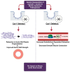
Potential impact of Cav1 ablation on MFS aeropathy. In MFS mice, Cav1 deletion results in improvement in aortic wall elastin structure and aortic wall strength. The deletion of Cav1 also increases endothelium-mediated vasorelaxation in the aorta, while decreasing smooth muscle contraction in response to vasoconstricting agent phenylephrine. The overall conclusion is that Cav1 deletion provides some levels of beneficial impacts on aortic structure and function in MFS mice. The image was generated by Biorender (Toronto, ON, Canada).

References

    1. Milewicz D.M., Braverman A.C., De Backer J., Morris S.A., Boileau C., Maumenee I.H., Jondeau G., Evangelista A., Pyeritz R.E. Marfan syndrome. Nat. Rev. Dis. Primers. 2021;7:64. doi: 10.1038/s41572-021-00298-7. - DOI - PMC - PubMed
    1. Spencer M. Marfan syndrome. Nursing. 2024;54:19–25. doi: 10.1097/01.NURSE.0001007604.09204.9a. - DOI - PubMed
    1. Adamo C.S., Zuk A.V., Sengle G. The fibrillin microfibril/elastic fibre network: A critical extracellular supramolecular scaffold to balance skin homoeostasis. Exp. Dermatol. 2021;30:25–37. doi: 10.1111/exd.14191. - DOI - PubMed
    1. Li L., Huang J., Liu Y. The extracellular matrix glycoprotein fibrillin-1 in health and disease. Front. Cell Dev. Biol. 2023;11:1302285. doi: 10.3389/fcell.2023.1302285. - DOI - PMC - PubMed
    1. Benke K., Ágg B., Szilveszter B., Tarr F., Nagy Z.B., Pólos M., Daróczi L., Merkely B., Szabolcs Z. The role of transforming growth factor-beta in Marfan syndrome. Cardiol. J. 2013;20:227–234. doi: 10.5603/CJ.2013.0066. - DOI - PubMed

MeSH terms

Grants and funding

LinkOut - more resources