. 2024 Nov 26;25(23):12718.
doi: 10.3390/ijms252312718.

Ascorbic Acid Ameliorates Molecular and Developmental Defects in Human-Induced Pluripotent Stem Cell and Cerebral Organoid Models of Fragile X Syndrome

Affiliations

Ascorbic Acid Ameliorates Molecular and Developmental Defects in Human-Induced Pluripotent Stem Cell and Cerebral Organoid Models of Fragile X Syndrome

Keith M Gunapala et al. Int J Mol Sci. .

Abstract

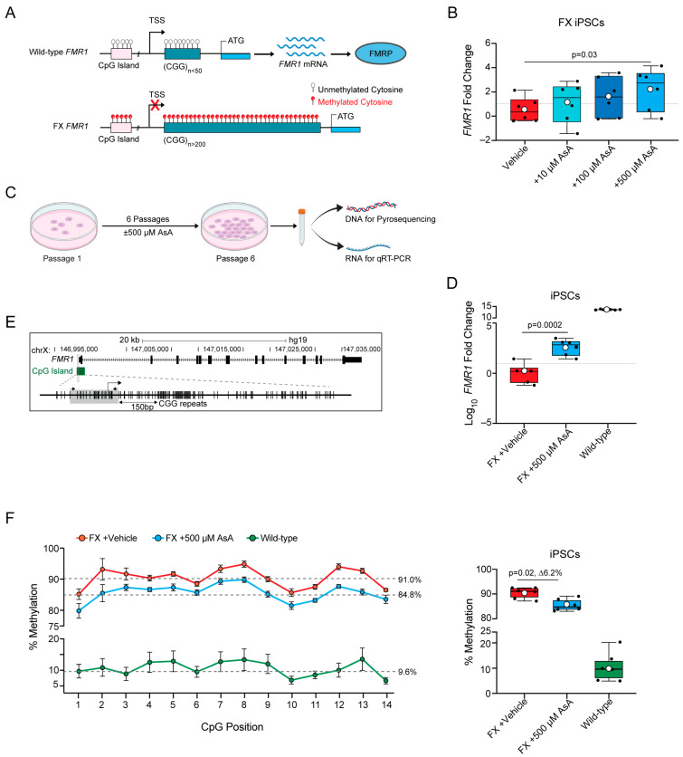
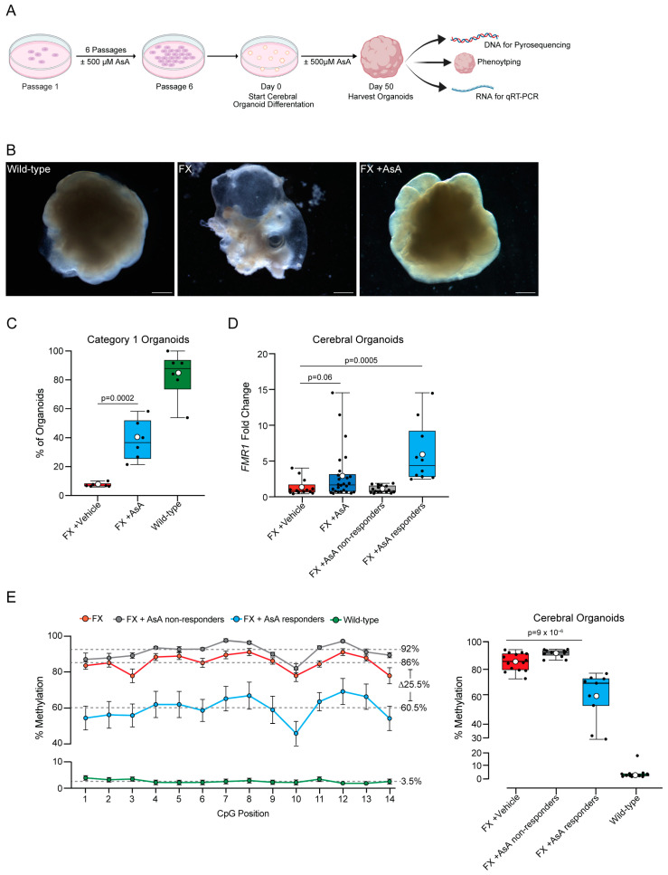
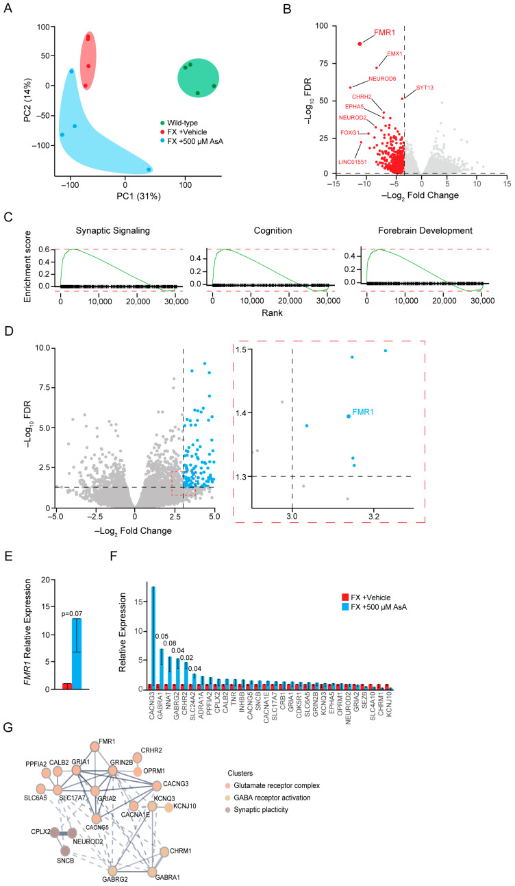
Fragile X Syndrome (FX) is the most common form of inherited cognitive impairment and falls under the broader category of Autism Spectrum Disorders (ASD). FX is caused by a CGG trinucleotide repeat expansion in the non-coding region of the X-linked Fragile X Messenger Ribonucleoprotein 1 (FMR1) gene, leading to its hypermethylation and epigenetic silencing. Animal models of FX rely on the deletion of the Fmr1 gene, which fails to replicate the epigenetic silencing mechanism of the FMR1 gene observed in human patients. Human stem cells carrying FX repeat expansions have provided a better understanding of the basis of epigenetic silencing of FMR1. Previous studies have found that 5-Azacytidine (5Azac) can reverse this methylation; however, 5Azac can be toxic, which may limit its therapeutic potential. Here, we show that the dietary factor Ascorbic Acid (AsA) can reduce DNA methylation in the FMR1 locus and lead to an increase in FMR1 gene expression in FX iPSCs and cerebral organoids. In addition, AsA treatment rescued neuronal gene expression and morphological defects observed in FX iPSC-derived cerebral organoids. Hence, we demonstrate that the dietary co-factor AsA can partially revert the molecular and morphological defects seen in human FX models in vitro. Our findings have implications for the development of novel therapies for FX in the future.

Keywords: Ascorbic Acid; Autism Spectrum Disorders (ASD); FMR1; Fragile X Syndrome; cerebral organoids; gene silencing; induced pluripotent stem cells; methylation; neurodevelopmental disorders.


Conflict of interest statement

The authors declare no conflicts of interest.

Figures

Figure 1
Figure 1
Ascorbic Acid restores FMR1 expression by reversing hypermethylation in FX iPSCs. (A) Schematic representation the gene structure of human FMR1 in wild-type and FX. In wild-type FMR1 (top section of the panel), there are less than 50 CGG repeats and no methylation of the CGG repeats and CpGs upstream; therefore, the gene is transcriptionally active, producing FMR1 mRNA and the FMRP protein product. In FX FMR1 (lower section of the panel), there are >200 CGG repeats that are hypermethylated; hypermethylation spreads upstream to the CpGs, and therefore, the gene is transcriptionally inactive. Unmethylated Cytosine is represented by transparent circular marks over the CpG and CGG sequence; methylated Cytosine is represented by red circular marks over the CpG and CGG sequence. (B) Dose−response curve for FX iPSCs treated for 1 passage (6 days) with Ascorbic Acid (AsA). FX vehicle control with 15 mM HEPES (red), 10 μM AsA (n = 6, turquoise), 100 μM AsA (n = 6, navy blue), or 500 μM AsA (n = 6, blue). Values plotted are fold changes of FMR1 expression normalized to the FX vehicle control. (C) Schematic representation the timeline and experimental setup of long-term exposure to AsA. Low-density iPSCs are plated and kept in culture for 6 days and passaged, repeating until the sixth passage when cells are then collected for either DNA extraction (to perform pyrosequencing) or RNA extraction (to perform qRT-PCR). (D) FMR1 gene expression fold change (in log10) from FX vehicle control (n = 6, red) and FX + 500 μM AsA (n = 6, blue), and wild-type (n = 6, green). (E) Schematic of the FMR1 gene structure with upstream CGG repeats and CpG island (in green) 150 base pairs upstream of the CGG. CpG island in green is used as a proxy to quantify methylation of the FMR1 locus. The gray box highlights the region of the CpG that was sequenced to quantify methylation levels by pyrosequencing. The arrows (black) show the locations of the forward and reverse primers. (F) Left panel: average methylation levels per CpG site across all samples of iPSCs. FX vehicle-control (n = 6, red); FX + 500 μM AsA (n = 6, blue) and wild-type (n = 6, green). Data are presented for 14 CpG sites, with error bars indicating the standard error of the mean (±SEM). Gray dashed lines represent the median percentage methylation across all CpGs for each group. Right panel, boxplot showing the percentage methylation level for each individual sample: FX vehicle control (n = 6, red), FX + 500 μM AsA (n = 6, blue), and wild-type (n = 6, green). All boxplots display the interquartile range (IQR) and median, with the mean indicated by white circles. P-values were calculated using the Wilcox rank-sum exact test.
Figure 2
Figure 2
Ascorbic Acid reactivates FMR1 and reduces methylation in FX cerebral organoids. (A) Schematic representation of the timeline and experimental setup for long-term exposure to AsA. Low-density iPSCs were plated and kept in culture for 6 days, passaged, and repeated until the sixth passage, where cells were then used to start the cerebral organoid differentiation protocol. Cerebral organoids are cultured until day 50, and they are collected for either DNA extraction (to perform pyrosequencing) or RNA extraction (to perform qRT-PCR) or imaged for phenotype assessment. (B) Representative images of wild-type (left panel), FX vehicle control (center panel), and FX + AsA (right panel) cerebral organoids. Scale bar for wild-type: 2200 μm, 3000 μm for FX, and 3000 μm for FX + AsA. (C) Percent of cerebral organoids per plate that had morphologically organized neuronal tissue by visual scoring of phenotype; FX vehicle control (n = 6, red), FX + 500 μM AsA (n = 6, blue), and wild-type (n = 6, green). n = 6 plates per genotype and condition; each plate contained 10–20 cerebral organoids. (D) FMR1 gene expression fold change between FX vehicle control cerebral organoids (n = 13, red), all FX + 500 μM AsA-treated cerebral organoids (n = 26, navy blue), and FX + 500 μM AsA-treated cerebral organoids stratified by the 95% CI of FX vehicle controls into FX + AsA non-responders (n = 16, gray) and FX + AsA responders (n = 10, blue). (E) Left panel: average percentage of methylation levels at each CpG site across all samples for FX (n = 14, red), non-responders (n = 11, gray), responders (n = 9, blue), and wild-type (n = 14, green). Data are presented for 14 CpG sites, with error bars indicating the standard error of the mean (± SEM). Gray dashed lines represent the median percentage methylation across all CpGs for each group. On the right panel is a boxplot showing the percentage methylation level for each sample. Individual samples are represented by black circles. The boxplot displays the interquartile range (IQR) and median, with the mean indicated by white circles. P-values were calculated using the Wilcoxon rank-sum exact test.
Figure 3
Figure 3
Ascorbic Acid treatment causes changes in gene expression related to neurodevelopment. (A) Principal Component Analysis (PCA) of the two most significant principal components (PC1 and PC2) identified by comparing FX vehicle cerebral organoids (n = 4, red), FX + 500 μM AsA cerebral organoids (n = 4, blue), and wild-type cerebral organoids (n = 4, green). (B) Volcano plot showing genes that are differentially expressed in FX vehicle cerebral organoids compared to wild-type cerebral organoids. Red indicates genes significantly downregulated in FX cerebral organoids compared with wild-type cerebral organoids. (C) Gene enrichment analysis showing families of genes downregulated in FX cerebral organoids compared to wild-type cerebral organoids. (D) Volcano plot showing genes that are differentially expressed in FX + 500 μM AsA cerebral organoids compared to FX vehicle cerebral organoids). Blue dots indicate genes with significant upregulation due to AsA treatment. (E) Relative expression levels of FMR1 in FX (red) and FX + 500 μM AsA (blue) cerebral organoids. (F) Change in the expression levels of genes found to be most downregulated in FX cerebral organoids compared to wild-type cerebral organoids. Comparison between FX cerebral organoids (red) and FX + 500 μM AsA cerebral organoids (blue). (G) String analysis of the most upregulated genes in FX + 500 μM AsA cerebral organoids showing gene families involved in neuronal development, synapse formation, and maturation. All statistical analyses were performed using unpaired one-tail t-tests between the FX vehicle control (red) and 500 μM AsA treatment (blue). Error bars are plotted as the ± standard error of the mean.

Similar articles

Cited by

References

    1. Crawford D.C., Acuna J.M., Sherman S.L. FMR1 and the fragile X syndrome: Human genome epidemiology review. Genet. Med. 2001;3:359–371. doi: 10.1097/00125817-200109000-00006. - DOI - PMC - PubMed
    1. Bhakar A.L., Dolen G., Bear M.F. The pathophysiology of fragile X (and what it teaches us about synapses) Annu. Rev. Neurosci. 2012;35:417–443. doi: 10.1146/annurev-neuro-060909-153138. - DOI - PMC - PubMed
    1. Davidson M., Sebastian S.A., Benitez Y., Desai S., Quinonez J., Ruxmohan S., Stein J.D., Cueva W. Behav-ioral Problems in Fragile X Syndrome: A Review of Clinical Management. Cureus. 2022;14:e21840. - PMC - PubMed
    1. Verkerk A.J., Pieretti M., Sutcliffe J.S., Fu Y.H., Kuhl D.P., Pizzuti A., Reiner O., Richards S., Victoria M.F., Zhang F.P., et al. Identification of a gene (FMR-1) containing a CGG repeat coincident with a breakpoint cluster region exhibiting length variation in fragile X syndrome. Cell. 1991;65:905–914. doi: 10.1016/0092-8674(91)90397-H. - DOI - PubMed
    1. Pieretti M., Zhang F.P., Fu Y.H., Warren S.T., Oostra B.A., Caskey C.T., Nelson D.L. Absence of expression of the FMR-1 gene in fragile X syndrome. Cell. 1991;66:817–822. doi: 10.1016/0092-8674(91)90125-I. - DOI - PubMed

MeSH terms

Substances

Grants and funding

LinkOut - more resources