. 2024 Nov 27;25(23):12745.
doi: 10.3390/ijms252312745.

Molecular Characteristics of Circ_002156 and Its Effects on Proliferation and Differentiation of Caprine Skeletal Muscle Satellite Cells

Affiliations

Molecular Characteristics of Circ_002156 and Its Effects on Proliferation and Differentiation of Caprine Skeletal Muscle Satellite Cells

Yuanhua Gu et al. Int J Mol Sci. .

Abstract

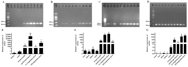
The proliferation and differentiation of skeletal muscle satellite cells (SMSCs) are responsible for the development of skeletal muscle. In our previous study, circ_002156 was found to be highly expressed in caprine Longissimus Dorsi muscle, but the regulatory role of the circular RNAs (circRNA) in goat SMSCs remains unclear. In this study, the authenticity of circ_002156 was validated, and its structurally characteristic and cellular localization as well as tissue expression of circ_002156 and its parent genes were investigated. Moreover, the effects of circ_002156 on the viability, proliferation, and differentiation of SMSCs were also studied. The circ_002156 is located on caprine chromosome 19 with a length of 36,478. The circRNA structurally originates from myosin heavy chain 2 (MYH2), MYH1, and MYH4 as well as intergenic sequences among the parent genes. RT-PCR and Sanger sequencing confirmed the authenticity of circ_002156. Most circ_002156 (55.5%) was expressed in the nuclei of SMSCs, while 44.5% of circ_002156 was located in the cytoplasm. The circ_002156 and its three parent genes had higher expression levels in the triceps brachii, quadriceps femoris, and longissimus dorsi muscle tissues than in the other five tissues. The expression of circ_002156 and its parent genes MYH1 and MYH4 reached the maximum on day 8 of differentiation, while MYH2 in expression reached the peak on day 4 after differentiation. The Pearson correlation coefficients revealed that circ_002156 had moderate or high positive correlations with the three parent genes in the expression of both quadriceps femoris muscle and SMSCs during different differentiation stages. The small interfering RNA circ_002156 (named si-circ_002156) remarkably increased the viability of the SMSCs. The si-circ_002156 also increased the number and parentage of Edu-labeled positive SMSCs as well as the expression levels of four cell proliferation marker genes. These suggest that circ_002156 inhibited the proliferation of SMSCs. Meanwhile, si-circ_002156 decreased the area of MyHC-labeled positive myotubes, the myotube fusion index, and myotube size as well as the expression of its three parent genes and four cell differentiation marker genes, suggesting a positive effect of circ_002156 on the differentiation of SMSCs. This study contributes to a better understanding of the roles of circ_002156 in the proliferation and differentiation of SMSCs.

Keywords: circ_002156; differentiation; goat; proliferation; skeletal muscle satellite cells.


Conflict of interest statement

We certify that there are no conflicts of interest with any financial organization regarding the material discussed in the manuscript.

Figures

Figure 1
Figure 1
Structure, identification, and cellular localization of circ_002156. (A) Structure diagram of circ_002156 derived from the parent genes myosin heavy chain 1(MYH1), MYH2, and MYH4. (B,C) The authenticity validation of circ_002156 using the RT-PCR assay and Sanger sequencing. (D) The cellular localization of circ_002156 in the goat skeletal muscle satellite cells (SMSCs) with GAPDH and U6 being the reference genes. M—marker. The head-to-tail splice junction site of circ_002156 was labeled using a red arrow.
Figure 2
Figure 2
The expression of circ_002156 (A,B) and GAPDH (C) in different tissues of goats detected using RT-PCR and RT-qPCR, respectively. M—marker; 1—heart; 2—liver; 3—spleen; 4—lungs; 5—kidneys; 6—testis; 7—triceps brachii; 8—quadriceps femoris; 9—longissimus dorsi of Liaoning cashmere goat (LC); 10—longissimus dorsi of Ziwuling black goat (ZB). Since circ_002156 was not expressed in the lung, it was not included in Figure 2. The different lowercase letters indicate significant differences (p < 0.05).
Figure 3
Figure 3
The expression of the parent genes MYH1 (A,E), MYH2 (B,F), MYH4 (C,G), and GAPDH (D) in different caprine tissues detected using RT-PCR and RT-qPCR, respectively. M—marker; 1—heart; 2—liver; 3—spleen; 4—lungs; 5—kidneys; 6—testis; 7: triceps brachii; 8—quadriceps femoris; 9—longissimus dorsi of Liaoning cashmere goat (LC); 10—longissimus dorsi of Ziwuling black goat (ZB). The different lowercase letters indicate significant differences (p < 0.05).
Figure 4
Figure 4
Pearson correlation analysis in the expression of quadriceps femoris between circ_002156 and the parent genes MYH1 (A), MYH2 (B), and MYH4 (C). Each circle represents the expression of the parent gene in different ZiwuLing black goats.
Figure 5
Figure 5
The expression levels of circ_002156 (A) and its parent genes MYH1 (B), MYH2 (C), and MYH4 (D) on days 0, 2, 4, 6, and 8 after skeletal muscle satellite cell (SMSC) differentiation. Different lowercase letters represent significant differences (p < 0.05).
Figure 6
Figure 6
Pearson correlation analysis between circ_002156 and its parent genes MYH1 (A), MYH2 (B), and MYH4 (C) in expression level at different differentiation stages of caprine skeletal muscle satellite cells (SMSCs). Each circle represents the expression of the parent gene in different ZiwuLing black goats.
Figure 7
Figure 7
Effect of si-circ_002156 on the viability of the caprine skeletal muscle satellite cells (SMSCs). (A) Schematic diagram of the si-circ_002156 sequence completely complementarily binding with the junction site sequences of circ_002156. (B) The expression level of circ_002156 detected in the SMSCs transfected with si-circ_002156. (C). Effect of si-circ_002156 on the viability of SMSCs investigated using the CCK-8 assay. * p < 0.05 and ** p < 0.01.
Figure 8
Figure 8
Effect of si-circ_002156 on the proliferation of skeletal muscle satellite cells (SMSCs). (A) The proliferation of SMSCs detected using an Edu assay. (B) The ratio of Edu-labeled positive SMSCs. (C) The expression levels of cell proliferation marker genes—cyclin dependent kinase 2 (CDK2), proliferating cell nuclear antigen (PCNA), CDK4, CyclinE, and p53 detected in SMSCs transfected with si-circ_002156. * p < 0.05 and ** p < 0.01.
Figure 9
Figure 9
Effect of circ_002156 on the differentiation of goat skeletal muscle satellite cells (SMSCs). (A) Transfection efficiency of si-circ_002156 detected. (B) The relative expression levels of its three parent genes and (C) four cell differentiation marker genes—myosin heavy chain (MyHC), myogenic differentiation (MyoD), myogenin (MyoG), and myocyte enhancer factor 2C (MEF2C) in SMSCs transfected with si-circ_002156. * p < 0.05 and ** p < 0.01.
Figure 10
Figure 10
Effect of circ_002156 on myotubes. (A) The immunofluorescence results of SMSCs when si-circ_002156 was transfected. (B) The area of MyHC-labeled positive myotubes. (C) Circ_002156 positively modulated the number of nuclei in the myotubes. ** p < 0.01.

Similar articles

References

    1. Sanger H.L., Klotz G., Riesner D., Gross H.J., Kleinschmidt A.K. Viroids are single-stranded covalently closed circular RNA molecules existing as highly base-paired rod-like structures. Proc. Natl. Acad. Sci USA. 1976;73:3852–3856. doi: 10.1073/pnas.73.11.3852. - DOI - PMC - PubMed
    1. Ebbesen K.K., Kjems J., Hansen T.B. Circular RNAs: Identification, biogenesis and function. Biochim. Biophys. Acta. 2016;1859:163–168. doi: 10.1016/j.bbagrm.2015.07.007. - DOI - PubMed
    1. Jeck W.R., Sharpless N.E. Detecting and characterizing circular RNAs. Nat. Biotechnol. 2014;32:453–461. doi: 10.1038/nbt.2890. - DOI - PMC - PubMed
    1. Kristensen L.S., Andersen M.S., Stagsted L.V.W., Ebbesen K.K., Hansen T.B., Kjems J. The biogenesis, biology and characterization of circular RNAs. Nat. Rev. Genet. 2019;20:675–691. doi: 10.1038/s41576-019-0158-7. - DOI - PubMed
    1. Li Z., Huang C., Bao C., Chen L., Lin M., Wang X., Zhong G., Yu B., Hu W., Dai L., et al. Exon-intron circular RNAs regulate transcription in the nucleus. Nat. Struct. Mol. Biol. 2015;22:256–264. doi: 10.1038/nsmb.2959. - DOI - PubMed

MeSH terms

LinkOut - more resources