. 2024 Nov 28;25(23):12789.
doi: 10.3390/ijms252312789.

The Neuroprotective Effect of the X Protein of Orthobornavirus Bornaense Type 1 in Amyotrophic Lateral Sclerosis

Affiliations

The Neuroprotective Effect of the X Protein of Orthobornavirus Bornaense Type 1 in Amyotrophic Lateral Sclerosis

Jeflie Tournezy et al. Int J Mol Sci. .

Abstract

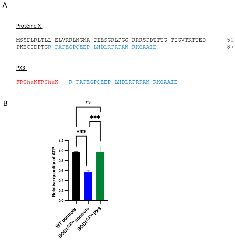
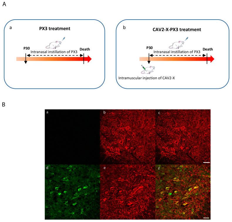
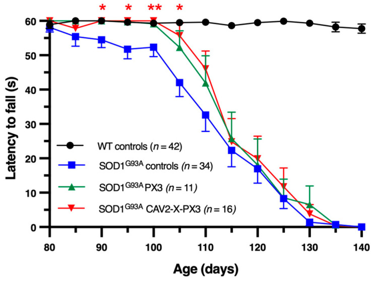
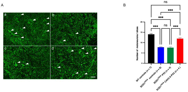
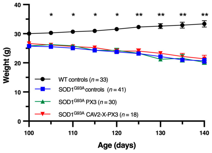
In amyotrophic lateral sclerosis (ALS), early mitochondrial dysfunction may contribute to progressive motor neuron loss. Remarkably, the ectopic expression of the Orthobornavirus bornaense type 1 (BoDV-1) X protein in mitochondria blocks apoptosis and protects neurons from degeneration. Therefore, this study examines the neuroprotective effects of X protein in an ALS mouse model. We first tested in vitro the effect of the X-derived peptide (PX3) on motoneurons primary cultures of SOD1G93A mice. The total intracellular adenosine triphosphate (ATP) content was measured after incubation of the peptide. We next tested in vivo the intramuscular injection of X protein using a canine viral vector (CAV2-X) and PX3 intranasal administrations in SOD1G93A mice. Disease onset and progression were assessed through rotarod performance, functional motor unit analysis via electrophysiology, and motor neuron survival by immunohistochemistry. The results showed that in vitro PX3 restored the ATP level in SOD1G93A motor neurons. In vivo, treated mice demonstrated better motor performance, preserved motor units, and higher motor neuron survival. Although life expectancy was not extended in this severe mouse model of motor neuron degeneration, the present findings clearly demonstrate the neuroprotective potential of X protein in a model of ALS. We are convinced that further studies may improve the therapeutic impact of X protein with optimized administration methods.

Keywords: ALS; BoDV-1; SOD1G93A mice; X protein; mitochondria.


Conflict of interest statement

The authors declare no conflicts of interest.

Figures

Figure 1
Figure 1
X-derived peptide PX3 restores ATP levels in primary cultures of SOD1G93A motor neurons. (A) Amino acid sequences of X protein and PX3 peptide. The X protein consists of 87 amino acids. The PX3 peptide corresponds to the carboxy-terminal 29 amino acids of X protein (highlighted in blue) to which a cell-permeable sequence (MPP, highlighted in red) is added. (B) The histogram bars represent the means of the relative amounts of ATP obtained in three independent cultures for each condition (WT controls, n = 17; SOD1G93A controls, n = 11; SOD1G93A PX3, n = 9). Error bars represent SEM. The data were analyzed by a Kruskal–Wallis test followed by a Conover test. ns: not significant; *** p < 0.001.
Figure 2
Figure 2
Administration of the CAV2-X and PX3 peptides (A) and expression of the X protein in SOD1G93A mice (B). (A): Animals received intranasal instillations of the PX3 peptide alone (a) or as part of a combination therapy that included a viral vector-expressing protein X (CAV2-X) together with intranasal PX3 instillations (b). A single injection of CAV2-X was carried out bilaterally in the triceps surae (Ts) muscles at the age of 30 days in wild-type and SOD1G93A mice. Intranasal instillations of the PX3 peptide were performed three times per week from the age of 30 days until the death of the animal. (B): Spinal cord sections of control mice (ac) and mice treated with CAV2-X-PX3 (df). (a,d) Ventral horn immunolabeled by an antibody directed against the X protein (b,e) Motor neurons immunolabeled with an anti-SMI32 antibody. (c,f) Superposition of (a,b) and of (d,e). In the later image (f), expression of the X protein in motor neurons is observed. Scale bar: 50 µm.
Figure 3
Figure 3
Delayed onset of motor symptoms in SOD1G93A-treated mice. After a training phase, the mice were tested from the age of 80 days. The test involved running on the cylinder of the rotarod rotating at a speed of eight revolutions per minute for 60 s. The test was carried out every 5 days. Each point represents the mean ± SEM. At each time point, the means were compared with a Kruskal–Wallis test followed by a post hoc Conover test. Red stars represent a significant difference between SOD1G93A mice treated with the coadministration of X and PX3 and SOD1G93A control mice (* p < 0.05; ** p < 0.01).
Figure 4
Figure 4
Increased maximum CMAP amplitude in SOD1G93A mice treated with X protein and PX3 peptide in the presymptomatic and symptomatic stages. CMAPs developed by Ts muscles were recorded at 70 days (A) and 90 days (B) in response to supramaximal stimulation of the tibial nerve in WT control (black bar), SOD1G93A control (blue bar), SOD1G93A PX3 (green bar), and SOD1G93AX-PX3 (red bar) mice. The histogram bars correspond to the means ± SEMs. The data were analyzed with a nonparametric statistical test (Kruskal–Wallis) followed by a multiple comparison test (Dunn test), ns: not significant, * p < 0.05, *** p <0.001.
Figure 5
Figure 5
Increased survival of lumbar motor neurons in SOD1G93A mice receiving cotreatment with X-PX3. (A): Lumbar spinal cord sections in the L4 segment in 90-day-old animals ((a): WT control mice; (b): SOD1G93A control mice; (c): SOD1G93A PX3 mice; (d): SOD1G93A X-PX3 mice). The motor neurons were revealed by an anti-SMI32 antibody. Scale bar: 50 µm. (B): The number of motor neurons in lumbar segments L3 and L5 was determined. A total of 20 ventral horns per animal were analyzed. Each bar represents the average number of motor neurons. Error bars represent SEM. The data were analyzed with a Kruskal–Wallis test followed by a Dunn’s test (ns: not significant; *** p < 0.001).
Figure 6
Figure 6
Body mass evolution between 100 days of age and death in SOD1G93A-treated mice. Mice were weighed every 5 days from 100 days of age until death. Each point represents the mean animal weight ± SEM. The means were compared with a two-way ANOVA test followed by a Tukey post hoc test. Significant differences were observed between WT controls (black rod symbols) and all groups of SOD mice; * p < 0.05; ** p < 0.01; no difference was revealed between controls (blue square symbols) and treated SOD mice (green and red triangle symbols). The disease end stage was defined as the time when the mouse could not right itself within 10 s when placed on its side.
Figure 7
Figure 7
Life expectancy of SOD1G93A mice was not improved by treatment with PX3 peptide alone or by the coadministration of PX3 peptide and X protein. (A): Kaplan–Meier survival plot of control SOD1G93A mice (blue) and mice treated with PX3 (green) and X-PX3 (red). Curves were compared with a log rank (Mantel–Cox) test (p = 0.838). The median survival was 133 days for control mice, 136.5 days for PX3 mice, and 135.5 days for X-PX3 mice. (B): Mean age of survival in different treated groups. Each bar represents the mean ± SEM. The means were compared with one-way ANOVA (ns: not significant). The disease end stage was defined as the time when the mouse could not right itself within 10 s when placed on its side.

References

    1. Grossman M. Amyotrophic lateral sclerosis—A multisystem neurodegenerative disorder. Nat. Rev. Neurol. 2019;15:5–6. doi: 10.1038/s41582-018-0103-y. - DOI - PubMed
    1. Shaw P.J. Molecular and cellular pathways of neurodegeneration in motor neurone disease. J. Neurol. Neurosurg. Psychiatry. 2005;76:1046–1057. doi: 10.1136/jnnp.2004.048652. - DOI - PMC - PubMed
    1. Mancuso R., Navarro X. Amyotrophic lateral sclerosis: Current perspectives from basic research to the clinic. Prog. Neurobiol. 2015;133:1–26. doi: 10.1016/j.pneurobio.2015.07.004. - DOI - PubMed
    1. Rosen D.R. Mutations in Cu/Zn superoxide dismutase gene are associated with familial amyotrophic lateral sclerosis. Nature. 1993;364:362. doi: 10.1038/364362c0. - DOI - PubMed
    1. Chia R., Chio A., Traynor B.J. Novel genes associated with amyotrophic lateral sclerosis: Diagnostic and clinical implications. Lancet Neurol. 2018;17:94–102. doi: 10.1016/S1474-4422(17)30401-5. - DOI - PMC - PubMed

LinkOut - more resources