. 2024 Dec 2;25(23):12962.
doi: 10.3390/ijms252312962.

2'-Hydroxycinnamaldehyde, a Natural Product from Cinnamon, Alleviates Ischemia/Reperfusion-Induced Microvascular Dysfunction and Oxidative Damage in Rats by Upregulating Cytosolic BAG3 and Nrf2/HO-1

Affiliations

2'-Hydroxycinnamaldehyde, a Natural Product from Cinnamon, Alleviates Ischemia/Reperfusion-Induced Microvascular Dysfunction and Oxidative Damage in Rats by Upregulating Cytosolic BAG3 and Nrf2/HO-1

Yu-Hsuan Cheng et al. Int J Mol Sci. .

Abstract

2'-Hydroxycinnamaldehyde (HCA), a natural product isolated from the bark of Cinnamomum cassia, has anti-inflammatory and anti-tumor activities. In this study, we explored whether HCA preconditioning could protect the heart against ischemia/reperfusion (I/R)-induced oxidative injury through cytosolic Bcl-2-associated athanogene 3 (BAG3) upregulation. In vivo HCA preconditioning was performed intraperitoneally in adult male Wistar rats (50 mg/kg body weight) three times/week for 2 weeks before cardiac I/R injury. The animals were divided into sham control (sham), I/R, and HCA preconditioning plus I/R (HCA+I/R) groups. We examined left ventricular pressure cardiac hemodynamics, the microcirculation, electrocardiograms, infarct size, and oxidative stress and performed Western blots, immunohistochemistry, and cytokine array assays. HCA pretreatment, via BAG3 overexpression, inhibited H2O2-induced H9c2 cell death. Cardiac I/R injury increased ST-segment elevation, left ventricular end-diastolic pressure, infarct size, myocardial disruption, tissue edema, erythrocyte accumulation, leukocyte infiltration, reactive oxygen species, malondialdehyde, 8-isoprostane, caspase 3-mediated apoptosis, 4HNE/GPX4-mediated ferroptosis, and fibrosis but decreased the microcirculation, cytosolic BAG3, and Beclin-1/LC3 II-mediated autophagy in the I/R hearts. HCA preconditioning significantly decreased these oxidative injuries by increasing cardiac cytosolic BAG3 and Nrf2/HO-1 signaling. HCA preconditioning significantly decreased cardiac I/R-enhanced mitochondrial fission DRP1 expression. Our data suggest that HCA preconditioning can efficiently improve myocardial I/R injury-induced cardiac dysfunction, apoptosis, ferroptosis, mitochondrial fission, and autophagy inhibition through cardiac BAG3 and Nrf2/HO-1 upregulation.

Keywords: 2′-Hydroxycinnamaldehyde; Bcl-2-associated athanogene 3; apoptosis; autophagy; ferroptosis; myocardial ischemia/reperfusion injury; natural product.


Conflict of interest statement

All the authors declare no potential conflicts of interest.

Figures
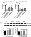
Figure 1
Figure 1
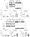
Effect of HCA pretreatment on H2O2-induced H9c2 cell death and BAG3 expression in H9c2 cells. HCA pretreatment (A) but not co-treatment (B) significantly inhibited H2O2-induced H9c2 cell death at a dose of 0.001–0.1 mg/mL. HCA pretreatment induced a dose-dependent upregulation of BAG3 expression in H9c2 cells (CE). All data are expressed as mean ± SEM (n = 3). * indicates significance vs. CON group (* p < 0.05; ** p < 0.01). # indicates significance vs. H2O2 group (# p < 0.05; ## p < 0.01).
Figure 2
Figure 2
Effect of HCA preconditioning on mitochondrial and cytosolic BAG3 expression in sham, HCA, I/R, and HCA+I/R groups. (A) Original graphs of cytosolic and mitochondrial BAG3 expression in sham and HCA hearts, determined via Western blot assay. Translocation of mitochondrial BAG3 (B) to cytosolic BAG3 (C) and translocation of mitochondrial cytochrome C (D) are noted between sham and HCA hearts. (E) Original traces of mitochondrial and cytosolic BAG3 expression. Significantly reduced cytosolic BAG3 expression is observed in I/R group vs. sham group, whereas significantly preserved cytosolic BAG3 is observed in HCA+I/R group vs. I/R group (G). Mitochondrial fractions of BAG3 expression (F) and cytochrome C expression (H) are not significantly different among sham, I/R, and HCA+I/R groups. Each symbol represents the respective individual. All data are expressed as mean ± SEM (n = 3–5). * indicates significance vs. sham group (* p < 0.05; ** p < 0.01). # indicates significance vs. I/R group (# p < 0.05).
Figure 3
Figure 3
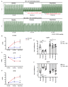
Effect of HCA preconditioning on cardiac I/R-induced changes in cardiac surface microcirculation. (A) Representative responses of cardiac microcirculation to cardiac I/R in the three groups of rats. (B) Statistical data of cardiac surface microcirculation. Significant decrease in cardiac microcirculation is observed in I/R group vs. sham group, whereas cardiac microcirculation is restored in HCA+I/R group. (C) Cardiac surface blood flow responses to baseline, 1 h of ischemia, and 4 h of reperfusion among the three groups. (D) Mean percentage change in cardiac surface blood flow during ischemia and reperfusion among the three groups. All data are expressed as mean ± SEM (n = 6). * indicates significance vs. sham group (** p < 0.01; *** p < 0.001). # indicates significance vs. I/R group (#### p < 0.0001).
Figure 4
Figure 4
Effect of HCA preconditioning on left ventricular pressure in I/R and HCA+I/R hearts. (A) Typical responses of left ventricular pressure during baseline, ischemia, and reperfusion phases in two groups. Statistical data for LVEDP (B), LVSP (C), LVDP (D), +dp/dt (E), and -dp/dt (F) between I/R and HCA+I/R groups. All data are expressed as mean ± SEM (n = 8). * indicates significance vs. sham group (* p < 0.05; ** p < 0.01; *** p < 0.001; **** p < 0.0001). # indicates significance vs. I/R group (# p < 0.05; #### p < 0.0001).
Figure 5
Figure 5
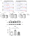
Effect of HCA preconditioning on cardiac I/R-affected EKG parameters and mitochondrial fission. (A) Representative EKG graphs in ST segment, with P-R and R-R intervals, in I/R and HCA+I/R groups. Statistical data for P-R intervals (B), R-R intervals (C), and heart rate (D). Expression of the mitochondrial fission marker DRP1 protein (E) in the three groups and statistical data (F). All data are expressed as mean ± SEM (n = 8). * indicates significance vs. sham group (** p < 0.01). # indicates significance vs. I/R group (# p < 0.05; ## p < 0.01; ### p < 0.001).
Figure 6
Figure 6
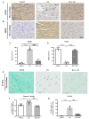
Effect of HCA preconditioning on cardiac I/R-induced pathologic changes, leukocyte infiltration, and fibrosis among sham, I/R, and HCA+I/R groups. (A) Structural alterations in response to cardiac I/R are demonstrated among the three groups via H&E staining. (B) Immunohistochemistry of CD45 staining (leukocyte biomarker) in the three groups. (C) Degree of fibrosis in heart tissue after cardiac I/R injury is indicated using Masson’s trichrome staining of the three groups. Percentage of myocyte content (D), percentage of erythrocyte extravasation (E), leukocyte infiltration (F), collagen volume fraction (G), high-sensitivity troponin-1 (H), and lactate dehydrogenase level (I) are compared among the three groups. All data are expressed as mean ± SEM. * indicates significance vs. sham group (** p < 0.01; *** p < 0.001; **** p < 0.0001). # indicates significance vs. I/R group (## p < 0.01; ### p < 0.001; #### p < 0.0001).
Figure 7
Figure 7
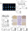
Effect of HCA preconditioning on cardiac I/R-induced infarct size, BAG3, and autophagy expression among the three groups. The typical infarct areas of I-VI sections among the three groups are displayed in (A). Percentages of six sections of infarct area in I/R and HCA+I/R groups are indicated in (B). Total infarct area (C), infarct area/AAR (D), and AAR/LV (E) are demonstrated. Cardiac BAG3 expression (F,G), Beclin-1 (H,J), and LC3II (H,K) examined via Western blot analysis are also shown. Immunohistochemistry of BAG3, Beclin-1, and LC3II is shown in (I). Statistical analysis of BAG3, Beclin-1, and LC3II in the three groups is shown in (LN), respectively. All data are expressed as mean ± SEM (n = 3). * indicates significant differences vs. sham group (** p < 0.01; *** p < 0.001; **** p < 0.0001). # indicates significant differences vs. I/R group (# p < 0.05; ### p < 0.001; #### p < 0.0001).
Figure 8
Figure 8
Effect of HCA preconditioning on cardiac I/R-induced 4HNE/GPX4-mediated ferroptosis and caspase 3-mediated apoptosis in the three groups. Typical GPX4 and 4HNE stains of the three groups are displayed in (A,B), respectively. The percentages of staining of 4HNE and GPX4 sections in the three groups are indicated in (C,D). Cardiac TUNEL expression in the three groups is shown in (E). Statistical analysis of caspase 3 activity is presented in (F). Statistical analysis of TUNEL-positive cells in the three groups is presented in (G). All data are expressed as mean ± SEM (n = 3). * indicates significant differences vs. sham group (** p < 0.01; *** p < 0.001; **** p < 0.0001). # indicates significant differences vs. I/R group (### p < 0.001; #### p < 0.0001).
Figure 9
Figure 9
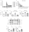
Effect of HCA on antioxidant activity in vitro and in vivo. ROS scavenging activity of HCA in (A,B) was determined using a CL analyzer. HCA dose-dependently and significantly inhibited ROS activity (B). Cardiac I/R significantly increased cardiac ROS (C), 8-isoprostane (D), and MDA (E) vs. sham group, whereas these oxidative stress markers significantly decreased in HCA+I/R vs. I/R group. Original Western blot shows that cardiac I/R decreased cardiac Nrf2 and HO-1 expression in I/R heart (F), whereas HCA preconditioning partially restored Nrf2 and HO-1 expression in HCA+I/R heart. The statistical data for Nrf2/GAPDH and HO-1/GAPDH are presented in (G,H), respectively. All data are expressed as mean ± SEM (n = 3–8). * indicates significant differences vs. sham group (*** p < 0.001; **** p < 0.0001). # indicates significant differences vs. I/R group (# p < 0.05; ## p < 0.01; ### p < 0.001; #### p < 0.0001).
Figure 10
Figure 10
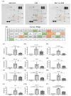
Effect of HCA preconditioning on cardiac I/R injury-induced expression of multiple cytokines in the three groups determined using cytokine array analysis. (A) Original cytokine expression. (B) Matching dot plot. The expression levels of TCK-1 (C), IL-1 R6 (D), ICAM-1 (E), MMP-8 (F), agrin (G), TNF-α (H), IL-2 (I), IL-4 (J), IL-10 (K), LIX (L), CINC-1 (M), and prolactin (N) are significantly elevated in I/R vs. sham group, whereas they are significantly decreased in HCA+I/R vs. I/R group. All data are expressed as mean ± SEM (n = 4). * indicates significant differences vs. sham group (** p < 0.01; *** p < 0.001; **** p < 0.0001). # indicates the significant differences vs. I/R group (# p < 0.05; ## p < 0.01; ### p < 0.001; #### p < 0.0001).

Similar articles

Cited by

References

    1. Luan F., Lei Z., Peng X., Chen L., Peng L., Liu Y., Rao Z., Yang R., Zeng N. Cardioprotective effect of cinnamaldehyde pretreatment on ischemia/reperfusion injury via inhibiting NLRP3 inflammasome activation and gasdermin D mediated cardiomyocyte pyroptosis. Chem. Interact. 2022;368:110245. doi: 10.1016/j.cbi.2022.110245. - DOI - PubMed
    1. Heusch G. Myocardial ischaemia–reperfusion injury and cardioprotection in perspective. Nat. Rev. Cardiol. 2020;17:773–789. doi: 10.1038/s41569-020-0403-y. - DOI - PubMed
    1. Hausenloy D.J., Yellon D.M. Myocardial ischemia-reperfusion injury: A neglected therapeutic target. J. Clin. Investig. 2013;123:92–100. doi: 10.1172/JCI62874. - DOI - PMC - PubMed
    1. Liu Y., Guo X., Zhang J., Han X., Wang H., Du F., Zeng X., Guo C. Protective Effects of the Soluble Receptor for Advanced Glycation End-Products on Pyroptosis during Myocardial Ischemia-Reperfusion. Oxid. Med. Cell. Longev. 2021;2021:9570971. doi: 10.1155/2021/9570971. - DOI - PMC - PubMed
    1. Shen Y.-C., Lee W.-S., Yang K.-T. Methyl palmitate protects heart against ischemia/reperfusion-induced injury through G-protein coupled receptor 40-mediated activation of the PI3K/AKT pathway. Eur. J. Pharmacol. 2021;905:174183. doi: 10.1016/j.ejphar.2021.174183. - DOI - PubMed

MeSH terms

LinkOut - more resources