Case Reports
. 2024 Dec 5;25(23):13059.
doi: 10.3390/ijms252313059.

Novel Mutation Lys30Glu in the TPM1 Gene Leads to Pediatric Left Ventricular Non-Compaction and Dilated Cardiomyopathy via Impairment of Structural and Functional Properties of Cardiac Tropomyosin

Affiliations
Case Reports

Novel Mutation Lys30Glu in the TPM1 Gene Leads to Pediatric Left Ventricular Non-Compaction and Dilated Cardiomyopathy via Impairment of Structural and Functional Properties of Cardiac Tropomyosin

Elena V Zaklyazminskaya et al. Int J Mol Sci. .

Abstract

Pediatric dilated cardiomyopathy (DCM) is a rare heart muscle disorder leading to the enlargement of all chambers and systolic dysfunction. We identified a novel de novo variant, c.88A>G (p.Lys30Glu, K30E), in the TPM1 gene encoding the major cardiac muscle tropomyosin (Tpm) isoform, Tpm1.1. The variant was found in a proband with DCM and left ventricular non-compaction who progressed to terminal heart failure at the age of 3 years and 8 months. To study the properties of the mutant protein, we produced recombinant K30E Tpm and used various biochemical and biophysical methods to compare its properties with those of WT Tpm. The K30E substitution decreased the thermal stability of Tpm and its complex with actin and significantly reduced the sliding velocity of the regulated thin filaments over a surface covered by ovine cardiac myosin in an in vitro motility assay across the entire physiological range of Ca2+ concentration. Our molecular dynamics simulations suggest that the charge reversal of the 30th residue of Tpm alters the actin monomer to which it is bound. We hypothesize that this rearrangement of the actin-Tpm interaction may hinder the transition of a myosin head attached to a nearby actin from a weakly to a strongly bound, force-generating state, thereby reducing myocardial contractility. The impaired myosin interaction with regulated actin filaments and the decreased thermal stability of the actin-Tpm complex at a near physiological temperature likely contribute to the pathogenicity of the variant and its causative role in progressive DCM.

Keywords: actin–myosin interaction; dilated cardiomyopathy; in vitro motility assay; left ventricular non-compaction; molecular dynamics; tropomyosin.


Conflict of interest statement

Natalia S. Ryabkova and Ivan A. Katrukha were employed by HyTest Ltd. The authors declare that this research was conducted in the absence of any commercial or financial relationships that could be construed as potential conflicts of interest. The funders had no role in the design of the study; in the collection, analyses, or interpretation of data; in the writing of the manuscript; or in the decision to publish the results.

Figures

Figure 1
Figure 1
Fragment of the EchoCG of the proband (II.2) with dilated cardiomyopathy at the age of 1 year and 8 months. The left atrium (LA, bottom right) and left ventricle (LV, upper right) are shown. The end systolic size (ESS) is 36 mm (z-score 5.43), and the end diastolic size (EDS LV) is 43 mm (z-score 3.83). The left ventricular ejection fraction (LV EF) is 30%, and mitral regurgitation is at stage 2 (blue spot in the middle of the figure). High trabeculation and finger-like protrusion of the LV apex are seen on the top (indicated by arrows).
Figure 2
Figure 2
Pedigree of the DCM84 family. Healthy family members are shown with open symbols, and the affected proband is indicated with a closed symbol and an arrow. The abbreviations are given in the text. The generation number and individual numbers within the generation are marked by Roman and Arabic numerals, respectively. To evaluate the pathogenicity of the K30E mutation in the TPM1 gene, we produced recombinant Tpm carrying the K30E substitution and applied various methods to investigate how this substitution affects the structural and functional properties of Tpm.
Figure 3
Figure 3
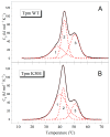
K30E substitution affects the structure of Tpm molecule. The plots show temperature dependences of excess heat capacity (Cp) obtained from DSC studies on Tpm WT (A) and Tpm with the K30E substitution (B) along with their deconvolution into distinct thermal transitions (calorimetric domains 1, 2, and 3). The calorimetric parameters derived from the DSC data for individual thermal transitions are provided in Table 1.
Figure 4
Figure 4
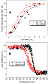
K30E substitution slightly enhances the actin-binding properties of Tpm but diminishes the thermal stability of the Tpm–F-actin complexes. The plots illustrate the effects of the K30E substitution on Tpm’s affinity for F-actin measured by the co-sedimentation assay in buffer containing 200 mM NaCl (A), and on the thermal stability of the Tpm–F-actin complexes, determined by light scattering measurements (B).
Figure 5
Figure 5
K30E substitution impairs the functional properties of Tpm. The plots display the effect of the K30E substitution in the Tpm molecule on the sliding velocity of regulated thin filaments along cardiac myosin in an in vitro motility assay depending on the Ca2+ concentration (pCa) at a saturating myosin concentration equal to 300 µg/mL (A), and on the myosin concentration in the flow cell at a saturating Ca2+ concentration (at pCa 4) (B). The experimental points represent the average values ± SD for the three experiments. Experimental data were fitted with the Hill equation. The sliding velocity parameters for WT Tpm and K30E Tpm derived from the data are presented in Table 2 and Table 3.
Figure 6
Figure 6
Top: snapshots of MD trajectories of actin–Tpm filaments with WT Tpm (A) and K30E Tpm (B). In the WT structure (A) Tpm residues Lys30 form hydrogen bonds with Glu241 residues of adjacent actin monomers, while in the K30E structure (B), the Glu30 residues of Tpm form hydrogen bonds with Lys326 of other actin monomers. The occupancies (1 indicates the presence; 0 indicates the absence) of hydrogen bonds between Tpm residue 30 and different actin monomers (A1, A2) for WT Tpm (C) and K30E Tpm (D). The green and blue actin monomers in A and B correspond to A1 and A2 in C and D, respectively, counting from the barbed end. Details of the MD simulations and the source of the initial atomic structure of the actin–Tpm complex are provided in Section 4.8.

References

    1. Mestroni L., Maisch B., McKenna W.J., Schwartz K., Charron P., Rocco C., Tesson F., Richter A., Wilke A., Komajda M. Guidelines for the study of familial dilated cardiomyopathies. Collaborative Research Group of the European Human and Capital Mobility Project on Familial Dilated Cardiomyopathy. Eur. Heart J. 1999;20:93–102. doi: 10.1053/euhj.1998.1145. - DOI - PubMed
    1. Solaro R.J., Goldspink P.H., Wolska B.M. Emerging concepts of mechanisms controlling cardiac tension: Focus on familial dilated cardiomyopathy (DCM) and sarcomere-directed therapies. Biomedicines. 2024;12:999. doi: 10.3390/biomedicines12050999. - DOI - PMC - PubMed
    1. Rampersaud E., Siegfried J.D., Norton N., Li D., Martin E., Hershberger R.E. Rare variant mutations identified in pediatric patients with dilated cardiomyopathy. Prog. Pediatr. Cardiol. 2011;31:39–47. doi: 10.1016/j.ppedcard.2010.11.008. - DOI - PMC - PubMed
    1. Peters S., Johnson R., Birch S., Zentner D., Hershberger R.E., Fatkin D. Familial dilated cardiomyopathy. Heart Lung Circ. 2020;29:566–574. doi: 10.1016/j.hlc.2019.11.018. - DOI - PubMed
    1. Hershberger R.E., Givertz M.M., Ho C.Y., Judge D.P., Kantor P.F., McBride K.L., Morales A., Taylor M.R., Vatta M., Ware S.M. Genetic evaluation of cardiomyopathy-A Heart Failure Society of America Practice Guideline. J. Card. Fail. 2018;24:281–302. doi: 10.1016/j.cardfail.2018.03.004. - DOI - PMC - PubMed

Publication types

LinkOut - more resources