. 2024 Dec 5;25(23):13082.
doi: 10.3390/ijms252313082.

Neuroprotective and Anti-Inflammatory Effects of Dimethyl Fumarate, Monomethyl Fumarate, and Cannabidiol in Neurons and Microglia

Affiliations

Neuroprotective and Anti-Inflammatory Effects of Dimethyl Fumarate, Monomethyl Fumarate, and Cannabidiol in Neurons and Microglia

Alicia Sánchez-Sanz et al. Int J Mol Sci. .

Abstract

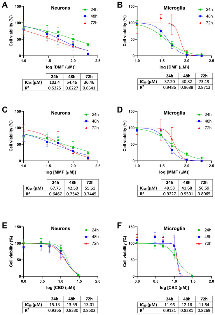
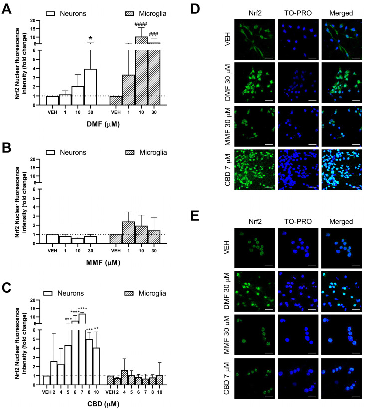
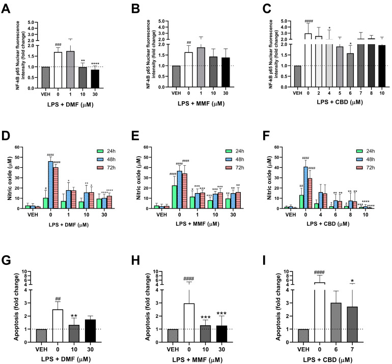
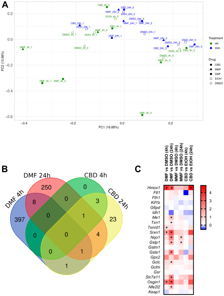
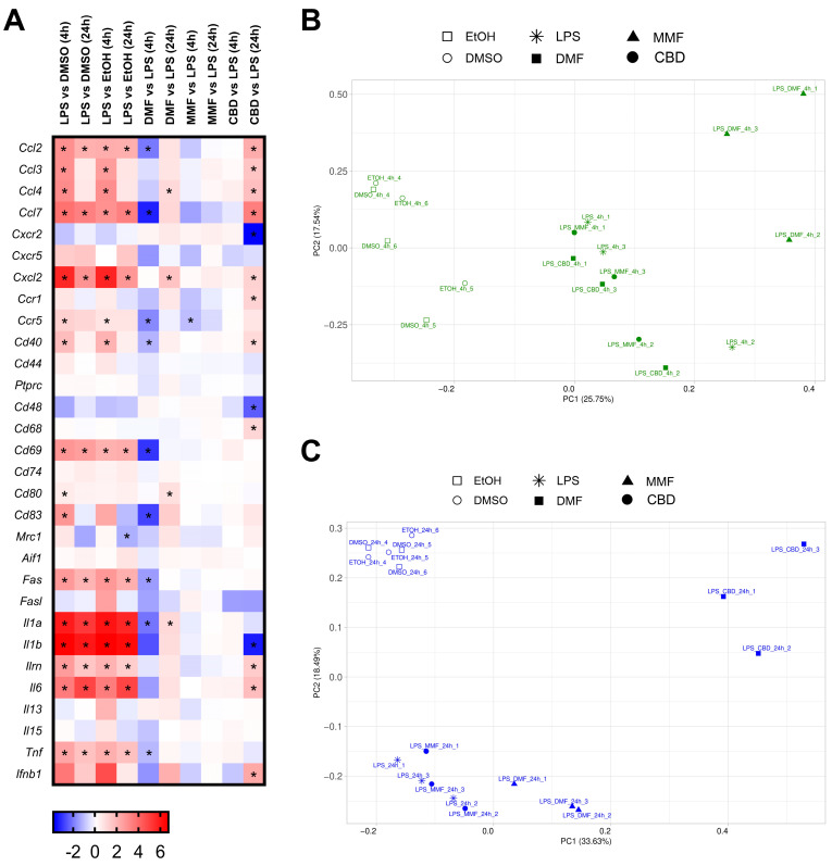
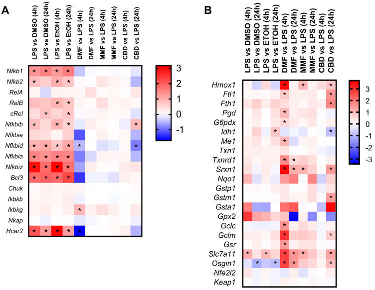
Dimethyl fumarate (DMF) is an immunomodulatory treatment for multiple sclerosis (MS) that can cross the blood-brain barrier, presenting neuroprotective potential. Its mechanism of action is not fully understood, and there is a need to characterize whether DMF or its bioactive metabolite monomethyl fumarate (MMF) exerts neuroprotective properties. Moreover, the combination of adjuvant agents such as cannabidiol (CBD) could be relevant to enhance neuroprotection. The aim of this study was to compare the neuroprotective and immunomodulatory effects of DMF, MMF, and CBD in neurons and microglia in vitro. We found that DMF and CBD, but not MMF, activated the Nrf2 antioxidant pathway in neurons. Similarly, only DMF and CBD, but not MMF, prevented the LPS-induced activation of the inflammatory pathway NF-kB in microglia. Additionally, the three drugs inhibited the production of nitric oxide in microglia and protected neurons against apoptosis. Transcriptomically, DMF modulated a greater number of inflammatory and Nrf2-related genes compared to MMF and CBD in both neurons and microglia. Our results show that DMF and MMF, despite being structurally related, present differences in their mechanisms of action that could be relevant for the achievement of neuroprotection in MS patients. Additionally, CBD shows potential as a neuroprotective agent.

Keywords: NF-kB; Nrf2; cannabidiol; dimethyl fumarate; microglia; monomethyl fumarate; multiple sclerosis; neuron; neuroprotection; transcriptome.


Conflict of interest statement

The authors declare no conflicts of interest.

Figures

Figure 1
Figure 1
Cell viability of the neurons and microglia cells treated with dimethyl fumarate (DMF), monomethyl fumarate (MMF), or cannabidiol (CBD). (A) Cell viability of neurons treated with DMF. (B) Cell viability of microglia cells treated with DMF. (C) Cell viability of neurons treated with MMF. (D) Cell viability of microglia cells treated with MMF. (E) Cell viability of neurons treated with CBD. (F) Cell viability of microglia cells treated with CBD. (AF) Dose–response relationships of each drug at 24, 48, and 72 h. The mean with standard deviation is represented of the percentage of cell viability with respect to the logarithm of the tested concentrations (10, 30, 50, 100, and 200 µM for DMF and MMF; 1, 3, 5, 10, and 30 µM for CBD). The percentage of cell viability was calculated relative to the vehicle-treated cells. The half-maximal inhibitory concentration (IC50) and the coefficient of determination (R2) values are indicated below each graph.
Figure 2
Figure 2
Effect of dimethyl fumarate (DMF), monomethyl fumarate (MMF), and cannabidiol (CBD) on the activation of Nrf2 in neurons and microglia. (A) Effect of DMF on Nrf2 activation in neurons and microglia. (B) Effect of MMF on Nrf2 activation in neurons and microglia. (C) Effect of CBD on Nrf2 activation in neurons and microglia. (D) Representative immunofluorescence images from neurons showing the Nrf2 protein location (green). TO-PRO (blue) was used as a nuclear counterstain. Scale bar: 20 µm, shown in the bottom-right corner of each image. (E) Representative immunofluorescence images from microglia showing Nrf2 protein location (green). TO-PRO (blue) was used as a nuclear counterstain. Scale bar: 20 µm, shown in the bottom-right corner of each image. (AC) White bars represent neurons and black dotted lines represent microglia. (AE) Neurons and microglia were treated for 4 h with either the vehicle (VEH) or drug. Bars represent the fold change of the mean nuclear fluorescence intensity of Nrf2 in the drug-treated cells compared to their respective vehicle. Data (mean ± standard deviation) are representative of three different experiments. The Kruskal–Wallis test was used to compare the different concentrations of each drug against their respective vehicle. * p < 0.05, ** p < 0.01, *** p < 0.001, **** p < 0.0001 in neurons; ### p < 0.001, #### p < 0.0001 in microglia.
Figure 3
Figure 3
Immunomodulatory effects of dimethyl fumarate (DMF), monomethyl fumarate (MMF), and cannabidiol (CBD) in microglia. (AC) Effect of DMF (A), MMF (B), and CBD (C) on the activation of NF-kB p65. Microglia cells were treated for 30 min with either vehicle (VEH), lipopolysaccharide (LPS), or LPS in combination with DMF, MMF, or CBD. Bars represent the fold change of the mean nuclear fluorescence intensity of NF-kB p65 in the drug-treated cells compared to the vehicle (represented by black dotted lines). (DF) Effect of DMF (D), MMF (E), and CBD (F) on the production of nitric oxide (NO). Microglia cells were treated for 24, 48, or 72 h with either the VEH, LPS, or LPS in combination with DMF, MMF, or CBD. Bars represent the concentration (µM) of NO. (GI) Effect of DMF (G), MMF (H), and CBD (I) on neuronal apoptosis. Neurons were cultured for 4 h with microglia-conditioned medium. Microglia had been previously treated with the VEH, LPS, or LPS in combination with DMF, MMF, or CBD for 48 h. Bars represent the fold change of the number of apoptotic cells (mean ± standard deviation) in the drug-treated cells compared to their respective vehicle. (AI) Data are representative of three different experiments. # p < 0.05, ## p < 0.01, ### p < 0.001, #### p < 0.0001 LPS compared to the vehicle (Mann–Whitney test); * p < 0.05, ** p < 0.01, *** p < 0.001, **** p < 0.0001 LPS + drug compared to LPS (Kruskal–Wallis test).
Figure 4
Figure 4
Transcriptomic profile of neurons treated with dimethyl fumarate (DMF), monomethyl fumarate (MMF), or cannabidiol (CBD). (A) Principal component analysis of the gene expression profile of neurons treated with 30 µM DMF, 30 µM MMF, 6 µM CBD, or their respective vehicles (DMSO or EtOH) for 4 and 24 h. Each condition presents experimental triplicates. The x-axis represents the first principal component (PC1), and the y-axis represents the second principal component (PC2). The percentages indicated in parentheses represent the percentages of variation explained by the principal components. (B) Venn diagram showing the number and overlap of the differentially expressed genes (DEGs) in neurons treated with DMF or CBD for 4 and 24 h. (C) Heat map representing the expression levels of genes from the Nrf2 signaling pathway. Gene expression levels are represented with the log2 of the fold change (FC), indicating the ratio of the gene expression in the drug-treated cells compared to their respective vehicle. * p-adj < 0.05.
Figure 5
Figure 5
Transcriptomic profile of the lipopolysaccharide (LPS)-activated microglia cells treated with dimethyl fumarate (DMF), monomethyl fumarate (MMF), or cannabidiol (CBD). (A) Heat map representing the expression levels of genes related to inflammation. Gene expression levels are represented with the log2 of the fold change (FC), indicating the ratio of the gene expression in the LPS-treated cells compared to the vehicle or drug-treated cells compared to LPS. * p-adj < 0.05. (B,C) Principal component analysis of the gene expression profile of the LPS-activated microglia cells treated with 30 µM DMF, 30 µM MMF, 6 µM CBD, or their respective vehicles (DMSO or EtOH) for 4 h (B) and 24 h (C). Each condition presents experimental triplicates. The x-axis represents the first principal component (PC1), and the y-axis represents the second principal component (PC2). The percentages indicated in parentheses represent the percentages of variation explained by the principal components.
Figure 6
Figure 6
Transcriptomic modulation of the NF-kB and Nrf2 pathways in the lipopolysaccharide (LPS)-activated microglia cells treated with dimethyl fumarate (DMF), monomethyl fumarate (MMF), or cannabidiol (CBD). (A) Heat map representing the expression levels of genes related to the NF-kB pathway. (B) Heat map representing the expression levels of genes related to the Nrf2 pathway. (A,B) LPS-activated microglia cells were treated with 30 µM DMF, 30 µM MMF, 6 µM CBD, or their respective vehicles (DMSO or EtOH) for 4 and 24 h. The gene expression levels are represented with the log2 of the fold change (FC), indicating the ratio of the gene expression in the LPS-treated cells compared to the vehicle or drug-treated cells compared to LPS. * p-adj < 0.05.

Similar articles

Cited by

References

    1. Saidu N.E.B., Kavian N., Leroy K., Jacob C., Nicco C., Batteux F., Alexandre J. Dimethyl fumarate, a two-edged drug: Current status and future directions. Med. Res. Rev. 2019;39:1923–1952. doi: 10.1002/med.21567. - DOI - PubMed
    1. Mrowietz U., Barker J., Boehncke W.H., Iversen L., Kirby B., Naldi L., Reich K., Tanew A., van de Kerkhof P.C.M., Warren R.B. Clinical use of dimethyl fumarate in moderate-to-severe plaque-type psoriasis: A European expert consensus. J. Eur. Acad. Dermatol. Venereol. 2018;32:3–14. doi: 10.1111/jdv.15218. - DOI - PubMed
    1. European Medicines Agency Tecfidera 120 mg Gastro-Resistant Hard Capsules. [(accessed on 30 October 2024)]. Available online: https://www.ema.europa.eu/documents/product-information/tecfidera-epar-p....
    1. Charabati M., Wheeler M.A., Weiner H.L., Quintana F.J. Multiple sclerosis: Neuroimmune crosstalk and therapeutic targeting. Cell. 2023;186:1309–1327. doi: 10.1016/j.cell.2023.03.008. - DOI - PMC - PubMed
    1. Yang J.H., Rempe T., Whitmire N., Dunn-Pirio A., Graves J.S. Therapeutic Advances in Multiple Sclerosis. Front. Neurol. 2022;13:824926. doi: 10.3389/fneur.2022.824926. - DOI - PMC - PubMed

MeSH terms

LinkOut - more resources