. 2024 Dec 6;25(23):13136.
doi: 10.3390/ijms252313136.

A 0.2 T-0.4 T Static Magnetic Field Improves the Bone Quality of Mice Subjected to Hindlimb Unloading and Reloading Through the Dual Regulation of BMSCs via Iron Metabolism

Affiliations

A 0.2 T-0.4 T Static Magnetic Field Improves the Bone Quality of Mice Subjected to Hindlimb Unloading and Reloading Through the Dual Regulation of BMSCs via Iron Metabolism

Jianping Wang et al. Int J Mol Sci. .

Abstract

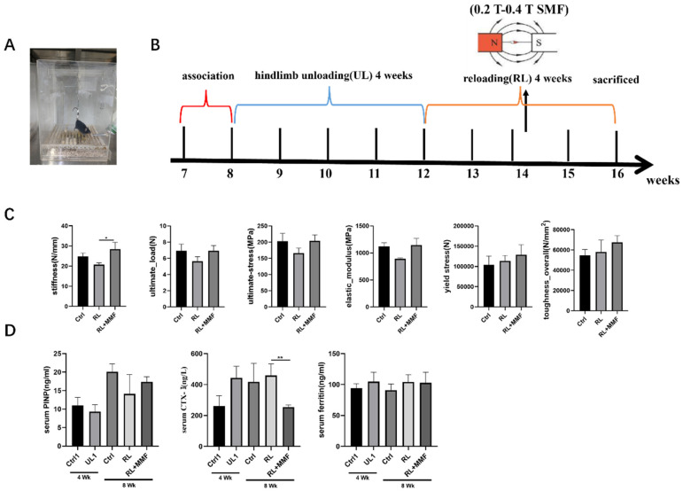
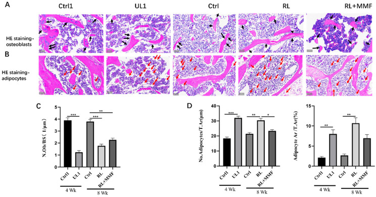
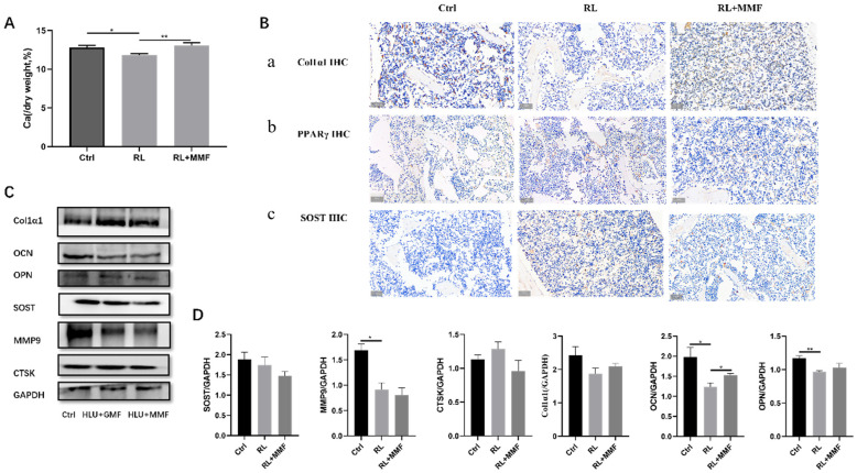
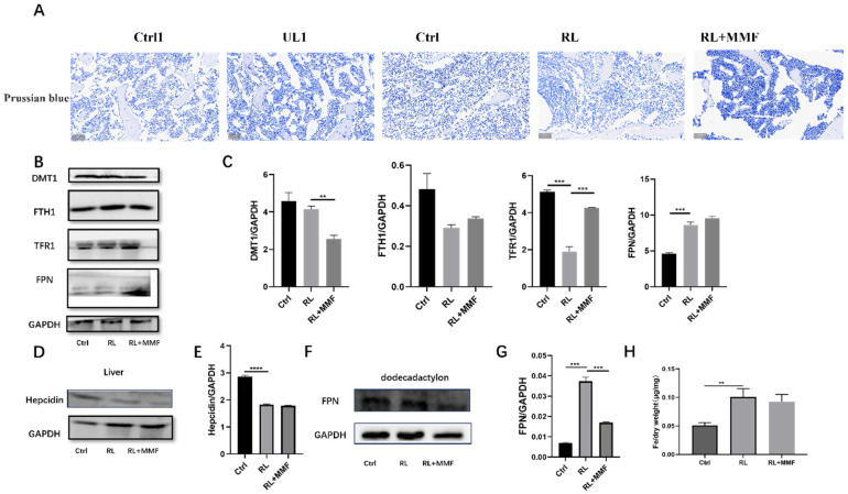
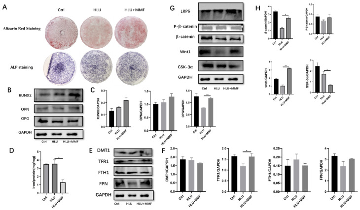
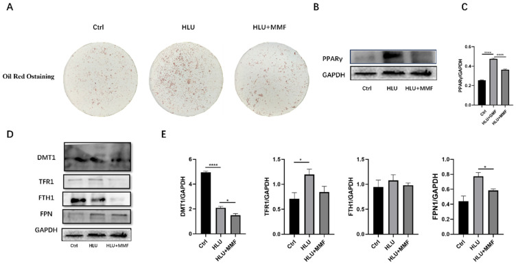
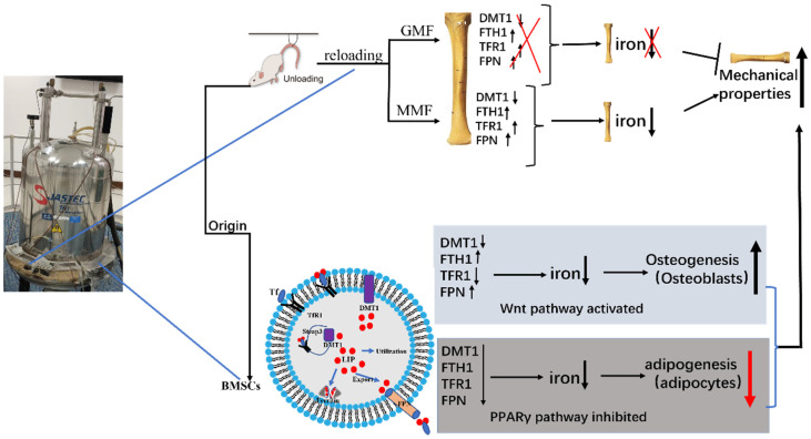
Osteoporosis is the most prevalent metabolic bone disease, especially when aggravated by aging and long-term bed rest of various causes and also when coupled with astronauts' longer missions in space. Research on the use of static magnetic fields (SMFs) has been progressing as a noninvasive method for osteoporosis due to the complexity of the disease, the inconsistency of the effects of SMFs, and the ambiguity of the mechanism. This paper studied the effects of mice subjected to hindlimb unloading (UL, HLU) and reloading by the 0.2 T-0.4 T static magnetic field (MMF). Primary bone marrow mesenchymal stem cells (BMSCs) were extracted to explore the mechanism. Eight-week-old male C57BL/6 mice were used as an osteoporosis model by HLU for four weeks. The HLU recovery period (reloading, RL) was carried out on all FVEs and recovered in the geomagnetic field (45-64 μT, GMF) and MMF, respectively, for 12 h/d for another 4 weeks. The tibia and femur of mice were taken; also, the primary BMSCs were extracted. MMF promoted the recovery of mechanical properties after HLU, increased the number of osteoblasts, and decreased the number of adipocytes in the bone marrow. MMF decreased the total iron content and promoted the total calcium content in the tibia. In vitro experiments showed that MMF promoted the osteogenic differentiation of BMSCs and inhibited adipogenic differentiation, which is related to iron metabolism, the Wnt/β-catenin pathway, and the PPARγ pathway. MMF accelerated the improvement in bone metabolism and iron metabolism in RL mice to a certain extent, which improved the bone quality of mice. MMF mainly promoted osteogenic differentiation and reduced the adipogenic differentiation of BMSCs, which provides a reliable research direction and transformation basis for the osteoporosis of elderly, bedridden patients and astronauts.

Keywords: 0.2 T–0.4 T SMF; BMSCs; HLU reloading; bone quality; iron metabolism.


Conflict of interest statement

The authors have no competing interests or other interests that might be perceived to influence the results and/or discussion reported in this paper.

Figures

Figure 1
Figure 1
Effects of MMF on mechanical properties and markers of bone metabolism in mice. (A) mouse tail suspension model, (B) experimental flow chart, (C) mechanical properties of mouse tibia, including stiffness, elastic modulus, ultimate strain, ultimate stress, ultimate load, and ultimate displacement, (D) measurement of serum biochemical indexes. n = 4–6. Data are shown as the mean ± SEM. * p < 0.05, ** p < 0.01.
Figure 2
Figure 2
The number of osteoblasts and adipocytes in the bone marrow of femur were evaluated. (A,B) The femoral sections were subjected to H&E staining to visualize osteoblasts and adipocytes. Black arrows indicate osteoblasts. Red arrows indicate adipocytes. Scale bar = 50 μm. Osteoblastogenesis were evaluated by the osteoblast number per bone surface (N.Ob/BS) in the trabecular bone; (C) Adipogenesis was evaluated by the adipocyte number per bone surface (No. Adipocytes/T.Ar) and adipocyte surface per bone surface (Adipocyte.Ar/T.Ar) in the bone marrow (D). n = 3. All the data are shown as mean ± SEM. * p < 0.05, ** p < 0.01, *** p < 0.001.
Figure 3
Figure 3
Bone metabolism of MMF on the femur of HLU reloading mice. (A) Total calcium content in the tibia of HLU reloading mice. (B) Immunohistochemical staining of Col1α1, PPARγ, and SOST. Scar bar = 50 μm. Brown color indicates positive expression. (C) Protein expression of osteogenic and osteoclast-related proteins in the tibia of HLU reloading mice; (D) Quantitative statistics of protein expression of (A). n = 4–6. Data are shown as the mean ± SEM. * p < 0.05, ** p < 0.01.
Figure 4
Figure 4
Effects of MMF on iron metabolism in HLU reloading mice. (A) Prussian blue staining of bone tissue in HLU reloading mice, Scar bar = 50 μm; (B) Protein expression related to iron metabolism in the tibia of HLU reloading mice; (C) Quantitative statistics of the protein expression of (B); (D) Protein expression of liver tissue in HLU reloading mice; (E) Quantitative statistics of the protein expression of (D); (F) Protein expression of the duodenum in HLU reloading mice; (G) Quantitative statistics of the protein expression of (F); (H) Total iron content in the tibia of HLU reloading mice. n = 3. Data are shown as the mean ± SEM. ** p < 0.01, *** p < 0.001, **** p < 0.0001.
Figure 5
Figure 5
MMF promotes osteogenic differentiation of primary BMSCs and mechanism. (A) ARS and ALP staining after 14 d of osteogenic differentiation of BMSCs, 10×; (B) Protein expression of osteogenic related molecules after 14 d of osteogenic differentiation of BMSCs; (C) Quantitative statistics of the protein expression of (B); (D) Total iron content of BMSCs cells after osteogenic differentiation for 14 d; (E) Expression of iron-metabolism-related proteins of BMSCs after osteogenic differentiation for 14 d; (F) Quantitative statistics of the protein expression of (E); (G) Protein expression of the Wnt/β-catenin signaling pathway after 14 d of osteogenic differentiation of BMSCs; (H) Quantitative statistics of the protein expression of (G). n = 3. Data are shown as the mean ± SEM. * p < 0.05, ** p < 0.01.
Figure 6
Figure 6
MMF inhibits adipogenic differentiation of primary BMSCs derived from HLU model mice. (A) Oil Red O staining after 14 d of adipogenic differentiation of BMSCs, 10×; (B) Protein expression of PPARγ after 14 d of adipogenic differentiation of BMSCs; (C) Quantitative statistics of the protein expression of (B); (D) Expression of iron-metabolism-related proteins of BMSCs after adipogenic differentiation for 14 d; (E) Quantitative statistics of the protein expression of (D). n = 3. Data are shown as the mean ± SEM. * p < 0.05, **** p < 0.0001.
Figure 7
Figure 7
Schematic diagram of results summarized in this paper. The use of 0.2 T–0.4 T SMF (MMF) improved the bone quality of HLU reloading mice by affecting bone metabolism and iron metabolism. Through study of the osteogenic and adipogenic differentiation of primary BMSCs, it was proved that MMF promoted osteogenic differentiation and activated the Wnt/β-catenin signaling pathway, inhibiting adipogenic differentiation. Upward arrows represent an increase; downward arrows represent a decrease. All the colors have no real meaning, just for beauty.

References

    1. Yang T.L., Shen H., Liu A., Dong S.S., Zhang L., Deng F.Y., Zhao Q., Deng H.W. A road map for understanding molecular and genetic determinants of osteoporosis. Nat. Rev. Endocrinol. 2020;16:91–103. doi: 10.1038/s41574-019-0282-7. - DOI - PMC - PubMed
    1. Wang Q., Dong J.F., Fang X., Chen Y. Application and modification of bone cement in vertebroplasty: A literature review. Jt. Dis. Relat. Surg. 2022;33:467–478. doi: 10.52312/jdrs.2022.628. - DOI - PMC - PubMed
    1. Carpenter R.D., LeBlanc A.D., Evans H., Sibonga J.D., Lang T.F. Long-term changes in the density and structure of the human hip and spine after long-duration spaceflight. Acta Astronaut. 2010;67:71–81. doi: 10.1016/j.actaastro.2010.01.022. - DOI
    1. Morgan E.F., Unnikrisnan G.U., Hussein A.I. Bone Mechanical Properties in Healthy and Diseased States. Annu. Rev. Biomed. Eng. 2018;20:119–143. doi: 10.1146/annurev-bioeng-062117-121139. - DOI - PMC - PubMed
    1. Ibrahim A., Magliulo N., Groben J., Padilla A., Akbik F., Abdel Hamid Z. Hardness, an Important Indicator of Bone Quality, and the Role of Collagen in Bone Hardness. J. Funct. Biomater. 2020;11:85. doi: 10.3390/jfb11040085. - DOI - PMC - PubMed

LinkOut - more resources