. 2024 Dec 7;25(23):13184.
doi: 10.3390/ijms252313184.

ADAM10 Expression by Ameloblasts Is Essential for Proper Enamel Formation

Affiliations

ADAM10 Expression by Ameloblasts Is Essential for Proper Enamel Formation

Shifa Shahid et al. Int J Mol Sci. .

Abstract

ADAM10 is a multi-functional proteinase that can cleave approximately 100 different substrates. Previously, it was demonstrated that ADAM10 is expressed by ameloblasts, which are required for enamel formation. The goal of this study was to determine if ADAM10 is necessary for enamel development. Deletion of Adam10 in mice is embryonically lethal and deletion of Adam10 from epithelia is perinatally lethal. We therefore deleted Adam10 from ameloblasts. Ameloblast-specific expression of the Tg(Amelx-iCre)872pap construct was confirmed. These mice were crossed with Adam10 floxed mice to generate Amelx-iCre; Adam10fl/fl mice (Adam10 cKO). The Adam10 cKO mice had discolored teeth with softer than normal enamel. Notably, the Adam10 cKO enamel density and volume were significantly reduced in both incisors and molars. Moreover, the incisor enamel rod pattern became progressively more disorganized, moving from the DEJ to the outer enamel surface, and this disorganized rod structure created gaps and S-shaped rods. ADAM10 cleaves proteins essential for cell signaling and for enamel formation such as RELT and COL17A1. ADAM10 also cleaves cell-cell contacts such as E- and N-cadherins that may support ameloblast movement necessary for normal rod patterns. This study shows, for the first time, that ADAM10 expressed by ameloblasts is essential for proper enamel formation.

Keywords: Cre Lox recombination; ameloblast migration; cervical enamel; density; hardness; mouse; occlusal enamel; volume.


Conflict of interest statement

The authors declare no conflicts of interest. The funders had no role in the design of the study; in the collection, analyses, or interpretation of data; in the writing of the manuscript; or in the decision to publish the results.

Figures
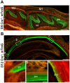
Figure 1
Figure 1
Amelx-iCre mice express Cre recombinase only in ameloblasts. Amelx-iCre mice were crossed with the mouse mT/mG reporter strain so that Cre expression would cause tissues to stain with green fluorescent protein (GFP). (A) GFP staining was restricted to ameloblasts in molars at postnatal day 5 whereas areas of red florescence indicate no Cre recombination. Scale bar 200 µm. (B) In postnatal day 12 maxillary incisor sections, amelogenin promoter driven iCre expression starts at the onset of the secretory stage (a) and persists throughout the length of the incisor where ameloblasts are present (b,c). Scale bar 1000 µm. M1, first molar; M2, second molar; am, ameloblasts.
Figure 2
Figure 2
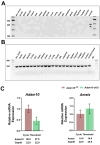
Genomic PCR analysis to detect the presence or absence of Amelx-iCre mediated recombination in multiple mouse tissues and quantification of Adam10 and amelogenin expression. (A) A 376 base pair mG product results from the Cre-recombined construct, while a (B) 200 base pair product results from the mT region that was not recombined by Amelx-iCre. Of the 20 tissues assessed, only the whole incisor demonstrated Amelx-iCre mediated recombination. (C) Quantification of Adam10 and amelogenin (AmelX) expression by qPCR analyses of 5-day old first molar enamel organs. The Adam10 cKO was expressed at significantly lower levels than was the Adam10fl/fl control, demonstrating that the preponderance of the Adam10 genes in the ameloblast layer were successfully excised. In contrast, no significant difference was observed in native amelogenin gene expression between the Adam10 cKO and Adam10fl/fl control mice, indicating that the Amelx-iCre construct did not affect native amelogenin expression. Dots along the error bars represent the results of individual samples. Dots along the error bars represent the results of individual samples.
Figure 3
Figure 3
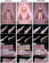
Phenotypic assessment of Amelx-iCre, Adam10fl/fl, and Adam10 cKO incisors plus molars from mice at 7 weeks of age. (A) Frontal view of maxillary and mandibular incisors. The Adam10 cKO incisor enamel appears to be dull, rough, pigmented, and abraded. (B) Mesial (upper left), lateral (upper right), lingual (lower right), and labial (lower left) views of mandibular incisors. The incisal edge of Adam10 cKO incisors were blunted due to attrition. (C) Buccal, occlusal, and lingual views of mandibular molars. Adam10 cKO molars appear rough, discolored, and show distinct chalky banding near the cervical edge. These molar cusps are rounded and show signs of attrition. Teeth from Amelx-iCre and from Adam10fl/fl mice appear normal in shape and color.
Figure 4
Figure 4
Quantification of incisor enamel hardness, volume, density and thickness. (A) Shown are nanohardness indents of enamel, dentin and alveolar bone that were performed on Amelx-iCre, Adam10fl/fl, and Adam10 cKO mice. Each sample is indented at 3 positions in bone (positions 0–2), 12 positions in dentin (3–14) and 6 positions in enamel (15–20). (B) Bar graph of the mean nanohardness (GPa) values show the Adam10 cKO mice had significantly reduced enamel hardness compared to controls. In contrast, dentin and bone nanohardness levels were not significantly different among the genotypes tested. (CE) 2D renderings from µCT analyses of adult hemi-mandibles in sagittal view. The mineralized enamel appeared less dense in Adam10 cKO incisors compared to controls. (FH) Compared to Amelx-iCre and Adam10fl/fl controls, Adam10 cKO mouse incisors exhibit 10% reduced enamel mineral density and 10% reduced enamel thickness Dots along the error bars represent the results of individual samples (*** p < 0.001, ** p < 0.01, ns; non-significant).
Figure 5
Figure 5
2D and 3D renderings from µCT analyses of erupted and unerupted mandibular first molars in Adam10 cKO mice and controls. (A) Compared to controls, deletion of Adam10 leads to a poorly defined and under mineralized enamel layer. Sagittal and coronal views reveal flattening of Adam10 cKO cusps suggesting that these molars have undergone attrition. Occlusal views of Adam10 cKO molars show a severely hypomineralized enamel layer with reduced density as observed by the worn (3D) and darker (2D) enamel. (B) Quantitative analyses of total molar enamel volume and density show approximately a 27% decrease in volume and a 14% reduction in mineral density compared to controls. Segmentation of molars into occlusal and cervical regions reveal a greater reduction of volume and density at the cervical region of Adam10 cKO molars when compared to the Amelx-iCre, and Adam10fl/fl molars. (C) 2D and 3D renderings from µCT analyses of unerupted mandibular molars at postnatal day 15 (P15). Like erupted molars, Adam10 cKO unerupted molars exhibit significantly reduced enamel volume and density. Enamel in the cervical region is present but is severely undermineralized. (D) For the unerupted molars, quantitative measurements confirmed an overall 16% and 15% reduction of total enamel volume and density respectively. A greater decrease in cervical enamel volume versus occlusal volume (17% versus 14%) and cervical density versus occlusal density (17% versus 13%) was also confirmed in Adam10 cKO unerupted molars. Note that the total decrease in enamel volume for the Adam10 cKO erupted molars was 27% while that for the unerupted molars was 16%. This difference may represent the level of attrition in the 7-week-old erupted molars. Dots along the error bars represent the results of individual samples (** p < 0.01, * p < 0.05).
Figure 6
Figure 6
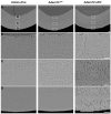
Backscatter SEM of 7-week mandibular incisor cross sections from Amelx-iCre, Adam10fl/fl, and Adam10 cKO mice. (A) Panels show cross-sections of incisor enamel at the buccal alveolar crest where the enamel layer is near to erupting into the oral cavity. Normal enamel thickness is present in Amelx-iCre, and Adam10fl/fl incisors, but the Adam10 cKO enamel is darker indicative of increased protein content and appears thinner than normal. Scale bar 10 µm. (BD) Higher magnification (2000×) reveals distinct enamel rod decussation patterns. Compared to the Amelx-iCre and Adam10fl/fl enamel, the rod/interrod pattern of Adam10 cKO enamel is somewhat disorganized near the dentin-enamel junction, but the enamel rods become progressively more S-shaped and loosely packed (porous) toward the outer enamel edge. Scale bar 10 µm.
Figure 7
Figure 7
Histological comparison of incisor ameloblasts (Am) and stratum intermedium (Si) from Adam10fl/fl (control) and Adam10 cKO (experimental) mice. Compared to the Adam10fl/fl incisor ameloblasts, the Adam10 cKO ameloblasts appeared to be normally aligned and did not significantly differ from those present in the Adam10fl/fl mice. Perhaps this is not unexpected since some level of ameloblast alignment would likely be necessary to generate a rod pattern even if that pattern was disorganized. Both the ameloblast and stratum intermedium tissue layers appeared normal. Od, odontoblast layer.

References

    1. Bartlett J.D., Smith C.E., Hu Y., Ikeda A., Strauss M., Liang T., Hsu Y.H., Trout A.H., McComb D.W., Freeman R.C., et al. MMP20-generated amelogenin cleavage products prevent formation of fan-shaped enamel malformations. Sci. Rep. 2021;11:10570. doi: 10.1038/s41598-021-90005-z. - DOI - PMC - PubMed
    1. Simmer J.P., Richardson A.S., Hu Y.Y., Smith C.E., Ching-Chun Hu J. A post-classical theory of enamel biomineralization and why we need one. Int. J. Oral Sci. 2012;4:129–134. doi: 10.1038/ijos.2012.59. - DOI - PMC - PubMed
    1. Bartlett J.D. Dental enamel development: Proteinases and their enamel matrix substrates. ISRN Dent. 2013;2013:684607. doi: 10.1155/2013/684607. - DOI - PMC - PubMed
    1. Wu K., Li X., Bai Y., Heng B.C., Zhang X., Deng X. The circadian clock in enamel development. Int. J. Oral Sci. 2024;16:56. doi: 10.1038/s41368-024-00317-9. - DOI - PMC - PubMed
    1. Kim J.W., Simmer J.P., Hart T.C., Hart P.S., Ramaswami M.D., Bartlett J.D., Hu J.C. MMP-20 mutation in autosomal recessive pigmented hypomaturation amelogenesis imperfecta. J. Med. Genet. 2005;42:271–275. doi: 10.1136/jmg.2004.024505. - DOI - PMC - PubMed

MeSH terms

LinkOut - more resources