. 2024 Dec 8;25(23):13205.
doi: 10.3390/ijms252313205.

A Role for Periostin Pathological Variants and Their Interaction with HSP70-1a in Promoting Pancreatic Cancer Progression and Chemoresistance

Affiliations

A Role for Periostin Pathological Variants and Their Interaction with HSP70-1a in Promoting Pancreatic Cancer Progression and Chemoresistance

Yasuo Tsunetoshi et al. Int J Mol Sci. .

Abstract

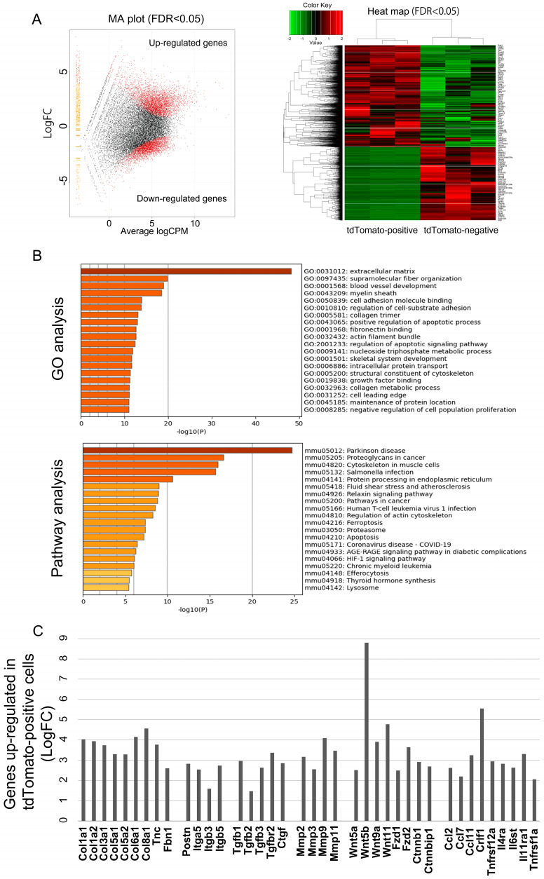
Pancreatic ductal adenocarcinoma (PDAC) characterized by an abundant cancer stroma is an aggressive malignancy with a poor prognosis. Periostin (Pn) is a key extracellular matrix (ECM) protein in various tumor progression. Previously, we described the role of Pn alternative splicing variants (ASVs) with specific functional features in breast cancer. Pn is known to associate with a chemoresistance of PDAC, but the functions of the Pn-ASVs remain largely unknown. In this study, we focused on physiological and pathological Pn-ASVs, and examined the characteristics of Pn-expressing cells and the difference in function of each ASV. We found that cancer-associated fibroblasts (CAFs) are a main source of Pn synthesis, which selectively secrete pathological Pn-ASVs with exon 21 both in mouse and human samples. RNA sequencing identified a gene signature of Pn-positive CAFs associated with ECM-related genes and chemokines, factors that shape the chemoresistance tumor microenvironment (TME). Additionally, only pathological Pn-ASVs interacted with heat shock protein 70-1a (HSP70-1a), leading to significant rescue of gemcitabine-induced PDAC apoptosis. In silico analysis revealed that the presence or absence of exon 21 changes the tertiary structure of Pn and the binding sites for HSP70-1a. Altogether, Pn-ASVs with exon 21 secreted from CAFs play a key role in supporting tumor growth by interacting with cancer cell-derived HSP70-1a, indicating that Pn-ASVs with exon 21 might be a potential therapeutic and diagnostic target in PDAC patients with rich stroma.

Keywords: alternative splicing variants; extracellular matrix protein; pancreatic cancers; periostin.


Conflict of interest statement

R. Morishita received honoraria, consulting fees and funds from Novartis, Takeda, Shionogi, Astellas, Boehringer Ingelheim, Daiichi-Sankyo and Pfizer. F. Sanada, Y. Taniyama and K. Shibata are members of Periotherapia Co., Ltd.

Figures

Figure 1
Figure 1
The expression of total Pn is elevated in PDAC. (A) Representative Pn immunohistochemical staining of normal pancreas and PDAC. S: stroma, C: cancer cells. (B) % of Pn-positive area in PDAC (n = 20) and normal pancreas (n = 4). (C) Average % of Pn-positive area in PDAC and normal pancreas. * p < 0.05 vs. normal pancreas.
Figure 2
Figure 2
Pn-ASVs expression in PDAC and CAFs. (A) Human Pn-ASV structures. In addition to four major variants, Pn 1, Pn 2-1, Pn 3 and Pn 4-1, four other isoforms have been reported. Red arrows indicate the position of primers for Pn-ASV detection. The EMI domain, the four FAS-1 domains and the N- and C-terminal end of the carboxyl-terminal domain are depicted. (B) Pn-ASVs mRNA expression in PDAC cell lines (Panc 1, AsPC 1 and BxPC 3) and CAFs (hPSC5 and hPSC14). N = 4, * p < 0.05 vs. Panc 1, AsPC 1 and BxPC 3, ** p < 0.05 vs. Panc 1, AsPC 1, BxPC 3 and hPSC5. p < 0.05 vs. Pn 1, Pn 3 and Pn 4, p < 0.05 vs. Pn 1 and Pn 2-1 and Pn 3. (C) Total Pn protein secreted from PDAC cell lines (Panc 1, AsPC 1 and BxPC 3) and CAFs (hPSC5 and hPSC14). (D) Pn-ASVs protein secreted from CAF. Pn-ASV proteins immunoprecipitated from a culture supernatant of CAFs using the specific antibodies against Pn exon 1, 17 and 21 were analyzed by Western blotting with Pn antibody for exon 12.
Figure 3
Figure 3
Restricted distribution of Pn-ASVs with exon 21 and sequencing analysis of Pn-positive cells. (A) A representative in situ hybridization image of total Pn (RNAscope) and Pn-ASVs with exon 21 (Basescope) in PDAC specimen. The red color indicates a positive signal for Pn mRNA. Both total Pn and Pn-ASVs with exon 21 mRNA was expressed in fibroblasts surrounding cancer. S: stroma, C: cancer cells. Bar indicates 100 μm. (B) Pn expression and prognosis in patients with PDAC. Pn expression and PDAC prognostic analysis was examined using the Kaplan–Meier plotter. It was found that the prognosis was worse in the group with high Pn expression. (C) Single-cell RNA-sequence analysis in different cell types show that the fibroblast and smooth muscle cell clusters are the ones that show high Pn expression. (D) Violin plot for Pn expression in different cell types in PDAC on the data from the Single Cell Portal.
Figure 4
Figure 4
PDAC tumors activate Pn expression in CAFs. (A) KPC mice-derived YFP+ PDAC cells were subcutaneously implanted in the back of 8-week-old male C57BL6 mice and sacrificed on day 35. (B) YFP-positive and negative cells isolation by fluorescence-activated cell sorting (FACS). (C) Total Pn (amplicon: exon 9–10) and Pn-ASV with exon 21 (amplicon: exon 21–22) expression was higher in YFP-negative cells containing CAFs. N = 4, * p < 0.05 vs. pre-transplantation cancer cell and YFP+ cell. (D) YFP+ PDAC cells were subcutaneously implanted into the back of 8-week-old male Postn-tdTomato mice and sacrificed on day 35. Tamoxifen was administered intraperitoneally 5 days prior to sacrifice. (E) Tdtomato, a Pn-positive signal, was identified in the stroma of the excised tumor. TFP-positive ODAC cells were shown in green. White bar indicates 100 μm. (F) CAFs were isolated from excised tumors by FACS using GFP-negative and CD90-positive sorting. Tdtomato was further used to divide the CAFs to Pn-positive and negative groups. (G) Tdtomato-positive CAFs had higher expression of total Pn (exon 9–10 amplicon) and exon 21-containing ASVs (exon 21–22 amplicon). N = 3, * p < 0.05 vs. CD90+ Tdtomato-CAFs.
Figure 5
Figure 5
The transcriptome of Pn-positive (CD90+ tdTomato+) and negative (CD90+ tdTomato-) CAFs. RNA sequence analysis was performed using Pn-positive and Pn-negative CAFs isolated from a mouse PDAC syngeneic model. RNA-seq analysis comparing Pn-positive and Pn-negative CAFs. (A) RNA-seq analysis showing a volcano plot and heat map of differentially expressed genes (DEGs) in Pn-positive and Pn-negative CAFs. The data represent three biological replicates. It revealed 7624 differentially expressed genes (DEGs, red dots in MA plot) when comparing Pn-positive CAFs from Pn-negative CAFs (FDR < 0.05). Among 7624 DEGs, 4418 were up-regulated, while 3206 were up-regulated in Pn-positive CAFs. (B) Gene ontology enrichment and KEGG analysis of up-regulated genes in the Pn-positive CAFs from the RNA-seq data. (C) Representative genes whose expression was increased in the Pn-positive CAFs group. Up-regulated genes in Pn-positive CAFs include several genes related to cancer progression and chemoresistance. Data are the log2 of fold change (LogFC). Relative expression pattern analysis of up-regulated genes in Pn-positive CAFs by qRT-PCR analysis to validate the RNA-seq data is shown in Supplementary Figure S4.
Figure 6
Figure 6
Pn-ASVs with exon 21 interacts with HSP70 and promotes gemcitabine resistance in pancreatic cancer. (A) Pull-down solutions were electrophoresed on gels and silver stained. Protein analysis of bands that appeared specifically in the pull-down solution of Pn2-1 was performed (red arrows). (B) Amino acid sequence of HSP70 was detected by LC-MS/MS analysis of gel bands analysis. Western blotting with primary antibody for HSP70 in post-pull-down solution detected a strong band in lane Pn2-1 but not Pn4-1. (C) HSPA1A and HSPA8 expression in PDAC cells and CAFs. HSP70 was higher in PDAC cell lines as compared to CAFs. N = 4, * p < 0.05 vs. hPSC 5, ** p < 0.05 vs. BxPC 3. (D) The 3D predicted steric structures of Pn2-1 and Pn4-1, and predicted binding sites with HSPA1A are shown. Colored blue to red from n-terminus to c-terminus. (E) GEM significantly reducing proliferation of PDAC and CAFs cell lines. N = 6, * p < 0.05 vs. 0 μM. (F) Pn-ASV with exon 21 suppresses GEM-induced PDAC cell line cell death. However, knockdown of HSPA1A prevents the rescue benefit. N = 12, * p < 0.05 vs. CTRL siRNA.

References

    1. Rawla P., Sunkara T., Gaduputi V. Epidemiology of Pancreatic Cancer: Global Trends, Etiology and Risk Factors. World J. Oncol. 2019;10:10–27. doi: 10.14740/wjon1166. - DOI - PMC - PubMed
    1. Park W., Chawla A., O’Reilly E.M. Pancreatic Cancer. A Review. JAMA. 2021;326:851–862. doi: 10.1001/jama.2021.13027. - DOI - PMC - PubMed
    1. Perez V.M., Kearney J.F., Yeh J.J. The PDAC Extracellular Matrix: A Review of the ECM Protein Composition, Tumor Cell Interaction, and Therapeutic Strategies. Front. Oncol. 2021;11:751311. doi: 10.3389/fonc.2021.751311. - DOI - PMC - PubMed
    1. Geng X., Chen H., Zhao L., Hu J., Yang W., Li G., Cheng C., Zhao Z., Zhang T., Li L., et al. Cancer-Associated Fibroblast (CAF) Heterogeneity and Targeting Therapy of CAFs in Pancreatic Cancer. Front. Cell Dev. Biol. 2021;9:655152. doi: 10.3389/fcell.2021.655152. - DOI - PMC - PubMed
    1. Neuzillet C., Nicolle R., Raffenne J., Tijeras-Raballand A., Brunel A., Astorgues-Xerri L., Vacher S., Arbateraz F., Fanjul M., Hilmi M., et al. Periostin- and podoplanin-positive cancer-associated fibroblast subtypes cooperate to shape the inflamed tumor microenvironment in aggressive pancreatic adenocarcinoma. J. Pathol. 2022;258:408–425. doi: 10.1002/path.6011. - DOI - PMC - PubMed

MeSH terms

LinkOut - more resources